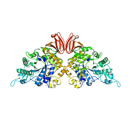
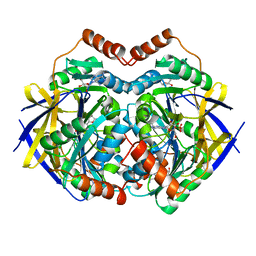
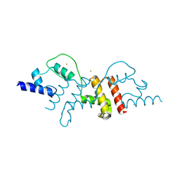
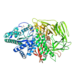
3B5B
 
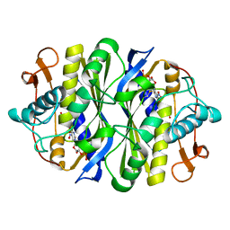 | | Crystal structure of the thymidylate synthase k48q | | 分子名称: | 2'-DEOXY-5-NITROURIDINE 5'-(DIHYDROGEN PHOSPHATE), 2'-DEOXY-5-NITROURIDINE 5'-MONOPHOSPHATE, FORMIC ACID, ... | | 著者 | Sotelo-Mundo, R.R, Arreola, R, Maley, F, Montfort, W.R. | | 登録日 | 2007-10-25 | | 公開日 | 2007-12-25 | | 最終更新日 | 2023-08-30 | | 実験手法 | X-RAY DIFFRACTION (2.7 Å) | | 主引用文献 | Role of an invariant lysine residue in folate binding on Escherichia coli thymidylate synthase: Calorimetric and crystallographic analysis of the K48Q mutant.
Int.J.Biochem.Cell Biol., 40, 2008
|
|
2ZHY
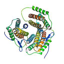 | | Crystal structure of a pduO-type ATP:cobalamin adenosyltransferase from Burkholderia thailandensis | | 分子名称: | ATP:cob(I)alamin adenosyltransferase, putative | | 著者 | Moon, J.H, Park, A.K, Jang, E.H, Kim, H.S, Chi, Y.M. | | 登録日 | 2008-02-11 | | 公開日 | 2008-07-29 | | 最終更新日 | 2024-03-13 | | 実験手法 | X-RAY DIFFRACTION (1.8 Å) | | 主引用文献 | Crystal structure of a PduO-type ATP:cobalamin adenosyltransferase from Burkholderia thailandensis.
Proteins, 72, 2008
|
|
2H1G
 | | ResA C74A/C77A | | 分子名称: | Thiol-disulfide oxidoreductase resA | | 著者 | Lewin, A, Crow, A, Oubrie, A, Le Brun, N.E. | | 登録日 | 2006-05-16 | | 公開日 | 2006-09-19 | | 最終更新日 | 2023-08-30 | | 実験手法 | X-RAY DIFFRACTION (3.1 Å) | | 主引用文献 | Molecular Basis for Specificity of the Extracytoplasmic Thioredoxin ResA.
J.Biol.Chem., 281, 2006
|
|
2H2S
 | |
2ZUV
 | | Crystal structure of Galacto-N-biose/Lacto-N-biose I phosphorylase in complex with GlcNAc, Ethylene glycol, and nitrate | | 分子名称: | 1,2-ETHANEDIOL, 2-acetamido-2-deoxy-alpha-D-glucopyranose, Lacto-N-biose phosphorylase, ... | | 著者 | Hidaka, M, Nishimoto, M, Kitaoka, M, Wakagi, T, Shoun, H, Fushinobu, S. | | 登録日 | 2008-10-28 | | 公開日 | 2008-12-30 | | 最終更新日 | 2024-04-03 | | 実験手法 | X-RAY DIFFRACTION (1.85 Å) | | 主引用文献 | The crystal structure of galacto-N-biose/lacto-N-biose I phosphorylase: A large deformation of a tim barrel scaffold
J.Biol.Chem., 284, 2009
|
|
2GXB
 | |
2GYY
 | |
2H74
 | |
2H7F
 | | Structure of variola topoisomerase covalently bound to DNA | | 分子名称: | 5'-D(*TP*AP*AP*TP*AP*AP*GP*GP*GP*CP*GP*AP*CP*A)-3', 5'-D(*TP*TP*GP*TP*CP*GP*CP*CP*CP*TP*T)-3', DNA topoisomerase 1 | | 著者 | Perry, K, Hwang, Y, Bushman, F.D, Van Duyne, G.D. | | 登録日 | 2006-06-02 | | 公開日 | 2006-08-15 | | 最終更新日 | 2021-10-20 | | 実験手法 | X-RAY DIFFRACTION (2.7 Å) | | 主引用文献 | Structural basis for specificity in the poxvirus topoisomerase.
Mol.Cell, 23, 2006
|
|
2H1T
 | |
4TVU
 | | Crystal structure of trehalose synthase from Deinococcus radiodurans reveals a closed conformation for catalysis of the intramolecular isomerization | | 分子名称: | 2-AMINO-2-HYDROXYMETHYL-PROPANE-1,3-DIOL, CALCIUM ION, MAGNESIUM ION, ... | | 著者 | Wang, Y.L, Chow, S.Y, Lin, Y.T, Liaw, S.H. | | 登録日 | 2014-06-28 | | 公開日 | 2014-12-17 | | 最終更新日 | 2023-11-08 | | 実験手法 | X-RAY DIFFRACTION (2.7 Å) | | 主引用文献 | Structures of trehalose synthase from Deinococcus radiodurans reveal that a closed conformation is involved in catalysis of the intramolecular isomerization.
Acta Crystallogr.,Sect.D, 70, 2014
|
|
2ZC2
 | | Crystal structure of DnaD-like replication protein from Streptococcus mutans UA159, gi 24377835, residues 127-199 | | 分子名称: | DnaD-like replication protein, ZINC ION | | 著者 | Duke, N.E.C, Clancy, S, Duggan, E, Joachimiak, A, Midwest Center for Structural Genomics (MCSG) | | 登録日 | 2007-11-02 | | 公開日 | 2007-12-25 | | 最終更新日 | 2017-10-11 | | 実験手法 | X-RAY DIFFRACTION (2.1 Å) | | 主引用文献 | Crystal Structure of DnaD-like replication protein from Streptococcus mutans UA159.
To be Published
|
|
2HBX
 | | Crystal Structure of alpha-Amino-beta-Carboxymuconate-epsilon-Semialdehyde-Decarboxylase (ACMSD) | | 分子名称: | 2-amino-3-carboxymuconate 6-semialdehyde decarboxylase, COBALT (II) ION | | 著者 | Martynowski, D, Eyobo, Y, Li, T, Yang, K, Liu, A, Zhang, H. | | 登録日 | 2006-06-14 | | 公開日 | 2006-09-19 | | 最終更新日 | 2024-02-14 | | 実験手法 | X-RAY DIFFRACTION (2.5 Å) | | 主引用文献 | Crystal Structure of alpha-Amino-beta-carboxymuconate-epsilon-semialdehyde Decarboxylase: Insight into the Active Site and Catalytic Mechanism of a Novel Decarboxylation Reaction.
Biochemistry, 45, 2006
|
|
2GJL
 | | Crystal Structure of 2-nitropropane dioxygenase | | 分子名称: | FLAVIN MONONUCLEOTIDE, hypothetical protein PA1024 | | 著者 | Suh, S.W. | | 登録日 | 2006-03-31 | | 公開日 | 2006-05-16 | | 最終更新日 | 2024-03-13 | | 実験手法 | X-RAY DIFFRACTION (2 Å) | | 主引用文献 | Crystal Structure of 2-Nitropropane Dioxygenase Complexed with FMN and Substrate: identification of the catalytic base
J.Biol.Chem., 281, 2006
|
|
2ZUS
 | | Crystal structure of Galacto-N-biose/Lacto-N-biose I phosphorylase | | 分子名称: | Lacto-N-biose phosphorylase, MAGNESIUM ION | | 著者 | Hidaka, M, Nishimoto, M, Kitaoka, M, Wakagi, T, Shoun, H, Fushinobu, S. | | 登録日 | 2008-10-28 | | 公開日 | 2008-12-30 | | 最終更新日 | 2024-03-13 | | 実験手法 | X-RAY DIFFRACTION (2.11 Å) | | 主引用文献 | The crystal structure of galacto-N-biose/lacto-N-biose I phosphorylase: A large deformation of a tim barrel scaffold
J.Biol.Chem., 284, 2009
|
|
2GL0
 | | Structure of PAE2307 in complex with adenosine | | 分子名称: | ADENOSINE, PHOSPHATE ION, conserved hypothetical protein | | 著者 | Lott, J.S, Paget, B, Johnston, J.M, Baker, E.N. | | 登録日 | 2006-04-04 | | 公開日 | 2006-06-06 | | 最終更新日 | 2023-08-30 | | 実験手法 | X-RAY DIFFRACTION (2.25 Å) | | 主引用文献 | The Structure of an Ancient Conserved Domain Establishes a Structural Basis for Stable Histidine Phosphorylation and Identifies a New Family of Adenosine-specific Kinases.
J.Biol.Chem., 281, 2006
|
|
2GIY
 | |
2GM7
 | | TenA Homolog/Thi-4 Thiaminase from Pyrobaculum Aerophilum | | 分子名称: | 2-{2-[2-(2-{2-[2-(2-ETHOXY-ETHOXY)-ETHOXY]-ETHOXY}-ETHOXY)-ETHOXY]-ETHOXY}-ETHANOL, GLYCEROL, PHOSPHATE ION, ... | | 著者 | Sawaya, M.R, Chan, S, Han, G.W, Perry, L.J. | | 登録日 | 2006-04-06 | | 公開日 | 2006-04-18 | | 最終更新日 | 2023-08-30 | | 実験手法 | X-RAY DIFFRACTION (2.8 Å) | | 主引用文献 | Crystal Structure of a Ten A Homolog/Thi-4 Thiaminase from Pyrobaculum Aerophilum
To be Published
|
|
2GMR
 | | Photosynthetic reaction center mutant from Rhodobacter sphaeroides with Asp L210 replaced with Asn | | 分子名称: | BACTERIOCHLOROPHYLL A, BACTERIOPHEOPHYTIN A, FE (II) ION, ... | | 著者 | Stachnik, J.M, Hermes, S, Gerwert, K, Hofmann, E. | | 登録日 | 2006-04-07 | | 公開日 | 2006-11-21 | | 最終更新日 | 2023-08-30 | | 実験手法 | X-RAY DIFFRACTION (2.5 Å) | | 主引用文献 | Proton uptake in the reaction center mutant L210DN from Rhodobacter sphaeroides via protonated water molecules.
Biochemistry, 45, 2006
|
|
2Z7J
 | | Structural insights into de multifunctional VP3 protein of birnaviruses:gold derivative | | 分子名称: | Capsid assembly protein VP3, GOLD ION | | 著者 | Casanas, A, Navarro, A, Ferrer-Orta, C, Gonzalez, D, Rodriguez, J.F, Verdaguer, N. | | 登録日 | 2007-08-23 | | 公開日 | 2008-02-05 | | 最終更新日 | 2024-02-21 | | 実験手法 | X-RAY DIFFRACTION (2.4 Å) | | 主引用文献 | Structural insights into the multifunctional protein VP3 of birnaviruses.
Structure, 16, 2008
|
|
3BXW
 | |
3BON
 | |
3C6Q
 | |
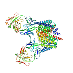
3BUB
 | | Golgi alpha-mannosidase II with an empty active site | | 分子名称: | (4S)-2-METHYL-2,4-PENTANEDIOL, 2-acetamido-2-deoxy-beta-D-glucopyranose, Alpha-mannosidase 2, ... | | 著者 | Kuntz, D.A, Rose, D.R. | | 登録日 | 2008-01-02 | | 公開日 | 2008-07-01 | | 最終更新日 | 2023-08-30 | | 実験手法 | X-RAY DIFFRACTION (1.38 Å) | | 主引用文献 | Probing the substrate specificity of Golgi alpha-mannosidase II by use of synthetic oligosaccharides and a catalytic nucleophile mutant.
J.Am.Chem.Soc., 130, 2008
|
|
3BKS
 | |


