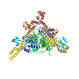
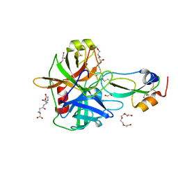
4IV8
 
 | |
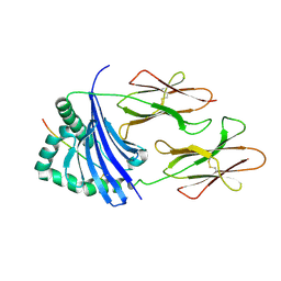
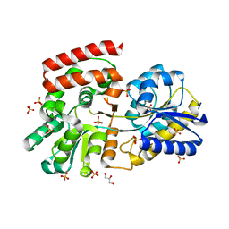
3FC0
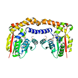
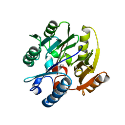
 | | 1.8 A crystal structure of murine GITR ligand dimer expressed in Drosophila melanogaster S2 cells | | 分子名称: | ACETATE ION, GITR ligand | | 著者 | Chattopadhyay, K, Ramagopal, U.A, Nathenson, S.G, Almo, S.C. | | 登録日 | 2008-11-20 | | 公開日 | 2008-12-30 | | 最終更新日 | 2023-09-06 | | 実験手法 | X-RAY DIFFRACTION (1.76 Å) | | 主引用文献 | 1.8 A structure of murine GITR ligand dimer expressed in Drosophila melanogaster S2 cells.
Acta Crystallogr.,Sect.D, 65, 2009
|
|
4L8U
 | | X-ray study of human serum albumin complexed with 9 amino camptothecin | | 分子名称: | (2S)-2-[1-amino-8-(hydroxymethyl)-9-oxo-9,11-dihydroindolizino[1,2-b]quinolin-7-yl]-2-hydroxybutanoic acid, MYRISTIC ACID, Serum albumin | | 著者 | Wang, Z, Ho, J.X, Ruble, J, Rose, J.P, Carter, D.C. | | 登録日 | 2013-06-17 | | 公開日 | 2013-07-24 | | 最終更新日 | 2017-11-15 | | 実験手法 | X-RAY DIFFRACTION (2.01 Å) | | 主引用文献 | Structural studies of several clinically important oncology drugs in complex with human serum albumin.
Biochim.Biophys.Acta, 1830, 2013
|
|
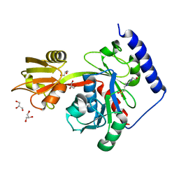
3OT7
 | | Escherichia coli apo-manganese superoxide dismutase | | 分子名称: | Superoxide dismutase [Mn] | | 著者 | Whittaker, M.M, Lerch, T.F, Kirillova, O, Chapman, M.S, Whittaker, J.W. | | 登録日 | 2010-09-10 | | 公開日 | 2010-12-22 | | 最終更新日 | 2023-09-06 | | 実験手法 | X-RAY DIFFRACTION (1.901 Å) | | 主引用文献 | Subunit dissociation and metal binding by Escherichia coli apo-manganese superoxide dismutase.
Arch.Biochem.Biophys., 505, 2011
|
|
3FF6
 | | Human ACC2 CT domain with CP-640186 | | 分子名称: | (3R)-1'-(9-ANTHRYLCARBONYL)-3-(MORPHOLIN-4-YLCARBONYL)-1,4'-BIPIPERIDINE, Acetyl-CoA carboxylase 2 | | 著者 | Williams, S.P, Madauss, K.P, Burkhart, W.A. | | 登録日 | 2008-12-02 | | 公開日 | 2009-05-19 | | 最終更新日 | 2024-04-03 | | 実験手法 | X-RAY DIFFRACTION (3.19 Å) | | 主引用文献 | The human ACC2 CT-domain C-terminus is required for full functionality and has a novel twist.
Acta Crystallogr.,Sect.D, 65, 2009
|
|
4LA0
 | | X-ray study of human serum albumin complexed with bicalutamide | | 分子名称: | R-BICALUTAMIDE, SERUM ALBUMIN | | 著者 | Wang, Z, Ho, J.X, Ruble, J, Rose, J.P, Carter, D.C. | | 登録日 | 2013-06-18 | | 公開日 | 2013-07-24 | | 最終更新日 | 2014-02-19 | | 実験手法 | X-RAY DIFFRACTION (2.4 Å) | | 主引用文献 | Structural studies of several clinically important oncology drugs in complex with human serum albumin.
Biochim.Biophys.Acta, 1830, 2013
|
|
4IX1
 | | Crystal structure of hypothetical protein OPAG_01669 from Rhodococcus Opacus PD630, Target 016205 | | 分子名称: | PHOSPHATE ION, hypothetical protein | | 著者 | Malashkevich, V.N, Bhosle, R, Toro, R, Hillerich, B, Gizzi, A, Garforth, S, Kar, A, Chan, M.K, Lafluer, J, Patel, H, Matikainen, B, Chamala, S, Lim, S, Celikgil, A, Villegas, G, Evans, B, Zenchek, W, Love, J, Fiser, A, Khafizov, K, Seidel, R, Bonanno, J.B, Almo, S.C, New York Structural Genomics Research Consortium (NYSGRC) | | 登録日 | 2013-01-24 | | 公開日 | 2013-02-06 | | 最終更新日 | 2013-05-08 | | 実験手法 | X-RAY DIFFRACTION (2.8 Å) | | 主引用文献 | Crystal structure of hypothetical protein OPAG_01669 from Rhodococcus Opacus PD630, Target 016205
To be Published
|
|
4LAK
 | | Crystal structure of Cordyceps militaris IDCase D323N mutant in apo form | | 分子名称: | Uracil-5-carboxylate decarboxylase, ZINC ION | | 著者 | Xu, S, Li, W, Zhu, J, Wang, R, Li, Z, Xu, G.L, Ding, J. | | 登録日 | 2013-06-20 | | 公開日 | 2013-10-02 | | 最終更新日 | 2023-11-08 | | 実験手法 | X-RAY DIFFRACTION (2.41 Å) | | 主引用文献 | Crystal structures of isoorotate decarboxylases reveal a novel catalytic mechanism of 5-carboxyl-uracil decarboxylation and shed light on the search for DNA decarboxylase.
Cell Res., 23, 2013
|
|
3P8P
 | | Crystal Structure of Human Dimethylarginine Dimethylaminohydrolase-1 (DDAH-1) variant C274S bound with N5-(1-iminopentyl)-L-ornithine | | 分子名称: | N(G),N(G)-dimethylarginine dimethylaminohydrolase 1, N~5~-[(1E)-pentanimidoyl]-L-ornithine | | 著者 | Monzingo, A.F, Lluis, M, Wang, Y, Fast, W, Robertus, J.D. | | 登録日 | 2010-10-14 | | 公開日 | 2010-11-10 | | 最終更新日 | 2023-09-06 | | 実験手法 | X-RAY DIFFRACTION (2.5 Å) | | 主引用文献 | Characterization of C-Alkyl Amidines as Bioavailable Covalent Reversible Inhibitors of Human DDAH-1.
Chemmedchem, 6, 2011
|
|
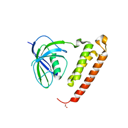
4IYL
 | | 30S ribosomal protein S15 from Campylobacter jejuni | | 分子名称: | 30S ribosomal protein S15 | | 著者 | Osipiuk, J, Nocek, B, Gu, M, Kwon, K, Anderson, W.F, Joachimiak, A, Center for Structural Genomics of Infectious Diseases (CSGID) | | 登録日 | 2013-01-28 | | 公開日 | 2013-02-06 | | 最終更新日 | 2023-09-20 | | 実験手法 | X-RAY DIFFRACTION (2.36 Å) | | 主引用文献 | 30S ribosomal protein S15 from Campylobacter jejuni
To be Published
|
|
4LB4
 | | Crystal structure of human AR complexed with NADP+ and {2-[(4-bromo-2,3,5,6-tetrafluorobenzyl)carbamoyl]-5-chlorophenoxy}acetic acid | | 分子名称: | Aldose reductase, NADP NICOTINAMIDE-ADENINE-DINUCLEOTIDE PHOSPHATE, {2-[(4-bromo-2,3,5,6-tetrafluorobenzyl)carbamoyl]-5-chlorophenoxy}acetic acid | | 著者 | Cousido-Siah, A, Mitschler, A, Ruiz, F.X, Fanfrlik, J, Kolar, M, Hobza, P, Podjarny, A. | | 登録日 | 2013-06-20 | | 公開日 | 2014-04-30 | | 最終更新日 | 2023-09-20 | | 実験手法 | X-RAY DIFFRACTION (0.8 Å) | | 主引用文献 | Modulation of aldose reductase inhibition by halogen bond tuning.
Acs Chem.Biol., 8, 2013
|
|
4IZ9
 | |
4ISL
 | | Crystal Structure of the inactive Matriptase in complex with its inhibitor HAI-1 | | 分子名称: | GLUTATHIONE, GLYCEROL, Kunitz-type protease inhibitor 1, ... | | 著者 | Huang, M.D, Zhao, B.Y, Yuan, C, Li, R. | | 登録日 | 2013-01-16 | | 公開日 | 2013-03-06 | | 最終更新日 | 2023-09-20 | | 実験手法 | X-RAY DIFFRACTION (2.29 Å) | | 主引用文献 | Crystal structures of matriptase in complex with its inhibitor hepatocyte growth factor activator inhibitor-1.
J.Biol.Chem., 288, 2013
|
|
4IS6
 | | Crystal structure of HLA-DR4 bound to GP100 peptide | | 分子名称: | HLA class II histocompatibility antigen, DR alpha chain, DRB1-4 beta chain, ... | | 著者 | Li, Y. | | 登録日 | 2013-01-16 | | 公開日 | 2013-10-23 | | 最終更新日 | 2013-11-20 | | 実験手法 | X-RAY DIFFRACTION (2.5 Å) | | 主引用文献 | Structure-Based Design of Altered MHC Class II-Restricted Peptide Ligands with Heterogeneous Immunogenicity.
J.Immunol., 191, 2013
|
|
4ITD
 | | Structures of DNA duplexes containing O6-carboxymethylguanine, a lesion associated with gastrointestinal cancer, reveal a mechanism for inducing transition mutation | | 分子名称: | 2'-(4-HYDROXYPHENYL)-5-(4-METHYL-1-PIPERAZINYL)-2,5'-BI-BENZIMIDAZOLE, DNA (5'-D(*CP*GP*CP*GP*(C6G)P*AP*TP*TP*CP*GP*CP*G)-3'), MAGNESIUM ION | | 著者 | Zhang, F, Suzuki, K, Tsunoda, M, Wilkinson, O, Millington, C.L, Williams, D.M, Morishita, E.C, Takenaka, A. | | 登録日 | 2013-01-18 | | 公開日 | 2013-05-08 | | 最終更新日 | 2024-03-20 | | 実験手法 | X-RAY DIFFRACTION (1.94 Å) | | 主引用文献 | Structures of DNA duplexes containing O6-carboxymethylguanine, a lesion associated with gastrointestinal cancer, reveal a mechanism for inducing pyrimidine transition mutations
Nucleic Acids Res., 41, 2013
|
|
4LFB
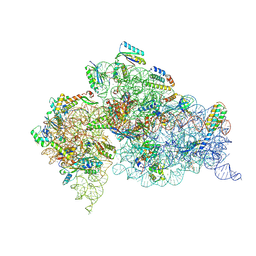 | | Crystal Structure of 30S ribosomal subunit from Thermus thermophilus | | 分子名称: | 16S rRNA, MAGNESIUM ION, NEOMYCIN, ... | | 著者 | Demirci, H, Belardinelli, R, Carr, J, Murphy IV, F, Jogl, G, Dahlberg, A.E, Gregory, S.T. | | 登録日 | 2013-06-26 | | 公開日 | 2014-07-02 | | 実験手法 | X-RAY DIFFRACTION (3.009 Å) | | 主引用文献 | Crystal Structure of 30S ribosomal subunit from Thermus thermophilus
to be published, 2013
|
|
4IT4
 | |
3OOA
 | |
4ITN
 | | Crystal structure of "compact P-loop" LpxK from Aquifex aeolicus in complex with chloride at 2.2 angstrom resolution | | 分子名称: | 4-(2-HYDROXYETHYL)-1-PIPERAZINE ETHANESULFONIC ACID, CHLORIDE ION, GLYCEROL, ... | | 著者 | Emptage, R.P, Pemble IV, C.W, York, J.D, Raetz, C.R.H, Zhou, P. | | 登録日 | 2013-01-18 | | 公開日 | 2013-04-03 | | 最終更新日 | 2023-09-20 | | 実験手法 | X-RAY DIFFRACTION (2.1912 Å) | | 主引用文献 | Mechanistic Characterization of the Tetraacyldisaccharide-1-phosphate 4'-Kinase LpxK Involved in Lipid A Biosynthesis.
Biochemistry, 52, 2013
|
|
3FO8
 | | Crystal structure of the bacteriophage T4 tail sheath protein, protease resistant fragment gp18PR | | 分子名称: | ACETATE ION, Tail sheath protein Gp18 | | 著者 | Aksyuk, A.A, Leiman, P.G, Kurochkina, L.P, Shneider, M.M, Kostyuchenko, V.A, Mesyanzhinov, V.V, Rossmann, M.G. | | 登録日 | 2008-12-29 | | 公開日 | 2009-03-10 | | 最終更新日 | 2024-02-21 | | 実験手法 | X-RAY DIFFRACTION (1.8 Å) | | 主引用文献 | The tail sheath structure of bacteriophage T4: a molecular machine for infecting bacteria.
Embo J., 28, 2009
|
|
4IUZ
 | | High resolution crystal structure of racemic ester insulin | | 分子名称: | DI(HYDROXYETHYL)ETHER, GLYCEROL, Insulin A chain, ... | | 著者 | Avital-Shmilovici, M, Mandal, K, Gates, Z.P, Phillips, N, Weiss, M.A, Kent, S.B.H. | | 登録日 | 2013-01-22 | | 公開日 | 2013-02-13 | | 最終更新日 | 2023-09-20 | | 実験手法 | X-RAY DIFFRACTION (1.6 Å) | | 主引用文献 | Fully Convergent Chemical Synthesis of Ester Insulin: Determination of the High Resolution X-ray Structure by Racemic Protein Crystallography.
J.Am.Chem.Soc., 135, 2013
|
|
4LKS
 | | Structure of CBM32-3 from a family 31 glycoside hydrolase from Clostridium perfringens in complex with galactose | | 分子名称: | CALCIUM ION, Glycosyl hydrolase, family 31/fibronectin type III domain protein, ... | | 著者 | Grondin, J.M, Duan, D, Kirlin, A.C, Furness, H.S, Allingham, J.S, Smith, S.P. | | 登録日 | 2013-07-08 | | 公開日 | 2014-12-24 | | 最終更新日 | 2023-09-20 | | 実験手法 | X-RAY DIFFRACTION (1.5 Å) | | 主引用文献 | Diverse modes of galacto-specific carbohydrate recognition by a family 31 glycoside hydrolase from Clostridium perfringens.
Plos One, 12, 2017
|
|
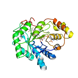
3FQ0
 | | Staphylococcus aureus dihydrofolate reductase complexed with NADPH and 2,4-diamino-5-(3-(2,5-dimethoxyphenyl)prop-1-ynyl)-6-ethylpyrimidine (UCP120B) | | 分子名称: | 5-[3-(2,5-dimethoxyphenyl)prop-1-yn-1-yl]-6-ethylpyrimidine-2,4-diamine, NADPH DIHYDRO-NICOTINAMIDE-ADENINE-DINUCLEOTIDE PHOSPHATE, Trimethoprim-sensitive dihydrofolate reductase | | 著者 | Anderson, A.C, Frey, K.M, Liu, J, Lombardo, M.N. | | 登録日 | 2009-01-06 | | 公開日 | 2009-03-24 | | 最終更新日 | 2024-04-03 | | 実験手法 | X-RAY DIFFRACTION (1.69 Å) | | 主引用文献 | Crystal structures of wild-type and mutant methicillin-resistant Staphylococcus aureus dihydrofolate reductase reveal an alternate conformation of NADPH that may be linked to trimethoprim resistance.
J.Mol.Biol., 387, 2009
|
|
3EXS
 | | Crystal structure of KGPDC from Streptococcus mutans in complex with D-R5P | | 分子名称: | RIBULOSE-5-PHOSPHATE, RmpD (Hexulose-6-phosphate synthase) | | 著者 | Li, G.L, Liu, X, Wang, K.T, Li, L.F, Su, X.D. | | 登録日 | 2008-10-17 | | 公開日 | 2009-08-25 | | 最終更新日 | 2023-11-01 | | 実験手法 | X-RAY DIFFRACTION (2.5 Å) | | 主引用文献 | Open-closed conformational change revealed by the crystal structures of 3-keto-L-gulonate 6-phosphate decarboxylase from Streptococcus mutans
Biochem.Biophys.Res.Commun., 381, 2009
|
|
3OTM
 | |


