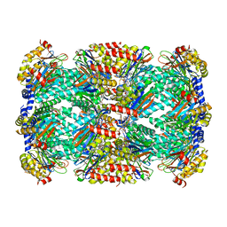
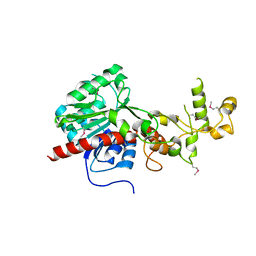
1FJX
 
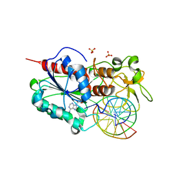 | | STRUCTURE OF TERNARY COMPLEX OF HHAI METHYLTRANSFERASE MUTANT (T250G) IN COMPLEX WITH DNA AND ADOHCY | | 分子名称: | DNA (5'-D(*TP*GP*AP*TP*AP*GP*CP*GP*CP*TP*AP*TP*C)-3'), HHAI DNA METHYLTRANSFERASE, S-ADENOSYL-L-HOMOCYSTEINE, ... | | 著者 | Vilkaitis, G, Dong, A, Weinhold, E, Cheng, X, Klimasauskas, S. | | 登録日 | 2000-08-08 | | 公開日 | 2000-12-15 | | 最終更新日 | 2024-02-07 | | 実験手法 | X-RAY DIFFRACTION (2.26 Å) | | 主引用文献 | Functional roles of the conserved threonine 250 in the target recognition domain of HhaI DNA methyltransferase.
J.Biol.Chem., 275, 2000
|
|
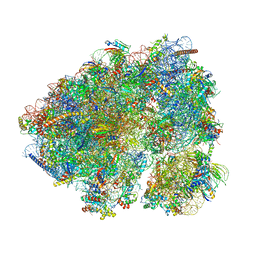
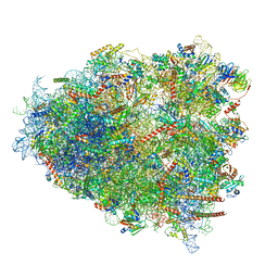
8JDK
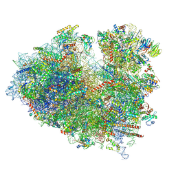 | | Structure of the Human cytoplasmic Ribosome with human tRNA Asp(ManQ34) and mRNA(GAU) | | 分子名称: | 18S ribosomal RNA, 28S ribosomal RNA, 40S ribosomal protein S10, ... | | 著者 | Ishiguro, K, Yokoyama, T, Shirouzu, M, Suzuki, T. | | 登録日 | 2023-05-14 | | 公開日 | 2023-12-06 | | 最終更新日 | 2023-12-27 | | 実験手法 | ELECTRON MICROSCOPY (2.26 Å) | | 主引用文献 | Glycosylated queuosines in tRNAs optimize translational rate and post-embryonic growth.
Cell, 186, 2023
|
|
7RK8
 | |
7A8F
 | |
4W6U
 | | Crystal Structure of Full-Length Split GFP Mutant E115H/T118H With Nickel Mediated Crystal Contacts, P 21 21 21 Space Group | | 分子名称: | 1,2-ETHANEDIOL, CITRIC ACID, NICKEL (II) ION, ... | | 著者 | Leibly, D.J, Waldo, G.S, Yeates, T.O. | | 登録日 | 2014-08-20 | | 公開日 | 2015-02-18 | | 最終更新日 | 2023-11-15 | | 実験手法 | X-RAY DIFFRACTION (2.28 Å) | | 主引用文献 | A Suite of Engineered GFP Molecules for Oligomeric Scaffolding.
Structure, 23, 2015
|
|
6WNK
 | | Macrocyclic peptides TDI5575 that selectively inhibit the Mycobacterium tuberculosis proteasome | | 分子名称: | (12S,15S)-N-[(2-fluorophenyl)methyl]-10,13-dioxo-12-{2-oxo-2-[(2R)-2-phenylpyrrolidin-1-yl]ethyl}-2-oxa-11,14-diazatricyclo[15.2.2.1~3,7~]docosa-1(19),3(22),4,6,17,20-hexaene-15-carboxamide, CITRIC ACID, DIMETHYLFORMAMIDE, ... | | 著者 | Hsu, H.C, Li, H. | | 登録日 | 2020-04-22 | | 公開日 | 2021-04-28 | | 最終更新日 | 2023-10-18 | | 実験手法 | X-RAY DIFFRACTION (2.28 Å) | | 主引用文献 | Macrocyclic Peptides that Selectively Inhibit the Mycobacterium tuberculosis Proteasome.
J.Med.Chem., 64, 2021
|
|
8CGN
 | | Non-rotated 80S S. cerevisiae ribosome with ligands | | 分子名称: | 18S ribosomal RNA, 25S ribosomal RNA, 40S ribosomal protein S0-A, ... | | 著者 | Milicevic, N, Jenner, L, Myasnikov, A, Yusupov, M, Yusupova, G. | | 登録日 | 2023-02-06 | | 公開日 | 2023-09-20 | | 最終更新日 | 2024-04-24 | | 実験手法 | ELECTRON MICROSCOPY (2.28 Å) | | 主引用文献 | mRNA reading frame maintenance during eukaryotic ribosome translocation.
Nature, 625, 2024
|
|
7JPI
 | | Crystal structure of EBOV glycoprotein with modified HR2 stalk at 2.3A resolution | | 分子名称: | 2-acetamido-2-deoxy-beta-D-glucopyranose, 2-acetamido-2-deoxy-beta-D-glucopyranose-(1-4)-2-acetamido-2-deoxy-beta-D-glucopyranose, Envelope glycoprotein, ... | | 著者 | Chaudhary, A, Stanfield, R.L, Wilson, I.A, Zhu, J. | | 登録日 | 2020-08-08 | | 公開日 | 2021-04-07 | | 最終更新日 | 2023-10-18 | | 実験手法 | X-RAY DIFFRACTION (2.28 Å) | | 主引用文献 | Single-component multilayered self-assembling nanoparticles presenting rationally designed glycoprotein trimers as Ebola virus vaccines.
Nat Commun, 12, 2021
|
|
6MRN
 | | Crystal Structure of ChlaDUB2 DUB domain | | 分子名称: | Deubiquitinase and deneddylase Dub2 | | 著者 | Hausman, J.M, Das, C. | | 登録日 | 2018-10-15 | | 公開日 | 2019-10-30 | | 最終更新日 | 2023-10-11 | | 実験手法 | X-RAY DIFFRACTION (2.29 Å) | | 主引用文献 | The Two Deubiquitinating Enzymes fromChlamydia trachomatisHave Distinct Ubiquitin Recognition Properties.
Biochemistry, 59, 2020
|
|
3LX6
 | |
8G5Y
 | | mRNA decoding in human is kinetically and structurally distinct from bacteria (IC state) | | 分子名称: | 1,4-DIAMINOBUTANE, 18S rRNA, 28S rRNA, ... | | 著者 | Holm, M, Natchiar, K.S, Rundlet, E.J, Myasnikov, A.G, Altman, R.B, Blanchard, S.C. | | 登録日 | 2023-02-14 | | 公開日 | 2023-04-19 | | 最終更新日 | 2023-11-15 | | 実験手法 | ELECTRON MICROSCOPY (2.29 Å) | | 主引用文献 | mRNA decoding in human is kinetically and structurally distinct from bacteria.
Nature, 617, 2023
|
|
2V4M
 | | The isomerase domain of human glutamine-fructose-6-phosphate transaminase 1 (GFPT1) in complex with fructose 6-phosphate | | 分子名称: | CHLORIDE ION, FRUCTOSE -6-PHOSPHATE, GLUCOSAMINE--FRUCTOSE-6-PHOSPHATE AMINOTRANSFERASE [ISOMERIZING] 1 | | 著者 | Moche, M, Lehtio, L, Andersson, J, Arrowsmith, C.H, Berglund, H, Collins, R, Dahlgren, L.G, Edwards, A.M, Flodin, S, Flores, A, Graslund, S, Hammarstrom, M, Johansson, A, Johansson, I, Karlberg, T, Kotenyova, T, Nilsson, M.E, Nyman, T, Persson, C, Sagemark, J, Svensson, S, Schueler, H, Thorsell, A.G, Tresaugues, L, Uppenberg, J, Van Den Berg, S, Welin, M, Wisniewska, M, Weigelt, J, Nordlund, P, Wikstrom, M. | | 登録日 | 2008-09-26 | | 公開日 | 2008-10-07 | | 最終更新日 | 2023-12-13 | | 実験手法 | X-RAY DIFFRACTION (2.29 Å) | | 主引用文献 | The Isomerase Domain of Human Gfpt1 in Complex with Fructose 6-Phosphate
To be Published
|
|
5HJQ
 | | Crystal structure of the TBC domain of Skywalker/TBC1D24 from Drosophila melanogaster in complex with inositol(1,4,5)triphosphate | | 分子名称: | D-MYO-INOSITOL-1,4,5-TRIPHOSPHATE, LD10117p | | 著者 | Fischer, B, Paesmans, J, Versees, W. | | 登録日 | 2016-01-13 | | 公開日 | 2016-09-21 | | 最終更新日 | 2024-01-10 | | 実験手法 | X-RAY DIFFRACTION (2.3 Å) | | 主引用文献 | Skywalker-TBC1D24 has a lipid-binding pocket mutated in epilepsy and required for synaptic function.
Nat.Struct.Mol.Biol., 23, 2016
|
|
5E8H
 | | Crystal structure of geranylfarnesyl pyrophosphate synthases 2 from Arabidopsis thaliana | | 分子名称: | Geranylgeranyl pyrophosphate synthase 3, chloroplastic | | 著者 | Wang, C, Chen, Q, Wang, G, Zhang, P. | | 登録日 | 2015-10-14 | | 公開日 | 2015-11-11 | | 最終更新日 | 2019-12-25 | | 実験手法 | X-RAY DIFFRACTION (2.3 Å) | | 主引用文献 | Structural Analyses of Short-Chain Prenyltransferases Identify an Evolutionarily Conserved GFPPS Clade in Brassicaceae Plants.
Mol Plant, 9, 2016
|
|
5IH6
 | | Human Casein Kinase 1 isoform delta (kinase domain) in complex with Epiblastin A derivative | | 分子名称: | 6-(3-bromophenyl)pteridine-2,4,7-triamine, Casein kinase I isoform delta, S,R MESO-TARTARIC ACID, ... | | 著者 | Ursu, A, Illich, D.J, Takemoto, Y, Porfetye, A.T, Zhang, M, Brockmeyer, A, Janning, P, Watanabe, N, Osada, H, Vetter, I.R, Ziegler, S, Schoeler, H.R, Waldmann, H. | | 登録日 | 2016-02-29 | | 公開日 | 2016-04-13 | | 最終更新日 | 2024-01-10 | | 実験手法 | X-RAY DIFFRACTION (2.3 Å) | | 主引用文献 | Epiblastin A Induces Reprogramming of Epiblast Stem Cells Into Embryonic Stem Cells by Inhibition of Casein Kinase 1.
Cell Chem Biol, 23, 2016
|
|
4PPJ
 | | Crystal structure of Phanta, a weakly fluorescent photochromic GFP-like protein. ON state | | 分子名称: | Monomeric Azami Green | | 著者 | Don Paul, C, Traore, D.A.K, Devenish, R.J, Close, D, Bell, T, Bradbury, A, Wilce, M.C.J, Prescott, M. | | 登録日 | 2014-02-27 | | 公開日 | 2015-04-08 | | 最終更新日 | 2024-04-03 | | 実験手法 | X-RAY DIFFRACTION (2.3 Å) | | 主引用文献 | X-Ray Crystal Structure and Properties of Phanta, a Weakly Fluorescent Photochromic GFP-Like Protein.
Plos One, 10, 2015
|
|
6Y1G
 | | Photoconverted HcRed in its optoacoustic state | | 分子名称: | 1,2-ETHANEDIOL, GFP-like non-fluorescent chromoprotein | | 著者 | Janowski, R, Fuenzalida-Werner, J.P, Mishra, K, Stiel, A.C, Niessing, D. | | 登録日 | 2020-02-12 | | 公開日 | 2020-07-29 | | 最終更新日 | 2024-01-24 | | 実験手法 | X-RAY DIFFRACTION (2.3 Å) | | 主引用文献 | Challenging a Preconception: Optoacoustic Spectrum Differs from the Optical Absorption Spectrum of Proteins and Dyes for Molecular Imaging.
Anal.Chem., 92, 2020
|
|
1EMM
 | |
1EML
 | |
5AB7
 | |
2DD9
 | | A mutant of GFP-like protein from Chiridius poppei | | 分子名称: | 3-CYCLOHEXYL-1-PROPYLSULFONIC ACID, CHLORIDE ION, green fluorescent protein | | 著者 | Suto, K, Masuda, H, Takenaka, Y, Mizuno, H. | | 登録日 | 2006-01-24 | | 公開日 | 2007-01-23 | | 最終更新日 | 2021-11-10 | | 実験手法 | X-RAY DIFFRACTION (2.3 Å) | | 主引用文献 | Structural basis for red-shifted emission of a GFP-like protein from the marine copepod Chiridius poppei
Genes Cells, 14, 2009
|
|
1EMC
 | |
6AA2
 | | X-ray structure of ReQy1 (oxidized form) | | 分子名称: | Green fluorescent protein | | 著者 | Sugiura, K, Yasuda, A, Tabushi, N, Tanaka, H, Kurisu, G, Hisabori, T. | | 登録日 | 2018-07-17 | | 公開日 | 2019-05-29 | | 最終更新日 | 2023-11-22 | | 実験手法 | X-RAY DIFFRACTION (2.3 Å) | | 主引用文献 | Multicolor redox sensor proteins can visualize redox changes in various compartments of the living cell.
Biochim Biophys Acta Gen Subj, 1863, 2019
|
|
2Z6X
 | | Crystal structure of 22G, the wild-type protein of the photoswitchable GFP-like protein Dronpa | | 分子名称: | photochromic protein Dronpa | | 著者 | Kikuchi, A, Jeyakanthan, J, Taka, J, Shiro, Y, Mizuno, H, Miyawaki, A. | | 登録日 | 2007-08-09 | | 公開日 | 2008-07-22 | | 最終更新日 | 2023-11-15 | | 実験手法 | X-RAY DIFFRACTION (2.3 Å) | | 主引用文献 | Light-dependent regulation of structural flexibility in a photochromic fluorescent protein.
Proc.Natl.Acad.Sci.Usa, 105, 2008
|
|
4GPB
 | |


