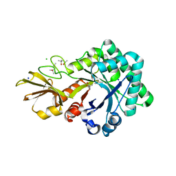
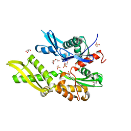
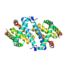
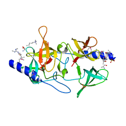
1O6Y
 
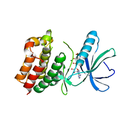 | | Catalytic domain of PknB kinase from Mycobacterium tuberculosis | | 分子名称: | MAGNESIUM ION, PHOSPHOMETHYLPHOSPHONIC ACID ADENYLATE ESTER, SERINE/THREONINE-PROTEIN KINASE PKNB | | 著者 | Ortiz-Lombardia, M, Pompeo, F, Boitel, B, Alzari, P.M, TB Structural Genomics Consortium (TBSGC) | | 登録日 | 2002-10-21 | | 公開日 | 2003-01-30 | | 最終更新日 | 2023-12-13 | | 実験手法 | X-RAY DIFFRACTION (2.2 Å) | | 主引用文献 | Crystal Structure of the Catalytic Domain of the Pknb Serine/Threonine Kinase from Mycobacterium Tuberculosis
J.Biol.Chem., 278, 2003
|
|
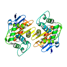
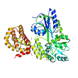
3LQS
 | | Complex Structure of D-Amino Acid Aminotransferase and 4-amino-4,5-dihydro-thiophenecarboxylic acid (ADTA) | | 分子名称: | 4-[({3-HYDROXY-2-METHYL-5-[(PHOSPHONOOXY)METHYL]PYRIDIN-4-YL}METHYL)AMINO]THIOPHENE-2-CARBOXYLIC ACID, ACETIC ACID, D-alanine aminotransferase | | 著者 | Lepore, B.W, Liu, D, Peng, Y, Fu, M, Yasuda, C, Manning, J.M, Silverman, R.B, Ringe, D. | | 登録日 | 2010-02-10 | | 公開日 | 2010-03-16 | | 最終更新日 | 2024-02-21 | | 実験手法 | X-RAY DIFFRACTION (1.9 Å) | | 主引用文献 | Chiral discrimination among aminotransferases: inactivation by 4-amino-4,5-dihydrothiophenecarboxylic acid.
Biochemistry, 49, 2010
|
|
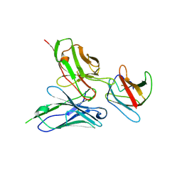
1NPI
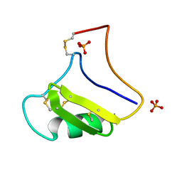 | | Tityus Serrulatus Neurotoxin (Ts1) at atomic resolution | | 分子名称: | PHOSPHATE ION, Toxin VII | | 著者 | Pinheiro, C.B, Marangoni, S, Toyama, M.H, Polikarpov, I. | | 登録日 | 2003-01-17 | | 公開日 | 2003-02-25 | | 最終更新日 | 2023-08-16 | | 実験手法 | X-RAY DIFFRACTION (1.16 Å) | | 主引用文献 | Structural analysis of Tityus serrulatus Ts1 neurotoxin at atomic resolution: insights into interactions with Na+ channels.
Acta Crystallogr.,Sect.D, 59, 2003
|
|
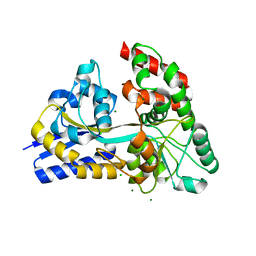
5OG0
 | | Crystal structure of human Alanine:Glyoxylate Aminotransferase major allele (AGT-Ma) at 2.5 Angstrom; internal aldimine with PLP in the active site | | 分子名称: | PYRIDOXAL-5'-PHOSPHATE, Serine--pyruvate aminotransferase | | 著者 | Giardina, G, Cutruzzola, F, Borri Voltattorni, C, Cellini, B, Montioli, R. | | 登録日 | 2017-07-11 | | 公開日 | 2017-09-27 | | 最終更新日 | 2024-01-17 | | 実験手法 | X-RAY DIFFRACTION (2.5 Å) | | 主引用文献 | Radiation damage at the active site of human alanine:glyoxylate aminotransferase reveals that the cofactor position is finely tuned during catalysis.
Sci Rep, 7, 2017
|
|
4UV6
 | |
4URI
 | | Crystal structure of chitinase-like agglutinin RobpsCRA from Robinia pseudoacacia | | 分子名称: | (4S)-2-METHYL-2,4-PENTANEDIOL, CHITINASE-RELATED AGGLUTININ, CHLORIDE ION, ... | | 著者 | Sulzenbacher, G, Roig-Zamboni, V, Peumans, W.J, Henrissat, B, van Damme, E.J.M, Bourne, Y. | | 登録日 | 2014-06-30 | | 公開日 | 2015-03-11 | | 最終更新日 | 2024-01-10 | | 実験手法 | X-RAY DIFFRACTION (1.85 Å) | | 主引用文献 | Structural Basis for Carbohydrate Binding Properties of a Plant Chitinase-Like Agglutinin with Conserved Catalytic Machinery.
J.Struct.Biol., 190, 2015
|
|
5OJI
 | | Crystal structure of the dehydrogenase/reductase SDR family member 4 (DHRS4) from Caenorhabditis elegans | | 分子名称: | Dehydrogenase/reductase SDR family member 4, ISATIN, NADP NICOTINAMIDE-ADENINE-DINUCLEOTIDE PHOSPHATE | | 著者 | Scheidig, A.J, Faust, A, Ebert, B, Maser, E, Kisiela, M. | | 登録日 | 2017-07-21 | | 公開日 | 2017-11-22 | | 最終更新日 | 2024-01-17 | | 実験手法 | X-RAY DIFFRACTION (1.6 Å) | | 主引用文献 | Crystal structure and catalytic characterization of the dehydrogenase/reductase SDR family member 4 (DHRS4) from Caenorhabditis elegans.
FEBS J., 285, 2018
|
|
5O6S
 | |
2RO5
 | | RDC-refined solution structure of the N-terminal DNA recognition domain of the Bacillus subtilis transition-state regulator SpoVT | | 分子名称: | Stage V sporulation protein T | | 著者 | Sullivan, D.M, Bobay, B.G, Kojetin, D.J, Thompson, R.J, Rance, M, Strauch, M.A, Cavanagh, J. | | 登録日 | 2008-03-08 | | 公開日 | 2008-11-11 | | 最終更新日 | 2024-05-01 | | 実験手法 | SOLUTION NMR | | 主引用文献 | Insights into the nature of DNA binding of AbrB-like transcription factors
Structure, 16, 2008
|
|
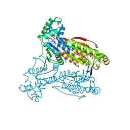
5OBX
 | | Mycoplasma genitalium DnaK-NBD | | 分子名称: | Chaperone protein DnaK, GLYCEROL, SULFATE ION, ... | | 著者 | Adell, M, Calisto, B, Fita, I, Martinelli, L. | | 登録日 | 2017-06-29 | | 公開日 | 2018-05-09 | | 最終更新日 | 2024-01-17 | | 実験手法 | X-RAY DIFFRACTION (1.78 Å) | | 主引用文献 | The nucleotide-bound/substrate-bound conformation of the Mycoplasma genitalium DnaK chaperone.
Protein Sci., 27, 2018
|
|
5OE0
 | | CRYSTAL STRUCTURE OF THE BETA-LACTAMASE OXA-181 | | 分子名称: | Beta-lactamase, CHLORIDE ION, SULFATE ION | | 著者 | Lund, B.A, Carlsen, T.J.O, Leiros, H.K.S, Thomassen, A.M. | | 登録日 | 2017-07-07 | | 公開日 | 2017-08-02 | | 最終更新日 | 2024-01-17 | | 実験手法 | X-RAY DIFFRACTION (2.0500083 Å) | | 主引用文献 | Structure, activity and thermostability investigations of OXA-163, OXA-181 and OXA-245 using biochemical analysis, crystal structures and differential scanning calorimetry analysis.
Acta Crystallogr F Struct Biol Commun, 73, 2017
|
|
2RKJ
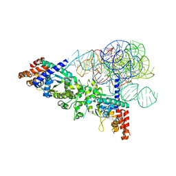 | | Cocrystal structure of a tyrosyl-tRNA synthetase splicing factor with a group I intron RNA | | 分子名称: | RNA (238-MER), RNA (5'-R(P*GP*CP*UP*U)-3'), Tyrosyl-tRNA synthetase | | 著者 | Paukstelis, P.J, Chen, J.-H, Chase, E, Lambowitz, A.M, Golden, B.L. | | 登録日 | 2007-10-16 | | 公開日 | 2008-01-08 | | 最終更新日 | 2023-08-30 | | 実験手法 | X-RAY DIFFRACTION (4.5 Å) | | 主引用文献 | Structure of a tyrosyl-tRNA synthetase splicing factor bound to a group I intron RNA.
Nature, 451, 2008
|
|
5A77
 | | Crystal structure of the homing endonuclease I-CvuI in complex with I- CreI target (C1221) in the presence of 2 mM Mg revealing DNA cleaved | | 分子名称: | 10MER DNA, 5'-D(*GP*AP*CP*GP*TP*TP*TP*TP* GP*AP*DGP*AP*CP*GP*TP*TP*TP*TP*GP*A)-3', 14MER DNA, ... | | 著者 | Molina, R, Redondo, P, LopezMendez, B, Villate, M, Merino, N, Blanco, F.J, Valton, J, Grizot, S, Duchateau, P, Prieto, J, Montoya, G. | | 登録日 | 2015-07-03 | | 公開日 | 2015-09-23 | | 最終更新日 | 2024-01-10 | | 実験手法 | X-RAY DIFFRACTION (2.5 Å) | | 主引用文献 | Crystal Structure of the Homing Endonuclease I-Cvui Provides a New Template for Genome Modification
J.Biol.Chem., 290, 2015
|
|
5AJK
 | |
4WGI
 | | A Single Diastereomer of a Macrolactam Core Binds Specifically to Myeloid Cell Leukemia 1 (MCL1) | | 分子名称: | (2S)-2-[(2S,3R)-10-{[(4-fluorophenyl)sulfonyl]amino}-3-methyl-2-[(methyl{[4-(trifluoromethyl)phenyl]carbamoyl}amino)methyl]-6-oxo-3,4-dihydro-2H-1,5-benzoxazocin-5(6H)-yl]propanoic acid, FORMIC ACID, MAGNESIUM ION, ... | | 著者 | Clifton, M.C, Fairman, J.W, Fang, C, D'Souza, B, Fulroth, B, Leed, A, McCarren, P, Wang, L, Wang, Y, Kaushik, V, Palmer, M, Wei, G, Golub, T.R, Hubbard, B.K, Serrano-Wu, M.H. | | 登録日 | 2014-09-18 | | 公開日 | 2014-11-19 | | 最終更新日 | 2023-09-27 | | 実験手法 | X-RAY DIFFRACTION (1.85 Å) | | 主引用文献 | Single Diastereomer of a Macrolactam Core Binds Specifically to Myeloid Cell Leukemia 1 (MCL1).
Acs Med.Chem.Lett., 5, 2014
|
|
5AAW
 | | Structure of a redesigned cross-reactive antibody to dengue virus with increased in vivo potency | | 分子名称: | DENGUE SEROTYPE 4 ENVELOPE PROTEIN DOMAIN 3, SCFV513 | | 著者 | Wong, Y, Robinson, L, Lescar, J, Sasisekharan, R. | | 登録日 | 2015-07-30 | | 公開日 | 2015-09-30 | | 最終更新日 | 2024-01-10 | | 実験手法 | X-RAY DIFFRACTION (3.27 Å) | | 主引用文献 | Structure-Guided Design of an Anti-Dengue Antibody Directed to a Non-Immunodominant Epitope.
Cell(Cambridge,Mass.), 162, 2015
|
|
1OCU
 | | Crystal structure of the yeast PX-domain protein Grd19p (sorting nexin 3) complexed to phosphatidylinosytol-3-phosphate. | | 分子名称: | 2-(BUTANOYLOXY)-1-{[(HYDROXY{[2,3,4,6-TETRAHYDROXY-5-(PHOSPHONOOXY)CYCLOHEXYL]OXY}PHOSPHORYL)OXY]METHYL}ETHYL BUTANOATE, SORTING NEXIN | | 著者 | Zhou, C.Z, Li de La Sierra-Gallay, I, Cheruel, S, Collinet, B, Minard, P, Blondeau, K, Henkes, G, Aufrere, R, Leulliot, N, Graille, M, Sorel, I, Savarin, P, de la Torre, F, Poupon, A, Janin, J, van Tilbeurgh, H. | | 登録日 | 2003-02-10 | | 公開日 | 2003-12-12 | | 最終更新日 | 2023-12-13 | | 実験手法 | X-RAY DIFFRACTION (2.3 Å) | | 主引用文献 | Crystal structure of the yeast Phox homology (PX) domain protein Grd19p complexed to phosphatidylinositol-3-phosphate.
J. Biol. Chem., 278, 2003
|
|
4ZZE
 | | Raffinose and panose binding protein from Bifidobacterium animalis subsp. lactis Bl-04, bound with panose | | 分子名称: | CHLORIDE ION, MAGNESIUM ION, Sugar binding protein of ABC transporter system, ... | | 著者 | Fredslund, F, Ejby, M, Andersen, J.M, Slotboom, D.J, Hachem, M.A. | | 登録日 | 2015-05-22 | | 公開日 | 2016-06-29 | | 最終更新日 | 2024-01-10 | | 実験手法 | X-RAY DIFFRACTION (1.76 Å) | | 主引用文献 | An ATP Binding Cassette Transporter Mediates the Uptake of alpha-(1,6)-Linked Dietary Oligosaccharides in Bifidobacterium and Correlates with Competitive Growth on These Substrates.
J. Biol. Chem., 291, 2016
|
|
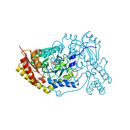
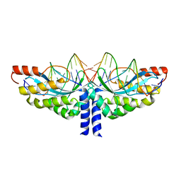
1NP3
 | | Crystal structure of class I acetohydroxy acid isomeroreductase from Pseudomonas aeruginosa | | 分子名称: | Ketol-acid reductoisomerase | | 著者 | Ahn, H.J, Eom, S.J, Yoon, H.-J, Lee, B.I, Cho, H, Suh, S.W. | | 登録日 | 2003-01-17 | | 公開日 | 2003-05-06 | | 最終更新日 | 2024-03-13 | | 実験手法 | X-RAY DIFFRACTION (2 Å) | | 主引用文献 | Crystal Structure of Class I Acetohydroxy Acid Isomeroreductase from Pseudomonas aeruginosa
J.Mol.Biol., 328, 2003
|
|
7ZJ5
 | | Unbound state of a brocolli-pepper aptamer FRET tile. | | 分子名称: | POTASSIUM ION, brocolli-pepper aptamer | | 著者 | McRae, E.K.S, Vallina, N.S, Hansen, B.K, Boussebayle, A, Andersen, E.S. | | 登録日 | 2022-04-08 | | 公開日 | 2023-04-19 | | 最終更新日 | 2024-07-24 | | 実験手法 | ELECTRON MICROSCOPY (4.55 Å) | | 主引用文献 | Structure determination of Pepper-Broccoli FRET pair by RNA origami scaffolding
To Be Published
|
|
1NUF
 | |
3MEV
 | | Crystal structure of SGF29 in complex with R2AK4me3 | | 分子名称: | GLYCEROL, Histone H3, SAGA-associated factor 29 homolog, ... | | 著者 | Bian, C.B, Xu, C, Lam, R, Bountra, C, Arrowsmith, C.H, Weigelt, J, Edwards, A.M, Bochkarev, A, Min, J, Structural Genomics Consortium (SGC) | | 登録日 | 2010-03-31 | | 公開日 | 2010-04-28 | | 最終更新日 | 2021-10-06 | | 実験手法 | X-RAY DIFFRACTION (1.83 Å) | | 主引用文献 | Sgf29 binds histone H3K4me2/3 and is required for SAGA complex recruitment and histone H3 acetylation.
Embo J., 30, 2011
|
|
3MEU
 | | Crystal structure of SGF29 in complex with H3R2me2sK4me3 | | 分子名称: | Histone H3, SAGA-associated factor 29 homolog, SULFATE ION | | 著者 | Bian, C.B, Xu, C, Bountra, C, Arrowsmith, C.H, Weigelt, J, Edwards, A.M, Bochkarev, A, Min, J, Structural Genomics Consortium (SGC) | | 登録日 | 2010-03-31 | | 公開日 | 2010-04-28 | | 最終更新日 | 2023-11-22 | | 実験手法 | X-RAY DIFFRACTION (1.28 Å) | | 主引用文献 | Sgf29 binds histone H3K4me2/3 and is required for SAGA complex recruitment and histone H3 acetylation.
Embo J., 30, 2011
|
|
5AJV
 | | Human PFKFB3 in complex with an indole inhibitor 1 | | 分子名称: | (2S)-2-amino-N-[4-[(2-amino-3-cyano-1H-indol-5-yl)oxy]phenyl]-3-hydroxy-propanamide, 2,6-di-O-phosphono-beta-D-fructofuranose, HUMAN PFKFB3, ... | | 著者 | Boyd, S, Brookfield, J.L, Critchlow, S.E, Cumming, I.A, Curtis, N.J, Debreczeni, J.E, Degorce, S.L, Donald, C, Evans, N.J, Groombridge, S, Hopcroft, P, Jones, N.P, Kettle, J.G, Lamont, S, Lewis, H.J, MacFaull, P, McLoughlin, S.B, Rigoreau, L.J.M, Smith, J.M, St-Gallay, S, Stock, J.K, Wheatley, E.R, Winter, J, Wingfield, J. | | 登録日 | 2015-02-27 | | 公開日 | 2015-04-22 | | 最終更新日 | 2024-05-08 | | 実験手法 | X-RAY DIFFRACTION (3.01 Å) | | 主引用文献 | Structure-Based Design of Potent and Selective Inhibitors of the Metabolic Kinase Pfkfb3.
J.Med.Chem., 58, 2015
|
|
5A6S
 | | Crystal structure of the CTP1L endolysin reveals how its activity is regulated by a secondary translation product | | 分子名称: | ENDOLYSIN, GLYCEROL, PENTAETHYLENE GLYCOL, ... | | 著者 | Dunne, M, Leicht, S, Krichel, B, Mertens, H.D.T, Thompson, A, Krijgsveld, J, Svergun, D.I, GomezTorres, N, Garde, S, Uetrecht, C, Narbad, A, Mayer, M.J, Meijers, R. | | 登録日 | 2015-07-01 | | 公開日 | 2015-12-30 | | 最終更新日 | 2024-01-10 | | 実験手法 | X-RAY DIFFRACTION (1.9 Å) | | 主引用文献 | Crystal Structure of the Ctp1L Endolysin Reveals How its Activity is Regulated by a Secondary Translation Product.
J.Biol.Chem., 291, 2016
|
|


