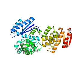
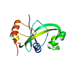
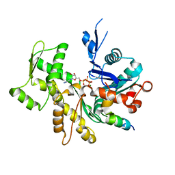
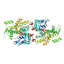
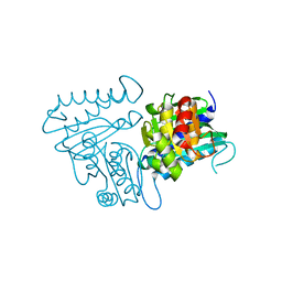
7KTY
 
 | | Data clustering and dynamics of chymotrypsinogen average structure | | 分子名称: | Chymotrypsinogen A, SULFATE ION | | 著者 | Nguyen, T, Phan, K.L, Kreitler, D.F, Andrews, L.C, Gabelli, S.B, Kozakov, D, Jakoncic, J, Shi, W, Sweet, R.M, Soares, A.S, Bernstein, H.J. | | 登録日 | 2020-11-24 | | 公開日 | 2021-03-10 | | 最終更新日 | 2023-10-18 | | 実験手法 | X-RAY DIFFRACTION (2 Å) | | 主引用文献 | A simple technique to classify diffraction data from dynamic proteins according to individual polymorphs.
Acta Crystallogr D Struct Biol, 78, 2022
|
|
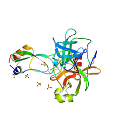
8GTY
 | |
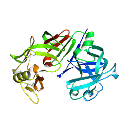
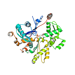
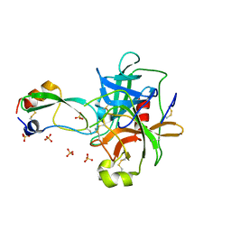
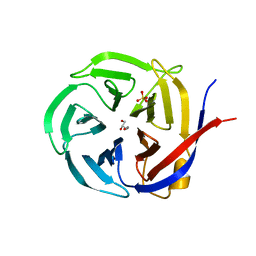
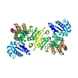
3VGC
 | | GAMMA-CHYMOTRYPSIN L-NAPHTHYL-1-ACETAMIDO BORONIC ACID ACID INHIBITOR COMPLEX | | 分子名称: | GAMMA CHYMOTRYPSIN, L-1-NAPHTHYL-2-ACETAMIDO-ETHANE BORONIC ACID, SULFATE ION | | 著者 | Stoll, V.S, Eger, B.T, Hynes, R.C, Martichonok, V, Jones, J.B, Pai, E.F. | | 登録日 | 1997-05-01 | | 公開日 | 1997-11-12 | | 最終更新日 | 2023-08-09 | | 実験手法 | X-RAY DIFFRACTION (1.67 Å) | | 主引用文献 | Differences in binding modes of enantiomers of 1-acetamido boronic acid based protease inhibitors: crystal structures of gamma-chymotrypsin and subtilisin Carlsberg complexes.
Biochemistry, 37, 1998
|
|
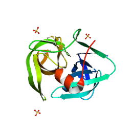
5EUJ
 | | PYRUVATE DECARBOXYLASE FROM ZYMOBACTER PALMAE | | 分子名称: | 1,2-ETHANEDIOL, MAGNESIUM ION, Pyruvate decarboxylase, ... | | 著者 | Buddrus, L, Crennell, S.J, Leak, D.J, Danson, M.J, Andrews, E.S.V, Arcus, V.L. | | 登録日 | 2015-11-18 | | 公開日 | 2016-09-14 | | 最終更新日 | 2024-01-10 | | 実験手法 | X-RAY DIFFRACTION (2.15 Å) | | 主引用文献 | Crystal structure of pyruvate decarboxylase from Zymobacter palmae.
Acta Crystallogr.,Sect.F, 72, 2016
|
|
5GUS
 | | Crystal structure of ASCH domain from Zymomonas mobilis | | 分子名称: | 3,6,9,12,15,18,21-HEPTAOXATRICOSANE-1,23-DIOL, CHLORIDE ION, Helix-turn-helix domain-containing protein, ... | | 著者 | Ha, S.C, Park, S.Y, Kim, J.S. | | 登録日 | 2016-08-31 | | 公開日 | 2017-08-30 | | 最終更新日 | 2024-03-20 | | 実験手法 | X-RAY DIFFRACTION (1.951 Å) | | 主引用文献 | Crystal structure of an ASCH protein from Zymomonas mobilis and its ribonuclease activity specific for single-stranded RNA.
Sci Rep, 7, 2017
|
|
5GUQ
 | |
4CMS
 | | X-RAY ANALYSES OF ASPARTIC PROTEINASES IV. STRUCTURE AND REFINEMENT AT 2.2 ANGSTROMS RESOLUTION OF BOVINE CHYMOSIN | | 分子名称: | CHYMOSIN B | | 著者 | Newman, M, Frazao, C, Khan, G, Tickle, I.J, Blundell, T.L, Safro, M, Andreeva, N, Zdanov, A. | | 登録日 | 1991-11-01 | | 公開日 | 1991-11-07 | | 最終更新日 | 2017-11-29 | | 実験手法 | X-RAY DIFFRACTION (2.2 Å) | | 主引用文献 | X-ray analyses of aspartic proteinases. IV. Structure and refinement at 2.2 A resolution of bovine chymosin.
J.Mol.Biol., 221, 1991
|
|
2GCT
 | |
3U9D
 | | Crystal Structure of a chimera containing the N-terminal domain (residues 8-24) of drosophila Ciboulot and the C-terminal domain (residues 13-44) of bovine Thymosin-beta4, bound to G-actin-ATP | | 分子名称: | ADENOSINE-5'-TRIPHOSPHATE, Actin, alpha skeletal muscle, ... | | 著者 | Renault, L, Husson, C, Carlier, M.F, Didry, D. | | 登録日 | 2011-10-18 | | 公開日 | 2012-01-25 | | 最終更新日 | 2023-09-13 | | 実験手法 | X-RAY DIFFRACTION (2.5 Å) | | 主引用文献 | How a single residue in individual beta-thymosin/WH2 domains controls their functions in actin assembly.
Embo J., 31, 2012
|
|
2CGA
 | |
4H4F
 | | Crystal structure of human chymotrypsin C (CTRC) bound to inhibitor eglin c from Hirudo medicinalis | | 分子名称: | Chymotrypsin-C, Eglin C, PHOSPHATE ION | | 著者 | Batra, J, Soares, A.S, Radisky, E.S. | | 登録日 | 2012-09-17 | | 公開日 | 2013-02-27 | | 最終更新日 | 2013-04-24 | | 実験手法 | X-RAY DIFFRACTION (1.9 Å) | | 主引用文献 | Long-range Electrostatic Complementarity Governs Substrate Recognition by Human Chymotrypsin C, a Key Regulator of Digestive Enzyme Activation.
J.Biol.Chem., 288, 2013
|
|
3SJH
 | | Crystal Structure of a chimera containing the N-terminal domain (residues 8-29) of drosophila Ciboulot and the C-terminal domain (residues 18-44) of bovine Thymosin-beta4, bound to G-actin-ATP-Latrunculin A | | 分子名称: | ADENOSINE-5'-TRIPHOSPHATE, Actin, alpha skeletal muscle, ... | | 著者 | Renault, L, Husson, C, Carlier, M.F, Didry, D. | | 登録日 | 2011-06-21 | | 公開日 | 2012-01-25 | | 最終更新日 | 2023-09-13 | | 実験手法 | X-RAY DIFFRACTION (1.75 Å) | | 主引用文献 | How a single residue in individual beta-thymosin/WH2 domains controls their functions in actin assembly
Embo J., 31, 2012
|
|
3U8X
 | | Crystal Structure of a chimera containing the N-terminal domain (residues 8-29) of drosophila Ciboulot and the C-terminal domain (residues 18-44) of bovine Thymosin-beta4, bound to G-actin-ATP | | 分子名称: | ADENOSINE-5'-TRIPHOSPHATE, Actin, alpha skeletal muscle, ... | | 著者 | Renault, L, Husson, C, Carlier, M.F, Didry, D. | | 登録日 | 2011-10-17 | | 公開日 | 2012-01-25 | | 最終更新日 | 2023-09-13 | | 実験手法 | X-RAY DIFFRACTION (2 Å) | | 主引用文献 | How a single residue in individual beta-thymosin/WH2 domains controls their functions in actin assembly
Embo J., 31, 2012
|
|
1MTN
 | | BOVINE ALPHA-CHYMOTRYPSIN:BPTI CRYSTALLIZATION | | 分子名称: | ALPHA-CHYMOTRYPSIN, BASIC PANCREATIC TRYPSIN INHIBITOR, SULFATE ION | | 著者 | Capasso, C, Rizzi, M, Menegatti, E, Ascenzi, P, Bolognesi, M. | | 登録日 | 1996-03-28 | | 公開日 | 1996-08-17 | | 最終更新日 | 2023-08-09 | | 実験手法 | X-RAY DIFFRACTION (2.8 Å) | | 主引用文献 | Crystal structure of the bovine alpha-chymotrypsin:Kunitz inhibitor complex. An example of multiple protein:protein recognition sites.
J.Mol.Recog., 10, 1997
|
|
1P2N
 | | Structural consequences of accommodation of four non-cognate amino-acid residues in the S1 pocket of bovine trypsin and chymotrypsin | | 分子名称: | Chymotrypsinogen A, Pancreatic trypsin inhibitor, SULFATE ION | | 著者 | Helland, R, Czapinska, H, Leiros, I, Olufsen, M, Otlewski, J, Smalaas, A.O. | | 登録日 | 2003-04-15 | | 公開日 | 2004-04-20 | | 最終更新日 | 2023-08-16 | | 実験手法 | X-RAY DIFFRACTION (1.8 Å) | | 主引用文献 | Structural consequences of accommodation of four non-cognate amino acid residues in the S1 pocket of bovine trypsin and chymotrypsin.
J.Mol.Biol., 333, 2003
|
|
4PL7
 | |
4PL8
 | |
1P2M
 | | Structural consequences of accommodation of four non-cognate amino-acid residues in the S1 pocket of bovine trypsin and chymotrypsin | | 分子名称: | Chymotrypsinogen A, Pancreatic trypsin inhibitor, SULFATE ION | | 著者 | Helland, R, Czapinska, H, Leiros, I, Olufsen, M, Otlewski, J, Smalaas, A.O. | | 登録日 | 2003-04-15 | | 公開日 | 2004-04-20 | | 最終更新日 | 2023-08-16 | | 実験手法 | X-RAY DIFFRACTION (1.75 Å) | | 主引用文献 | Structural consequences of accommodation of four non-cognate amino acid residues in the S1 pocket of bovine trypsin and chymotrypsin.
J.Mol.Biol., 333, 2003
|
|
1P2O
 | | Structural consequences of accommodation of four non-cognate amino-acid residues in the S1 pocket of bovine trypsin and chymotrypsin | | 分子名称: | Chymotrypsinogen A, Pancreatic trypsin inhibitor, SULFATE ION | | 著者 | Helland, R, Czapinska, H, Leiros, I, Olufsen, M, Otlewski, J, Smalaas, A.O. | | 登録日 | 2003-04-15 | | 公開日 | 2004-04-20 | | 最終更新日 | 2023-08-16 | | 実験手法 | X-RAY DIFFRACTION (2 Å) | | 主引用文献 | Structural consequences of accommodation of four non-cognate amino acid residues in the S1 pocket of bovine trypsin and chymotrypsin.
J.Mol.Biol., 333, 2003
|
|
2EA3
 | |
4N81
 | |
1XG6
 | |
3NOM
 | | Crystal Structure of Zymomonas mobilis Glutaminyl Cyclase (monoclinic form) | | 分子名称: | CALCIUM ION, GLYCEROL, Glutamine cyclotransferase, ... | | 著者 | Parthier, C, Carrillo, D.R, Stubbs, M.T. | | 登録日 | 2010-06-25 | | 公開日 | 2010-11-03 | | 最終更新日 | 2023-11-01 | | 実験手法 | X-RAY DIFFRACTION (2.4 Å) | | 主引用文献 | Kinetic and structural characterization of bacterial glutaminyl cyclases from Zymomonas mobilis and Myxococcus xanthus
Biol.Chem., 391, 2010
|
|
3NOL
 | | Crystal structure of Zymomonas mobilis Glutaminyl Cyclase (trigonal form) | | 分子名称: | CALCIUM ION, GLYCEROL, Glutamine cyclotransferase, ... | | 著者 | Parthier, C, Carrillo, D.R, Stubbs, M.T. | | 登録日 | 2010-06-25 | | 公開日 | 2010-11-03 | | 最終更新日 | 2023-11-01 | | 実験手法 | X-RAY DIFFRACTION (1.7 Å) | | 主引用文献 | Kinetic and structural characterization of bacterial glutaminyl cyclases from Zymomonas mobilis and Myxococcus xanthus
Biol.Chem., 391, 2010
|
|
1R0L
 | | 1-deoxy-D-xylulose 5-phosphate reductoisomerase from zymomonas mobilis in complex with NADPH | | 分子名称: | 1-deoxy-D-xylulose 5-phosphate reductoisomerase, NADPH DIHYDRO-NICOTINAMIDE-ADENINE-DINUCLEOTIDE PHOSPHATE | | 著者 | Ricagno, S, Grolle, S, Bringer-Meyer, S, Sahm, H, Lindqvist, Y, Schneider, G. | | 登録日 | 2003-09-22 | | 公開日 | 2004-07-13 | | 最終更新日 | 2023-08-23 | | 実験手法 | X-RAY DIFFRACTION (2.7 Å) | | 主引用文献 | Crystal structure of 1-deoxy-d-xylulose-5-phosphate reductoisomerase from Zymomonas mobilis at 1.9-A resolution.
Biochim.Biophys.Acta, 1698, 2004
|
|


