3CAL
 
 | |
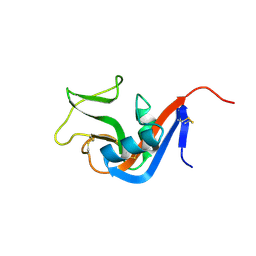
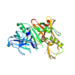
3CD0
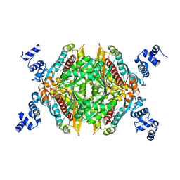 | | Thermodynamic and structure guided design of statin hmg-coa reductase inhibitors | | 分子名称: | (3R,5R)-7-{2-[(4-fluorobenzyl)carbamoyl]-4-(4-fluorophenyl)-1-(1-methylethyl)-1H-imidazol-5-yl}-3,5-dihydroxyheptanoic acid, 3-hydroxy-3-methylglutaryl-coenzyme A reductase | | 著者 | Pavlovsky, A, Sarver, R.W, Harris, M.S, Finzel, B.C. | | 登録日 | 2008-02-26 | | 公開日 | 2008-06-17 | | 最終更新日 | 2024-02-21 | | 実験手法 | X-RAY DIFFRACTION (2.4 Å) | | 主引用文献 | Thermodynamic and structure guided design of statin based inhibitors of 3-hydroxy-3-methylglutaryl coenzyme a reductase.
J.Med.Chem., 51, 2008
|
|
3CD7
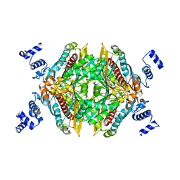 | | Thermodynamic and structure guided design of statin hmg-coa reductase inhibitors | | 分子名称: | (3R,5R)-7-[5-(ANILINOCARBONYL)-3,4-BIS(4-FLUOROPHENYL)-1-ISOPROPYL-1H-PYRROL-2-YL]-3,5-DIHYDROXYHEPTANOIC ACID, 3-hydroxy-3-methylglutaryl-coenzyme A reductase | | 著者 | Pavlovsky, A, Sarver, R.W, Harris, M.S, Finzel, B.C. | | 登録日 | 2008-02-26 | | 公開日 | 2008-06-17 | | 最終更新日 | 2024-02-21 | | 実験手法 | X-RAY DIFFRACTION (2.05 Å) | | 主引用文献 | Thermodynamic and structure guided design of statin based inhibitors of 3-hydroxy-3-methylglutaryl coenzyme a reductase.
J.Med.Chem., 51, 2008
|
|
3CDB
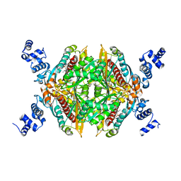 | | Thermodynamic and structure guided design of statin hmg-coa reductase inhibitors | | 分子名称: | (3R,5R)-7-{3-[(4-carbamoylphenyl)sulfamoyl]-4,5-bis(4-fluorophenyl)-2-(1-methylethyl)-1H-pyrrol-1-yl}-3,5-dihydroxyheptanoic acid, 3-hydroxy-3-methylglutaryl-coenzyme A reductase | | 著者 | Pavlovsky, A, Sarver, R.W, Harris, M.S, Finzel, B.C. | | 登録日 | 2008-02-26 | | 公開日 | 2008-06-17 | | 最終更新日 | 2024-02-21 | | 実験手法 | X-RAY DIFFRACTION (2.3 Å) | | 主引用文献 | Thermodynamic and structure guided design of statin based inhibitors of 3-hydroxy-3-methylglutaryl coenzyme a reductase.
J.Med.Chem., 51, 2008
|
|
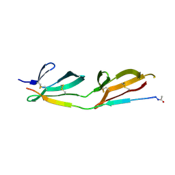
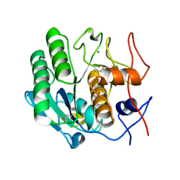
3CAD
 | | Crystal structure of Natural Killer Cell Receptor, Ly49G | | 分子名称: | Lectin-related NK cell receptor LY49G1 | | 著者 | Cho, S. | | 登録日 | 2008-02-19 | | 公開日 | 2008-04-08 | | 最終更新日 | 2011-07-13 | | 実験手法 | X-RAY DIFFRACTION (2.6 Å) | | 主引用文献 | Molecular Architecture of the Major Histocompatibility Complex Class I-binding Site of Ly49 Natural Killer Cell Receptors.
J.Biol.Chem., 283, 2008
|
|
2JK7
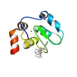 | | XIAP BIR3 bound to a Smac Mimetic | | 分子名称: | (3S,6S,7Z,10AS)-N-(DIPHENYLMETHYL)-6-{[(2S)-2-(METHYLIDENEAMINO)BUTANOYL]AMINO}-5-OXO-1,2,3,5,6,9,10,10A-OCTAHYDROPYRROLO[1,2-A]AZOCINE-3-CARBOXAMIDE, BACULOVIRAL IAP REPEAT-CONTAINING PROTEIN 4, ZINC ION | | 著者 | Saito, N.G, Meagher, J.L, Stuckey, J.A. | | 登録日 | 2008-08-21 | | 公開日 | 2008-11-04 | | 最終更新日 | 2024-05-01 | | 実験手法 | X-RAY DIFFRACTION (2.82 Å) | | 主引用文献 | Structure-Based Design, Synthesis, Evaluation, and Crystallographic Studies of Conformationally Constrained Smac Mimetics as Inhibitors of the X-Linked Inhibitor of Apoptosis Protein (Xiap).
J.Med.Chem., 51, 2008
|
|
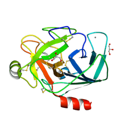
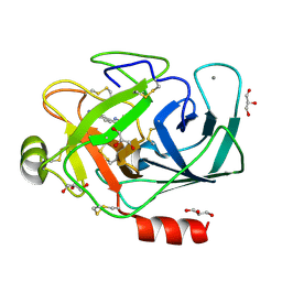
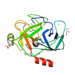
3CBK
 | | chagasin-cathepsin B | | 分子名称: | Cathepsin B, Chagasin | | 著者 | Redzynia, I, Bujacz, G.D, Abrahamson, M, Ljunggren, A, Jaskolski, M, Mort, J.S. | | 登録日 | 2008-02-22 | | 公開日 | 2008-05-27 | | 最終更新日 | 2023-11-01 | | 実験手法 | X-RAY DIFFRACTION (2.67 Å) | | 主引用文献 | Displacement of the occluding loop by the parasite protein, chagasin, results in efficient inhibition of human cathepsin B.
J.Biol.Chem., 283, 2008
|
|
2K2F
 | |
2JT0
 | | Solution structure of F104W cardiac troponin C | | 分子名称: | Troponin C, slow skeletal and cardiac muscles | | 著者 | Wang, X, Mercier, P, Letourneau, P.-J, Sykes, B.D. | | 登録日 | 2007-07-17 | | 公開日 | 2008-05-27 | | 最終更新日 | 2024-05-29 | | 実験手法 | SOLUTION NMR | | 主引用文献 | Effects of Phe-to-Trp mutation and fluorotryptophan incorporation on the solution structure of cardiac troponin C, and analysis of its suitability as a potential probe for in situ NMR studies.
Protein Sci., 14, 2005
|
|
3CDA
 | | Thermodynamic and structure guided design of statin hmg-coa reductase inhibitors | | 分子名称: | (3R,5R)-7-{3-(4-fluorophenyl)-1-(1-methylethyl)-4-phenyl-5-[(4-sulfamoylphenyl)carbamoyl]-1H-pyrrol-2-yl}-3,5-dihydroxyheptanoic acid, 3-hydroxy-3-methylglutaryl-coenzyme A reductase | | 著者 | Pavlovsky, A, Sarver, R.W, Harris, M.S, Finzel, B.C. | | 登録日 | 2008-02-26 | | 公開日 | 2008-06-17 | | 最終更新日 | 2024-02-21 | | 実験手法 | X-RAY DIFFRACTION (2.07 Å) | | 主引用文献 | Thermodynamic and structure guided design of statin based inhibitors of 3-hydroxy-3-methylglutaryl coenzyme a reductase.
J.Med.Chem., 51, 2008
|
|
3CDS
 | |
3DGZ
 | | Crystal Structure of Mouse Mitochondrial Thioredoxin Reductase, C-terminal 3-residue truncation | | 分子名称: | FLAVIN-ADENINE DINUCLEOTIDE, Thioredoxin reductase 2, [(2R,3R,4R,5R)-5-(6-AMINO-9H-PURIN-9-YL)-3-HYDROXY-4-(PHOSPHONOOXY)TETRAHYDROFURAN-2-YL]METHYL [(2R,3S,4S)-3,4-DIHYDROXYTETRAHYDROFURAN-2-YL]METHYL DIHYDROGEN DIPHOSPHATE | | 著者 | Eckenroth, B.E, Hondal, R.J, Everse, S.J. | | 登録日 | 2008-06-16 | | 公開日 | 2009-06-16 | | 最終更新日 | 2017-10-25 | | 実験手法 | X-RAY DIFFRACTION (2.25 Å) | | 主引用文献 | Crystal Structure of Mouse Mitochondrial Thioredoxin Reductase, C-terminal 3-residue truncation
To be Published
|
|
3DE7
 | |
2Z17
 | | Crystal structure of PDZ domain from human Pleckstrin homology, Sec7 | | 分子名称: | Pleckstrin homology Sec7 and coiled-coil domains-binding protein | | 著者 | Kishishita, S, Nishino, A, Murayama, K, Terada, T, Shirouzu, M, Yokoyama, S, RIKEN Structural Genomics/Proteomics Initiative (RSGI) | | 登録日 | 2007-05-08 | | 公開日 | 2008-05-13 | | 最終更新日 | 2024-03-13 | | 実験手法 | X-RAY DIFFRACTION (2.7 Å) | | 主引用文献 | Crystal structure of PDZ domain from human Pleckstrin homology, Sec7
To be Published
|
|
3DNZ
 | |
3DE4
 | |
3A60
 | | Crystal structure of unphosphorylated p70S6K1 (Form I) | | 分子名称: | Ribosomal protein S6 kinase beta-1, STAUROSPORINE | | 著者 | Sunami, T, Byrne, N, Diehl, R.E, Funabashi, K, Hall, D.L, Ikuta, M, Patel, S.B, Shipman, J.M, Smith, R.F, Takahashi, I, Zugay-Murphy, J, Iwasawa, Y, Lumb, K.J, Munshi, S.K, Sharma, S. | | 登録日 | 2009-08-17 | | 公開日 | 2009-10-27 | | 最終更新日 | 2023-11-01 | | 実験手法 | X-RAY DIFFRACTION (2.8 Å) | | 主引用文献 | Structural basis of human p70 ribosomal S6 kinase-1 regulation by activation loop phosphorylation.
J.Biol.Chem., 285, 2010
|
|
3A7X
 | |
3A86
 | |
3A81
 | |
3DUY
 | | Crystal structure of human beta-secretase in complex with NVP-AFJ144 | | 分子名称: | (2R,4S,5S)-N-butyl-4-hydroxy-2,7-dimethyl-5-{[N-(4-methylpentanoyl)-L-methionyl]amino}octanamide, Beta-secretase 1 | | 著者 | Rondeau, J.-M. | | 登録日 | 2008-07-18 | | 公開日 | 2009-02-24 | | 最終更新日 | 2023-08-30 | | 実験手法 | X-RAY DIFFRACTION (1.97 Å) | | 主引用文献 | Structure-based design and synthesis of macrocyclic peptidomimetic beta-secretase (BACE-1) inhibitors.
Bioorg.Med.Chem.Lett., 19, 2009
|
|
3DVS
 | |
2L40
 | | Mouse prion protein (121-231) containing the substitution Y169A | | 分子名称: | Major prion protein | | 著者 | Christen, B, Damberger, F.F, Perez, D.R, Hornemann, S, Wuthrich, K. | | 登録日 | 2010-09-28 | | 公開日 | 2011-08-10 | | 最終更新日 | 2023-06-14 | | 実験手法 | SOLUTION NMR | | 主引用文献 | Cellular prion protein conformation and function.
Proc.Natl.Acad.Sci.USA, 108, 2011
|
|
3DWD
 | | Crystal structure of the ArfGAP domain of human ARFGAP1 | | 分子名称: | ADP-ribosylation factor GTPase-activating protein 1, UNKNOWN ATOM OR ION, ZINC ION | | 著者 | Nedyalkova, L, Tong, Y, Tempel, W, Landry, R, Arrowsmith, C.H, Edwards, A.M, Bountra, C, Wilkstrom, M, Bochkarev, A, Park, H, Structural Genomics Consortium (SGC) | | 登録日 | 2008-07-22 | | 公開日 | 2008-08-05 | | 最終更新日 | 2023-08-30 | | 実験手法 | X-RAY DIFFRACTION (2.4 Å) | | 主引用文献 | Crystal structure of the ArfGAP domain of human ARFGAP1
To be Published
|
|
3DE3
 | |


