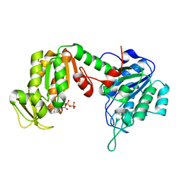
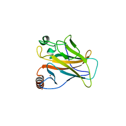
2ZW3
 
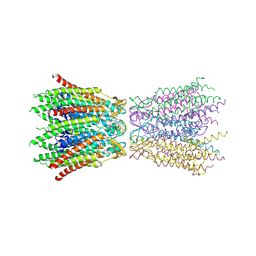 | | Structure of the connexin-26 gap junction channel at 3.5 angstrom resolution | | 分子名称: | Gap junction beta-2 protein | | 著者 | Maeda, S, Nakagawa, S, Suga, M, Yamashita, E, Oshima, A, Fujiyoshi, Y, Tsukihara, T. | | 登録日 | 2008-12-01 | | 公開日 | 2009-04-07 | | 最終更新日 | 2011-07-13 | | 実験手法 | X-RAY DIFFRACTION (3.5 Å) | | 主引用文献 | Structure of the connexin 26 gap junction channel at 3.5 A resolution
Nature, 458, 2009
|
|
2ZG1
 | |
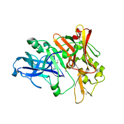
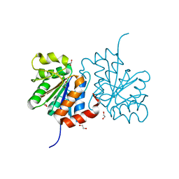
2IVS
 | | Crystal structure of non-phosphorylated RET tyrosine kinase domain | | 分子名称: | 2',3'- cyclic AMP, FORMIC ACID, PROTO-ONCOGENE TYROSINE-PROTEIN KINASE RECEPTOR RET | | 著者 | Knowles, P.P, Murray-Rust, J, McDonald, N.Q. | | 登録日 | 2006-06-16 | | 公開日 | 2006-08-14 | | 最終更新日 | 2023-12-13 | | 実験手法 | X-RAY DIFFRACTION (2 Å) | | 主引用文献 | Structure and Chemical Inhibition of the Ret Tyrosine Kinase Domain.
J.Biol.Chem., 281, 2006
|
|
2Z7G
 | |
2ZGV
 | | Crystal Structure of human phosphoglycerate kinase bound to D-ADP | | 分子名称: | ADENOSINE-5'-DIPHOSPHATE, Phosphoglycerate kinase 1 | | 著者 | Arold, S.T, Gondeau, C, Lionne, C, Chaloin, L. | | 登録日 | 2008-01-26 | | 公開日 | 2008-07-01 | | 最終更新日 | 2023-11-01 | | 実験手法 | X-RAY DIFFRACTION (2 Å) | | 主引用文献 | Molecular basis for the lack of enantioselectivity of human 3-phosphoglycerate kinase
Nucleic Acids Res., 36, 2008
|
|
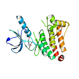
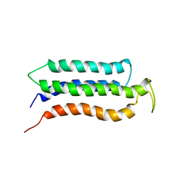
2J10
 | | p53 tetramerization domain mutant T329F Q331K | | 分子名称: | CELLULAR TUMOR ANTIGEN P53 | | 著者 | Carbajo, R.J, Mora, P, Sanchez del Pino, M.M, Perez-Paya, E, Pineda-Lucena, A. | | 登録日 | 2006-08-08 | | 公開日 | 2007-08-28 | | 最終更新日 | 2024-05-15 | | 実験手法 | SOLUTION NMR | | 主引用文献 | Solvent-exposed residues located in the beta-sheet modulate the stability of the tetramerization domain of p53--a structural and combinatorial approach.
Proteins, 71, 2008
|
|
2ZHR
 | |
2J5W
 | | Ceruloplasmin revisited: structural and functional roles of various metal cation binding sites | | 分子名称: | 2-acetamido-2-deoxy-beta-D-glucopyranose, CALCIUM ION, CERULOPLASMIN, ... | | 著者 | Bento, I, Peixoto, C, Zaitsev, V.N, Lindley, P.F. | | 登録日 | 2006-09-19 | | 公開日 | 2007-02-06 | | 最終更新日 | 2023-12-13 | | 実験手法 | X-RAY DIFFRACTION (2.8 Å) | | 主引用文献 | Ceruloplasmin Revisited: Structural and Functional Roles of Various Metal Cation-Binding Sites.
Acta Crystallogr.,Sect.D, 63, 2007
|
|
2VWV
 | | ephB4 kinase domain inhibitor complex | | 分子名称: | EPHRIN TYPE-B RECEPTOR 4, N'-(3-CHLORO-4-METHOXY-PHENYL)-N-(3,4,5-TRIMETHOXYPHENYL)-1,3,5-TRIAZINE-2,4-DIAMINE | | 著者 | Read, J, Brassington, C.A, Green, I, McCall, E.J, Valentine, A.L, Kettle, J.G, Leach, A.G. | | 登録日 | 2008-06-27 | | 公開日 | 2008-07-08 | | 最終更新日 | 2023-12-13 | | 実験手法 | X-RAY DIFFRACTION (1.9 Å) | | 主引用文献 | Inhibitors of the Tyrosine Kinase Ephb4. Part 1: Structure-Based Design and Optimization of a Series of 2,4-Bis-Anilinopyrimidines
Bioorg.Med.Chem.Lett., 18, 2008
|
|
2J1W
 | |
2J0Y
 | | L-ficolin complexed to b-1,3-D-glucan | | 分子名称: | 2-acetamido-2-deoxy-beta-D-glucopyranose, ACETATE ION, CALCIUM ION, ... | | 著者 | Garlatti, V, Gaboriaud, C. | | 登録日 | 2006-08-08 | | 公開日 | 2007-01-23 | | 最終更新日 | 2020-07-29 | | 実験手法 | X-RAY DIFFRACTION (2.35 Å) | | 主引用文献 | Structural Insights Into the Innate Immune Recognition Specificities of L- and H-Ficolins.
Embo J., 26, 2007
|
|
2W8N
 | |
3FZZ
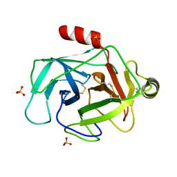 | | Structure of GrC | | 分子名称: | Granzyme C, SULFATE ION | | 著者 | Buckle, A.M, Kaiserman, D, Whisstock, J.C. | | 登録日 | 2009-01-27 | | 公開日 | 2009-03-17 | | 最終更新日 | 2023-11-01 | | 実験手法 | X-RAY DIFFRACTION (2.5 Å) | | 主引用文献 | Structure of granzyme C reveals an unusual mechanism of protease autoinhibition
Proc.Natl.Acad.Sci.USA, 106, 2009
|
|
2W3O
 | |
2W4J
 | | X-ray structure of a DAP-Kinase 2-277 | | 分子名称: | ACETATE ION, ADENOSINE-5'-DIPHOSPHATE, DEATH-ASSOCIATED PROTEIN KINASE 1, ... | | 著者 | De Diego, I, Kuper, J, Lehmann, F, Wilmanns, M. | | 登録日 | 2008-11-27 | | 公開日 | 2009-12-22 | | 最終更新日 | 2023-12-13 | | 実験手法 | X-RAY DIFFRACTION (1.3 Å) | | 主引用文献 | X-Ray Structure of a Dap-Kinase Calmodulin Complex
To be Published
|
|
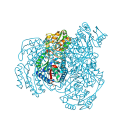
2WC0
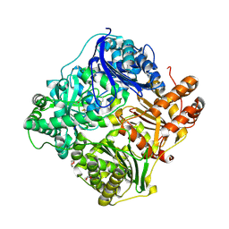 | | crystal structure of human insulin degrading enzyme in complex with iodinated insulin | | 分子名称: | 1,4-DIETHYLENE DIOXIDE, INSULIN A CHAIN, INSULIN B CHAIN, ... | | 著者 | Manolopoulou, M, Guo, Q, Malito, E, Schilling, A.B, Tang, W.J. | | 登録日 | 2009-03-06 | | 公開日 | 2009-03-24 | | 最終更新日 | 2023-12-13 | | 実験手法 | X-RAY DIFFRACTION (2.8 Å) | | 主引用文献 | Molecular Basis of Catalytic Chamber-Assisted Unfolding and Cleavage of Human Insulin by Human Insulin Degrading Enzyme.
J.Biol.Chem., 284, 2009
|
|
2KSR
 | |
2WGX
 | |
2KUM
 | | Solution structure of the human chemokine CCL27 | | 分子名称: | C-C motif chemokine 27 | | 著者 | Kirkpatrick, J.P, Jansma, A, Hsu, A, Handel, T.M, Nietlispach, D. | | 登録日 | 2010-02-22 | | 公開日 | 2010-03-02 | | 最終更新日 | 2022-03-16 | | 実験手法 | SOLUTION NMR | | 主引用文献 | NMR analysis of the structure, dynamics, and unique oligomerization properties of the chemokine CCL27.
J.Biol.Chem., 285, 2010
|
|
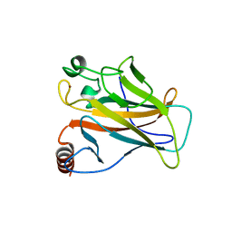
3B38
 | | Structure of A104V DJ-1 | | 分子名称: | 1,2-ETHANEDIOL, Protein DJ-1 | | 著者 | Lakshminarasimhan, M, Maldonado, M.T, Zhou, W, Fink, A.L, Wilson, M.A. | | 登録日 | 2007-10-19 | | 公開日 | 2008-01-15 | | 最終更新日 | 2023-08-30 | | 実験手法 | X-RAY DIFFRACTION (1.85 Å) | | 主引用文献 | Structural Impact of Three Parkinsonism-Associated Missense Mutations on Human DJ-1.
Biochemistry, 47, 2008
|
|
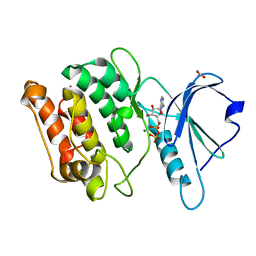
2W4R
 | | Crystal structure of the regulatory domain of human LGP2 | | 分子名称: | MERCURY (II) ION, PROBABLE ATP-DEPENDENT RNA HELICASE DHX58, SULFATE ION | | 著者 | Pippig, D.A, Hellmuth, J.C, Cui, S, Kirchhofer, A, Lammens, K, Lammens, A, Schmidt, A, Rothenfusser, S, Hopfner, K.P. | | 登録日 | 2008-12-01 | | 公開日 | 2009-02-24 | | 最終更新日 | 2023-12-13 | | 実験手法 | X-RAY DIFFRACTION (2.6 Å) | | 主引用文献 | The Regulatory Domain of the Rig-I Family ATPase Lgp2 Senses Double-Stranded RNA.
Nucleic Acids Res., 37, 2009
|
|
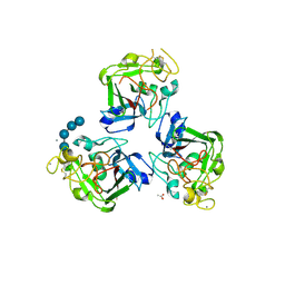
3B3W
 | | Crystal structure of the S228A mutant of the aminopeptidase from Vibrio proteolyticus in complex with leucine | | 分子名称: | Bacterial leucyl aminopeptidase, LEUCINE, SODIUM ION, ... | | 著者 | Ataie, N.J, Hoang, Q.Q, Zahniser, M.P.D, Milne, A, Petsko, G.A, Ringe, D. | | 登録日 | 2007-10-22 | | 公開日 | 2007-11-27 | | 最終更新日 | 2023-08-30 | | 実験手法 | X-RAY DIFFRACTION (1.75 Å) | | 主引用文献 | Zinc coordination geometry and ligand binding affinity: the structural and kinetic analysis of the second-shell serine 228 residue and the methionine 180 residue of the aminopeptidase from Vibrio proteolyticus.
Biochemistry, 47, 2008
|
|
3B4F
 | | Carbonic anhydrase inhibitors. Interaction of 2-(hydrazinocarbonyl)-3-phenyl-1H-indole-5-sulfonamide with twelve mammalian isoforms: kinetic and X-Ray crystallographic studies | | 分子名称: | 2-(hydrazinocarbonyl)-3-phenyl-1H-indole-5-sulfonamide, Carbonic anhydrase 2, MERCURY (II) ION, ... | | 著者 | Guzel, o, Temperini, c, Innocenti, a, Scozzafava, A, Salman, a, Supuran, c.t. | | 登録日 | 2007-10-24 | | 公開日 | 2008-01-22 | | 最終更新日 | 2023-08-30 | | 実験手法 | X-RAY DIFFRACTION (1.89 Å) | | 主引用文献 | Carbonic anhydrase inhibitors. Interaction of 2-(hydrazinocarbonyl)-3-phenyl-1H-indole-5-sulfonamide with 12 mammalian isoforms: kinetic and X-ray crystallographic studies.
Bioorg.Med.Chem.Lett., 18, 2008
|
|
2VOK
 | | Murine TRIM21 | | 分子名称: | 52 KDA RO PROTEIN | | 著者 | Keeble, A.H, Khan, Z, Forster, A, James, L.C. | | 登録日 | 2008-02-19 | | 公開日 | 2008-04-15 | | 最終更新日 | 2024-02-07 | | 実験手法 | X-RAY DIFFRACTION (1.3 Å) | | 主引用文献 | Trim21 is an Igg Receptor that is Structurally, Thermodynamically, and Kinetically Conserved.
Proc.Natl.Acad.Sci.USA, 105, 2008
|
|
2KR6
 | |


