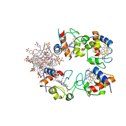
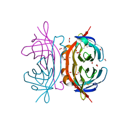
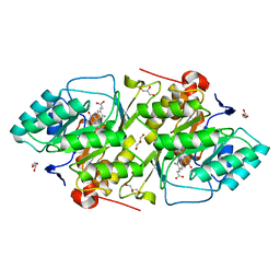
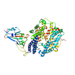
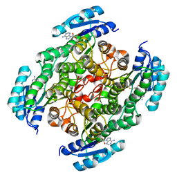
1M5X
 
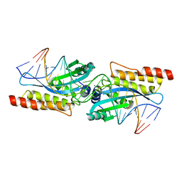 | | Crystal structure of the homing endonuclease I-MsoI bound to its DNA substrate | | 分子名称: | 5'-D(*CP*GP*GP*AP*AP*CP*TP*GP*TP*CP*TP*CP*AP*CP*GP*AP*CP*GP*TP*TP*CP*TP*GP*C)-3', 5'-D(*GP*CP*AP*GP*AP*AP*CP*GP*TP*CP*GP*TP*GP*AP*GP*AP*CP*AP*GP*TP*TP*CP*CP*G)-3', CALCIUM ION, ... | | 著者 | Chevalier, B, Turmel, M, Lemieux, C, Monnat, R.J, Stoddard, B.L. | | 登録日 | 2002-07-10 | | 公開日 | 2003-06-03 | | 最終更新日 | 2024-04-03 | | 実験手法 | X-RAY DIFFRACTION (2.25 Å) | | 主引用文献 | Flexible DNA Target Site Recognition by Divergent Homing Endonuclease Isoschizomers I-CreI and I-MsoI
J.Mol.Biol., 329, 2003
|
|
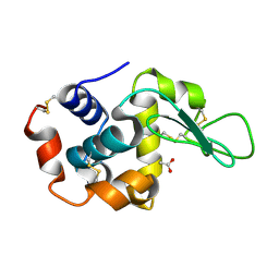
2HAL
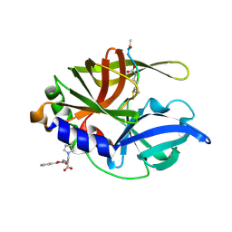 | | An episulfide cation (thiiranium ring) trapped in the active site of HAV 3C proteinase inactivated by peptide-based ketone inhibitors | | 分子名称: | Hepatitis A Protease 3C, N-ACETYL-LEUCYL-PHENYLALANYL-PHENYLALANYL-GLUTAMATE-FLUOROMETHYLKETONE INHIBITOR, N-[(BENZYLOXY)CARBONYL]-L-ALANINE | | 著者 | Yin, J, Cherney, M.M, Bergmann, E.M, James, M.N. | | 登録日 | 2006-06-13 | | 公開日 | 2006-08-08 | | 最終更新日 | 2023-08-30 | | 実験手法 | X-RAY DIFFRACTION (1.35 Å) | | 主引用文献 | An Episulfide Cation (Thiiranium Ring) Trapped in the Active Site of HAV 3C Proteinase Inactivated by Peptide-based Ketone Inhibitors.
J.Mol.Biol., 361, 2006
|
|
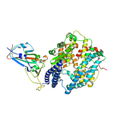
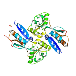
2F7C
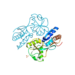 | | CatM effector binding domain with its effector cis,cis-muconate | | 分子名称: | (2Z,4Z)-HEXA-2,4-DIENEDIOIC ACID, HTH-type transcriptional regulator catM, SULFATE ION | | 著者 | Clark, T, Haddad, S, Ezezika, O, Neidle, E, Momany, C. | | 登録日 | 2005-11-30 | | 公開日 | 2006-10-31 | | 最終更新日 | 2023-08-23 | | 実験手法 | X-RAY DIFFRACTION (2.162 Å) | | 主引用文献 | Distinct Effector-binding Sites Enable Synergistic Transcriptional Activation by BenM, a LysR-type Regulator.
J.Mol.Biol., 367, 2007
|
|
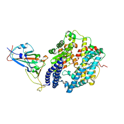
1JVJ
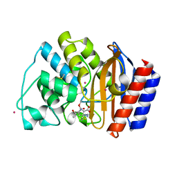 | | CRYSTAL STRUCTURE OF N132A MUTANT OF TEM-1 BETA-LACTAMASE IN COMPLEX WITH A N-FORMIMIDOYL-THIENAMYCINE | | 分子名称: | (5R)-5-[(1S,2R)-1-formyl-2-hydroxypropyl]-3-[(2-{[(E)-iminomethyl]amino}ethyl)sulfanyl]-4,5-dihydro-1H-pyrrole-2-carbox ylic acid, BETA-LACTAMASE TEM, POTASSIUM ION | | 著者 | Wang, X, Minasov, G, Shoichet, B.K. | | 登録日 | 2001-08-30 | | 公開日 | 2002-03-06 | | 最終更新日 | 2023-08-16 | | 実験手法 | X-RAY DIFFRACTION (1.73 Å) | | 主引用文献 | Noncovalent interaction energies in covalent complexes: TEM-1 beta-lactamase and beta-lactams.
Proteins, 47, 2002
|
|
7BBT
 | | Structure of cytochrome c in complex with a p-benzyl-sulfonato-calix[8]arene-PEG pseudorotaxane | | 分子名称: | 4-[[49,50,51,52,53,54,55,56-octahydroxy-11,17,23,29,35,41,47-heptakis[(4-sulfonatophenyl)methyl]-5-nonacyclo[43.3.1.13,7.19,13.115,19.121,25.127,31.133,37.139,43]hexapentaconta-1(49),3,5,7(56),9,11,13(55),15,17,19(54),21,23,25(53),27,29,31(52),33,35,37(51),39,41,43(50),45,47-tetracosaenyl]methyl]benzenesulfonate, Cytochrome c iso-1, HEME C, ... | | 著者 | Mockler, N.M, Ramberg, K, Guagnini, F, Raston, C.L, Crowley, P.B. | | 登録日 | 2020-12-18 | | 公開日 | 2021-10-20 | | 最終更新日 | 2024-01-31 | | 実験手法 | X-RAY DIFFRACTION (3.023 Å) | | 主引用文献 | Noncovalent Protein-Pseudorotaxane Assembly Incorporating an Extended Arm Calix[8]arene with alpha-Helical Recognition Properties.
Cryst.Growth Des., 21, 2021
|
|
4YNL
 | | Crystal structure of the hood domain of Anabaena HetR in complex with the hexapeptide ERGSGR derived from PatS | | 分子名称: | Heterocyst differentiation control protein, Heterocyst inhibition-signaling peptide | | 著者 | Hu, H.X, Jiang, Y.L, Zhao, M.X, Zhang, C.C, Chen, Y, Zhou, C.Z. | | 登録日 | 2015-03-10 | | 公開日 | 2015-12-02 | | 最終更新日 | 2023-11-08 | | 実験手法 | X-RAY DIFFRACTION (2.1 Å) | | 主引用文献 | Structural insights into HetR-PatS interaction involved in cyanobacterial pattern formation
Sci Rep, 5, 2015
|
|
1Z3V
 | | Structure of Phanerochaete chrysosporium cellobiohydrolase Cel7D (CBH58) in complex with lactose | | 分子名称: | 2-acetamido-2-deoxy-beta-D-glucopyranose, beta-D-galactopyranose-(1-4)-beta-D-glucopyranose, cellulase | | 著者 | Ubhayasekera, W, Munoz, I.G, Stahlberg, J, Mowbray, S.L. | | 登録日 | 2005-03-14 | | 公開日 | 2005-04-26 | | 最終更新日 | 2023-10-25 | | 実験手法 | X-RAY DIFFRACTION (1.61 Å) | | 主引用文献 | Structures of Phanerochaete chrysosporium Cel7D in complex with product and inhibitors
Febs J., 272, 2005
|
|
6BZM
 | | GFGNFGTS from low-complexity/FG repeat domain of Nup98, residues 116-123 | | 分子名称: | Nuclear pore complex protein Nup98-Nup96 | | 著者 | Hughes, M.P, Rodriguez, J.A, Sawaya, M.R, Cascio, D, Chong, L, Gonen, T, Eisenberg, D.S. | | 登録日 | 2017-12-24 | | 公開日 | 2018-04-04 | | 最終更新日 | 2024-03-13 | | 実験手法 | ELECTRON CRYSTALLOGRAPHY (0.9 Å) | | 主引用文献 | Atomic structures of low-complexity protein segments reveal kinked beta sheets that assemble networks.
Science, 359, 2018
|
|
2OF9
 | |
2AWW
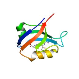 | | Synapse associated protein 97 PDZ2 domain variant C378G with C-terminal GluR-A peptide | | 分子名称: | 18-residue C-terminal peptide from glutamate receptor, ionotropic, AMPA1, ... | | 著者 | Von Ossowski, I, Oksanen, E, Von Ossowski, L, Cai, C, Sundberg, M, Goldman, A, Keinanen, K. | | 登録日 | 2005-09-02 | | 公開日 | 2006-08-29 | | 最終更新日 | 2024-05-29 | | 実験手法 | X-RAY DIFFRACTION (2.21 Å) | | 主引用文献 | Crystal structure of the second PDZ domain of SAP97 in complex with a GluR-A C-terminal peptide
Febs J., 273, 2006
|
|
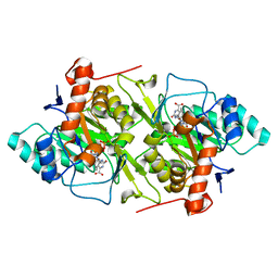
1JQX
 | | The R57A mutant of Lactococcus lactis dihydroorotate dehydrogenase A | | 分子名称: | FLAVIN MONONUCLEOTIDE, GLYCEROL, MAGNESIUM ION, ... | | 著者 | Norager, S, Arent, S, Bjornberg, O, Ottosen, M, Lo Leggio, L, Jensen, K.F, Larsen, S. | | 登録日 | 2001-08-09 | | 公開日 | 2003-09-09 | | 最終更新日 | 2023-10-25 | | 実験手法 | X-RAY DIFFRACTION (1.7 Å) | | 主引用文献 | Lactococcus lactis dihydroorotate dehydrogenase A mutants reveal important facets of the enzymatic function
J.Biol.Chem., 278, 2003
|
|
1JRC
 | | The N67A mutant of Lactococcus lactis dihydroorotate dehydrogenase A | | 分子名称: | FLAVIN MONONUCLEOTIDE, OROTIC ACID, dihydroorotate dehydrogenase A | | 著者 | Norager, S, Arent, S, Bjornberg, O, Ottosen, M, Lo Leggio, L, Jensen, K.F, Larsen, S. | | 登録日 | 2001-08-13 | | 公開日 | 2003-09-09 | | 最終更新日 | 2023-10-25 | | 実験手法 | X-RAY DIFFRACTION (1.8 Å) | | 主引用文献 | Lactococcus lactis dihydroorotate dehydrogenase A mutants reveal important facets of the enzymatic function
J.Biol.Chem., 278, 2003
|
|
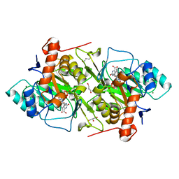
1JUE
 | | 1.8 A resolution structure of native lactococcus lactis dihydroorotate dehydrogenase A | | 分子名称: | ACETIC ACID, FLAVIN MONONUCLEOTIDE, GLYCEROL, ... | | 著者 | Norager, S, Arent, S, Bjornberg, O, Ottosen, M, Lo Leggio, L, Jensen, K.F, Larsen, S. | | 登録日 | 2001-08-24 | | 公開日 | 2003-09-09 | | 最終更新日 | 2023-10-25 | | 実験手法 | X-RAY DIFFRACTION (1.8 Å) | | 主引用文献 | Lactococcus lactis dihydroorotate dehydrogenase A mutants reveal important facets of the enzymatic function
J.Biol.Chem., 278, 2003
|
|
4Z98
 | | Crystal Structure of Hen Egg White Lysozyme using Serial X-ray Diffraction Data Collection | | 分子名称: | ACETATE ION, Lysozyme C | | 著者 | Murray, T.D, Lyubimov, A.Y, Ogata, C.M, Uervirojnangkoorn, M, Brunger, A.T, Berger, J.M. | | 登録日 | 2015-04-10 | | 公開日 | 2015-10-14 | | 最終更新日 | 2023-09-27 | | 実験手法 | X-RAY DIFFRACTION (1.55 Å) | | 主引用文献 | A high-transparency, micro-patternable chip for X-ray diffraction analysis of microcrystals under native growth conditions.
Acta Crystallogr. D Biol. Crystallogr., 71, 2015
|
|
7EKE
 | | Structure of SARS-CoV-2 spike receptor-binding domain F486L mutation complexed with human ACE2 | | 分子名称: | 2-acetamido-2-deoxy-beta-D-glucopyranose, Angiotensin-converting enzyme 2, Spike protein S1, ... | | 著者 | Han, P.C, Su, C, Zhang, Y.F, Qi, J.X, Gao, G.F. | | 登録日 | 2021-04-05 | | 公開日 | 2021-11-03 | | 最終更新日 | 2023-11-29 | | 実験手法 | X-RAY DIFFRACTION (2.7 Å) | | 主引用文献 | Molecular insights into receptor binding of recent emerging SARS-CoV-2 variants.
Nat Commun, 12, 2021
|
|
7EKG
 | | Structure of SARS-CoV-2 Beta variant spike receptor-binding domain complexed with human ACE2 | | 分子名称: | 2-acetamido-2-deoxy-beta-D-glucopyranose, 2-acetamido-2-deoxy-beta-D-glucopyranose-(1-4)-2-acetamido-2-deoxy-beta-D-glucopyranose, Angiotensin-converting enzyme 2, ... | | 著者 | Han, P.C, Su, C, Zhang, Y.F, Qi, J.X, Gao, G.F. | | 登録日 | 2021-04-05 | | 公開日 | 2021-11-03 | | 最終更新日 | 2023-11-29 | | 実験手法 | X-RAY DIFFRACTION (2.63 Å) | | 主引用文献 | Molecular insights into receptor binding of recent emerging SARS-CoV-2 variants.
Nat Commun, 12, 2021
|
|
7EKC
 | | Structure of SARS-CoV-2 Gamma variant spike receptor-binding domain complexed with human ACE2 | | 分子名称: | 2-acetamido-2-deoxy-beta-D-glucopyranose, 2-acetamido-2-deoxy-beta-D-glucopyranose-(1-4)-2-acetamido-2-deoxy-beta-D-glucopyranose, Angiotensin-converting enzyme 2, ... | | 著者 | Han, P.C, Su, C, Zhang, Y.F, Qi, J.X, Gao, G.F. | | 登録日 | 2021-04-05 | | 公開日 | 2021-11-03 | | 最終更新日 | 2023-11-29 | | 実験手法 | X-RAY DIFFRACTION (2.8 Å) | | 主引用文献 | Molecular insights into receptor binding of recent emerging SARS-CoV-2 variants.
Nat Commun, 12, 2021
|
|
7EKH
 | | Structure of SARS-CoV-2 spike receptor-binding domain Y453F mutation complexed with human ACE2 | | 分子名称: | 2-acetamido-2-deoxy-beta-D-glucopyranose, Angiotensin-converting enzyme 2, Spike protein S1, ... | | 著者 | Han, P.C, Su, C, Zhang, Y.F, Qi, J.X, Gao, G.F. | | 登録日 | 2021-04-05 | | 公開日 | 2021-11-03 | | 最終更新日 | 2023-11-29 | | 実験手法 | X-RAY DIFFRACTION (2.4 Å) | | 主引用文献 | Molecular insights into receptor binding of recent emerging SARS-CoV-2 variants.
Nat Commun, 12, 2021
|
|
7EKF
 | | Structure of SARS-CoV-2 Alpha variant spike receptor-binding domain complexed with human ACE2 | | 分子名称: | 2-acetamido-2-deoxy-beta-D-glucopyranose, 2-acetamido-2-deoxy-beta-D-glucopyranose-(1-4)-2-acetamido-2-deoxy-beta-D-glucopyranose, Angiotensin-converting enzyme 2, ... | | 著者 | Han, P.C, Su, C, Zhang, Y.F, Qi, J.X, Gao, G.F. | | 登録日 | 2021-04-05 | | 公開日 | 2021-11-03 | | 最終更新日 | 2023-11-29 | | 実験手法 | X-RAY DIFFRACTION (2.85 Å) | | 主引用文献 | Molecular insights into receptor binding of recent emerging SARS-CoV-2 variants.
Nat Commun, 12, 2021
|
|
2ZNB
 | |
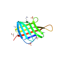
2F6G
 | | BenM effector binding domain | | 分子名称: | CHLORIDE ION, HTH-type transcriptional regulator benM, SULFATE ION | | 著者 | Clark, T, Haddad, S, Ezezika, O, Neidle, E, Momany, C. | | 登録日 | 2005-11-29 | | 公開日 | 2006-10-31 | | 最終更新日 | 2023-08-23 | | 実験手法 | X-RAY DIFFRACTION (1.908 Å) | | 主引用文献 | Distinct Effector-binding Sites Enable Synergistic Transcriptional Activation by BenM, a LysR-type Regulator.
J.Mol.Biol., 367, 2007
|
|
4ZJM
 | | Crystal Structure of Mycobacterium tuberculosis LpqH (Rv3763) | | 分子名称: | CHLORIDE ION, GLYCEROL, Lipoprotein LpqH, ... | | 著者 | Arbing, M.A, Chan, S, Kuo, E, Harris, L.R, Zhou, T.T, Eisenberg, D, TB Structural Genomics Consortium (TBSGC) | | 登録日 | 2015-04-29 | | 公開日 | 2015-05-13 | | 最終更新日 | 2023-09-27 | | 実験手法 | X-RAY DIFFRACTION (2.851 Å) | | 主引用文献 | Crystal Structure of Mycobacterium tuberculosis LpqH (Rv3763)
To Be Published
|
|
2F7B
 | | CatM effector binding domain | | 分子名称: | CHLORIDE ION, HTH-type transcriptional regulator catM, SULFATE ION | | 著者 | Clark, T, Haddad, S, Ezezika, O, Neidle, E, Momany, C. | | 登録日 | 2005-11-30 | | 公開日 | 2006-10-31 | | 最終更新日 | 2023-08-23 | | 実験手法 | X-RAY DIFFRACTION (1.9 Å) | | 主引用文献 | Distinct Effector-binding Sites Enable Synergistic Transcriptional Activation by BenM, a LysR-type Regulator.
J.Mol.Biol., 367, 2007
|
|
7XQM
 | |
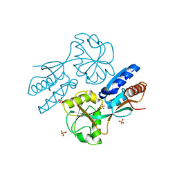
2A4N
 | | Crystal structure of aminoglycoside 6'-N-acetyltransferase complexed with coenzyme A | | 分子名称: | COENZYME A, SULFATE ION, aac(6')-Ii | | 著者 | Burk, D.L, Xiong, B, Breitbach, C, Berghuis, A.M. | | 登録日 | 2005-06-29 | | 公開日 | 2005-09-20 | | 最終更新日 | 2023-08-23 | | 実験手法 | X-RAY DIFFRACTION (2.2 Å) | | 主引用文献 | Structures of aminoglycoside acetyltransferase AAC(6')-Ii in a novel crystal form: structural and normal-mode analyses.
Acta Crystallogr.,Sect.D, 61, 2005
|
|


