Loading
PDBj
メニューPDBj@FacebookPDBj@TwitterPDBj@YouTubewwPDB FoundationwwPDB
RCSB PDBPDBeBMRBAdv. SearchSearch help

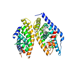
5I4V
DownloadVisualize
BU of 5i4v by Molmil
Discovery of novel, orally efficacious Liver X Receptor (LXR) beta agonists
分子名称: Oxysterols receptor LXR-beta,Nuclear receptor coactivator 2, Retinoic acid receptor RXR-beta,Nuclear receptor coactivator 2, {2-[(2R)-4-[4-(hydroxymethyl)-3-(methylsulfonyl)phenyl]-2-(propan-2-yl)piperazin-1-yl]-4-(trifluoromethyl)pyrimidin-5-yl}methanol
著者Chen, G, McKeever, B.M.
登録日2016-02-12
公開日2016-06-29
最終更新日2023-09-27
実験手法X-RAY DIFFRACTION (2.61 Å)
主引用文献Discovery of a Novel, Orally Efficacious Liver X Receptor (LXR) beta Agonist.
J.Med.Chem., 59, 2016
4G76
DownloadVisualize
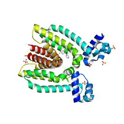
BU of 4g76 by Molmil
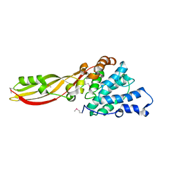
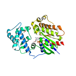
Structure of PaeM, a colicin M-like bacteriocin produced by Pseudomonas aeruginosa
分子名称: Phosphodiesterase
著者Touze, T, Graille, M, Mengin-Lecreulx, D.
登録日2012-07-20
公開日2012-09-12
最終更新日2023-12-06
実験手法X-RAY DIFFRACTION (2.385 Å)
主引用文献Functional and Structural Characterization of PaeM, a Colicin M-like Bacteriocin Produced by Pseudomonas aeruginosa.
J.Biol.Chem., 287, 2012
4GGK
DownloadVisualize
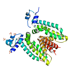
BU of 4ggk by Molmil
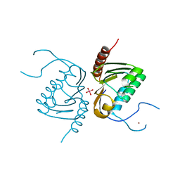
Crystal structure of Zucchini from mouse (mZuc / PLD6 / MitoPLD) bound to tungstate
分子名称: Mitochondrial cardiolipin hydrolase, TUNGSTATE(VI)ION, ZINC ION
著者Ipsaro, J.J, Haase, A.D, Hannon, G.J, Joshua-Tor, L.
登録日2012-08-06
公開日2012-10-10
最終更新日2023-09-13
実験手法X-RAY DIFFRACTION (2.1 Å)
主引用文献The structural biochemistry of Zucchini implicates it as a nuclease in piRNA biogenesis.
Nature, 491, 2012
1WVJ
DownloadVisualize
BU of 1wvj by Molmil
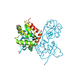
Exploring the GluR2 ligand-binding core in complex with the bicyclic AMPA analogue (S)-4-AHCP
分子名称: 3-(3-HYDROXY-7,8-DIHYDRO-6H-CYCLOHEPTA[D]ISOXAZOL-4-YL)-L-ALANINE, GLYCEROL, SULFATE ION, ...
著者Nielsen, B.B, Pickering, D.S, Greenwood, J.R, Brehm, L, Gajhede, M, Schousboe, A, Kastrup, J.S.
登録日2004-12-15
公開日2005-04-26
最終更新日2023-10-25
実験手法X-RAY DIFFRACTION (1.75 Å)
主引用文献Exploring the GluR2 ligand-binding core in complex with the bicyclical AMPA analogue (S)-4-AHCP
FEBS J., 272, 2005
3MRV
DownloadVisualize
BU of 3mrv by Molmil
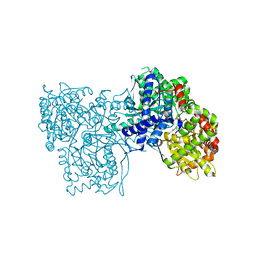
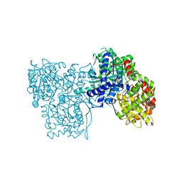
Glycogen phosphorylase complexed with 3-hydroxybenzaldehyde-4-(beta-D-glucopyranosyl) thiosemicarbazone
分子名称: DIMETHYL SULFOXIDE, Glycogen phosphorylase, muscle form, ...
著者Alexacou, K.-M.
登録日2010-04-29
公開日2011-01-05
最終更新日2023-11-22
実験手法X-RAY DIFFRACTION (1.94 Å)
主引用文献The binding of beta-D-glucopyranosyl-thiosemicarbazone derivatives to glycogen phosphorylase: a new class of inhibitors
Bioorg.Med.Chem., 18, 2010
3MT8
DownloadVisualize
BU of 3mt8 by Molmil
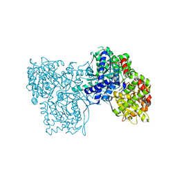
Glycogen phosphorylase complexed with 4-chlorobenzaldehyde-4-(beta-D-glucopyranosyl)-thiosemicarbazone
分子名称: Glycogen phosphorylase, muscle form, N-({(2E)-2-[(4-chlorophenyl)methylidene]hydrazino}carbonothioyl)-beta-D-glucopyranosylamine
著者Alexacou, K.-M.
登録日2010-04-30
公開日2011-01-05
最終更新日2023-11-22
実験手法X-RAY DIFFRACTION (2 Å)
主引用文献The binding of beta-D-glucopyranosyl-thiosemicarbazone derivatives to glycogen phosphorylase: a new class of inhibitors
Bioorg.Med.Chem., 18, 2010
3MT7
DownloadVisualize
BU of 3mt7 by Molmil
Glycogen phosphorylase complexed with 4-bromobenzaldehyde-4-(beta-D-glucopyranosyl)-thiosemicarbazone
分子名称: Glycogen phosphorylase, muscle form, N-({(2E)-2-[(4-bromophenyl)methylidene]hydrazino}carbonothioyl)-beta-D-glucopyranosylamine
著者Alexacou, K.-M.
登録日2010-04-30
公開日2011-01-05
最終更新日2023-11-22
実験手法X-RAY DIFFRACTION (2 Å)
主引用文献The binding of beta-D-glucopyranosyl-thiosemicarbazone derivatives to glycogen phosphorylase: a new class of inhibitors
Bioorg.Med.Chem., 18, 2010
1TVQ
DownloadVisualize
BU of 1tvq by Molmil
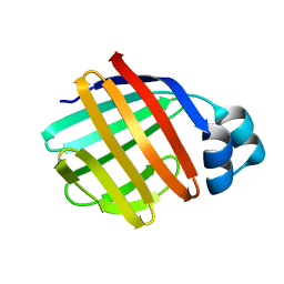
Crystal Structure of Apo Chicken Liver Basic Fatty Acid Binding Protein (or Bile Acid Binding Protein)
分子名称: Fatty acid-binding protein
著者Nichesola, D, Perduca, M, Capaldi, S, Carrizo, M.E, Righetti, P.G, Monaco, H.L.
登録日2004-06-30
公開日2004-11-23
最終更新日2024-03-13
実験手法X-RAY DIFFRACTION (2 Å)
主引用文献Crystal structure of chicken liver basic Fatty Acid-binding protein complexed with cholic acid
Biochemistry, 43, 2004
1TW4
DownloadVisualize
BU of 1tw4 by Molmil
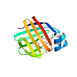
Crystal Structure of Chicken Liver Basic Fatty Acid Binding Protein (Bile Acid Binding Protein) Complexed With Cholic Acid
分子名称: CHOLIC ACID, Fatty acid-binding protein
著者Nichesola, D, Perduca, M, Capaldi, S, Carrizo, M.E, Righetti, P.G, Monaco, H.L.
登録日2004-06-30
公開日2004-11-23
最終更新日2024-03-13
実験手法X-RAY DIFFRACTION (2 Å)
主引用文献Crystal structure of chicken liver basic Fatty Acid-binding protein complexed with cholic acid
Biochemistry, 43, 2004
8HTG
DownloadVisualize
BU of 8htg by Molmil
Crystal structure of Golf in complex with GTP-gamma S and Mg
分子名称: 1,2-ETHANEDIOL, 5'-GUANOSINE-DIPHOSPHATE-MONOTHIOPHOSPHATE, Guanine nucleotide-binding protein G(olf) subunit alpha, ...
著者Kang, H, Choi, H.-J.
登録日2022-12-21
公開日2023-12-20
実験手法X-RAY DIFFRACTION (2.91 Å)
主引用文献Understanding the molecular mechanisms of odorant binding and activation of the human OR52 family.
Nat Commun, 14, 2023
3LV2
DownloadVisualize
BU of 3lv2 by Molmil
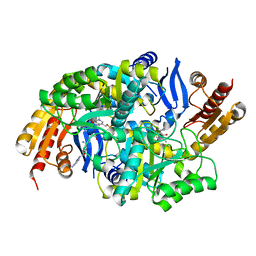
Crystal structure of Mycobacterium tuberculosis 7,8-diaminopelargonic acid synthase in complex with substrate analog sinefungin
分子名称: Adenosylmethionine-8-amino-7-oxononanoate aminotransferase, PYRIDOXAL-5'-PHOSPHATE, SINEFUNGIN
著者Dey, S, Sacchettini, J.C.
登録日2010-02-19
公開日2010-08-25
最終更新日2023-09-06
実験手法X-RAY DIFFRACTION (2.18 Å)
主引用文献Structural characterization of the Mycobacterium tuberculosis biotin biosynthesis enzymes 7,8-diaminopelargonic acid synthase and dethiobiotin synthetase .
Biochemistry, 49, 2010
4UYA
DownloadVisualize
BU of 4uya by Molmil
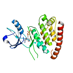
Structure of MLK4 kinase domain with ATPgammaS
分子名称: MAGNESIUM ION, MITOGEN-ACTIVATED PROTEIN KINASE KINASE KINASE MLK4, PHOSPHOTHIOPHOSPHORIC ACID-ADENYLATE ESTER
著者Read, J.A, Brassington, C, Pollard, H.K, Phillips, C, Green, I, Overmann, R, Collier, M.
登録日2014-08-29
公開日2015-09-30
最終更新日2024-01-10
実験手法X-RAY DIFFRACTION (2.8 Å)
主引用文献Recurrent Mlk4 Loss-of-Function Mutations Suppress Jnk Signaling to Promote Colon Tumorigenesis.
Cancer Res., 76, 2016
3BR1
DownloadVisualize
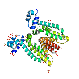
BU of 3br1 by Molmil
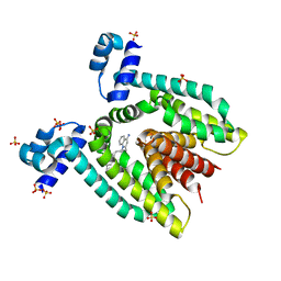
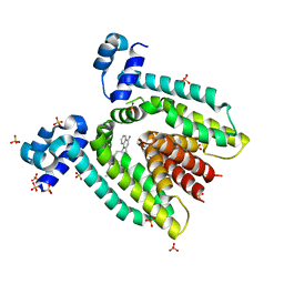
Crystal Structure of the Complex of Dequalinium Bound to QacR(E90Q), a Mutant of a Multidrug Binding Transcriptional Repressor
分子名称: DEQUALINIUM, HTH-type transcriptional regulator qacR, SULFATE ION
著者Brooks, B.E.
登録日2007-12-20
公開日2008-12-23
最終更新日2023-08-30
実験手法X-RAY DIFFRACTION (3.31 Å)
主引用文献Formal electrostatic interactions do not govern QacR-cation affinity
To be Published
2Q9E
DownloadVisualize
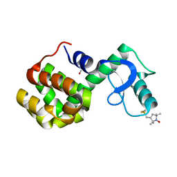
BU of 2q9e by Molmil
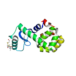
Structure of spin-labeled T4 lysozyme mutant S44R1
分子名称: 2-HYDROXYETHYL DISULFIDE, Lysozyme, S-[(1-oxyl-2,2,5,5-tetramethyl-2,5-dihydro-1H-pyrrol-3-yl)methyl] methanesulfonothioate
著者Guo, Z, Cascio, D, Hideg, K, Hubbell, W.L.
登録日2007-06-12
公開日2007-06-26
最終更新日2023-08-30
実験手法EPR (2.1 Å), X-RAY DIFFRACTION
主引用文献Structural determinants of nitroxide motion in spin-labeled proteins: Solvent-exposed sites in helix B of T4 lysozyme.
Protein Sci., 17, 2008
3BR2
DownloadVisualize
BU of 3br2 by Molmil
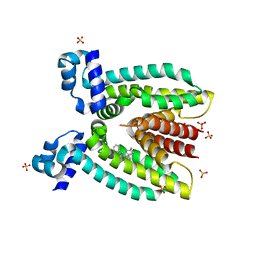
Crystal Structure of the Complex of Dequalinium Bound to QacR(E120Q), a Mutant of a Multidrug Binding Transcriptional Repressor
分子名称: DEQUALINIUM, HTH-type transcriptional regulator qacR, SULFATE ION
著者Brooks, B.E.
登録日2007-12-20
公開日2008-12-23
最終更新日2023-08-30
実験手法X-RAY DIFFRACTION (2.9 Å)
主引用文献Formal electrostatic interactions do not govern QacR-cation affinity
To be Published
3BR3
DownloadVisualize
BU of 3br3 by Molmil
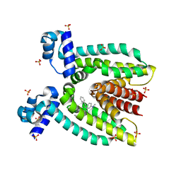
Crystal Structure of the Complex of Ethidium Bound to QacR(E90Q), a Mutant of a Multidrug Binding Transcriptional Repressor
分子名称: ETHIDIUM, HTH-type transcriptional regulator qacR, SULFATE ION
著者Brooks, B.E.
登録日2007-12-20
公開日2008-12-23
最終更新日2023-08-30
実験手法X-RAY DIFFRACTION (2.8 Å)
主引用文献Formal electrostatic interactions do not govern QacR-cation affinity
To be Published
3BR0
DownloadVisualize
BU of 3br0 by Molmil
Crystal Structure of the Complex of Malachite Green Bound to QacR(E120Q), a Mutant of a Multidrug Binding Transcriptional Repressor
分子名称: HTH-type transcriptional regulator qacR, IMIDAZOLE, MALACHITE GREEN, ...
著者Brooks, B.E.
登録日2007-12-20
公開日2008-12-23
最終更新日2023-08-30
実験手法X-RAY DIFFRACTION (2.42 Å)
主引用文献Formal electrostatic interactions do not govern QacR-cation affinity
To be Published
3BQZ
DownloadVisualize
BU of 3bqz by Molmil
Crystal Structure of the Complex of Malachite Green Bound to QacR(E90Q), a Mutant of a Multidrug Binding Transcriptional Repressor
分子名称: HTH-type transcriptional regulator qacR, IMIDAZOLE, MALACHITE GREEN, ...
著者Brooks, B.E.
登録日2007-12-20
公開日2008-12-23
最終更新日2023-08-30
実験手法X-RAY DIFFRACTION (2.17 Å)
主引用文献Formal electrostatic interactions do not govern QacR-cation affinity
To be Published
3BR6
DownloadVisualize
BU of 3br6 by Molmil
Crystal Structure of the Complex of Rhodamine 6G Bound to QacR(E120Q), a Mutant of a Multidrug Binding Transcriptional Repressor
分子名称: HTH-type transcriptional regulator qacR, RHODAMINE 6G, SULFATE ION
著者Brooks, B.E.
登録日2007-12-20
公開日2008-12-23
最終更新日2023-08-30
実験手法X-RAY DIFFRACTION (3.2 Å)
主引用文献Formal electrostatic interactions do not govern QacR-cation affinity
To be Published
3BR5
DownloadVisualize
BU of 3br5 by Molmil
Crystal Structure of the Complex of Rhodamine 6G Bound to QacR(E90Q), a Mutant of a Multidrug Binding Transcriptional Repressor
分子名称: HTH-type transcriptional regulator qacR, RHODAMINE 6G, SULFATE ION
著者Brooks, B.E.
登録日2007-12-20
公開日2008-12-23
最終更新日2023-08-30
実験手法X-RAY DIFFRACTION (2.9 Å)
主引用文献Formal electrostatic interactions do not govern QacR-cation affinity
To be Published
2Q9D
DownloadVisualize
BU of 2q9d by Molmil
Structure of spin-labeled T4 lysozyme mutant A41R1
分子名称: BETA-MERCAPTOETHANOL, Lysozyme, S-[(1-oxyl-2,2,5,5-tetramethyl-2,5-dihydro-1H-pyrrol-3-yl)methyl] methanesulfonothioate
著者Guo, Z, Cascio, D, Hideg, K, Hubbell, W.L.
登録日2007-06-12
公開日2007-06-26
最終更新日2023-08-30
実験手法EPR (1.4 Å), X-RAY DIFFRACTION
主引用文献Structural determinants of nitroxide motion in spin-labeled proteins: Solvent-exposed sites in helix B of T4 lysozyme.
Protein Sci., 17, 2008
3D1M
DownloadVisualize
BU of 3d1m by Molmil
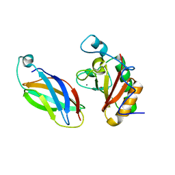
Crystal Structure of Sonic Hedgehog Bound to the third FNIII domain of CDO
分子名称: CALCIUM ION, Cell adhesion molecule, Sonic hedgehog protein, ...
著者McLellan, J.S, Leahy, D.J.
登録日2008-05-06
公開日2008-09-23
最終更新日2024-02-21
実験手法X-RAY DIFFRACTION (1.7 Å)
主引用文献The mode of Hedgehog binding to Ihog homologues is not conserved across different phyla.
Nature, 455, 2008
1SVJ
DownloadVisualize
BU of 1svj by Molmil
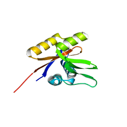
The solution structure of the nucleotide binding domain of KdpB
分子名称: Potassium-transporting ATPase B chain
著者Haupt, M, Bramkamp, M, Coles, M, Altendorf, K, Kessler, H.
登録日2004-03-29
公開日2004-09-21
最終更新日2024-05-22
実験手法SOLUTION NMR
主引用文献Inter-domain motions of the N-domain of the KdpFABC complex, a P-type ATPase, are not driven by ATP-induced conformational changes.
J.Mol.Biol., 342, 2004
4W1W
DownloadVisualize
BU of 4w1w by Molmil
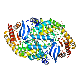
Crystal structure of 7,8-diaminopelargonic acid synthase (BioA) from Mycobacterium tuberculosis, complexed with 7-(diethylamino)-3-(thiophene-2-carbonyl)-2H-chromen-2-one
分子名称: 1,2-ETHANEDIOL, 7-(diethylamino)-3-(thiophen-2-ylcarbonyl)-2H-chromen-2-one, Adenosylmethionine-8-amino-7-oxononanoate aminotransferase, ...
著者Finzel, B.C, Ran, D.
登録日2014-08-13
公開日2015-02-04
最終更新日2023-12-27
実験手法X-RAY DIFFRACTION (1.9 Å)
主引用文献Target-Based Identification of Whole-Cell Active Inhibitors of Biotin Biosynthesis in Mycobacterium tuberculosis.
Chem.Biol., 22, 2015
1U7Q
DownloadVisualize
BU of 1u7q by Molmil
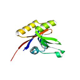
THE SOLUTION STRUCTURE OF THE NUCLEOTIDE BINDING DOMAIN OF KDPB
分子名称: Potassium-transporting ATPase B chain
著者Haupt, M, Bramkamp, M, Coles, M, Altendorf, K, Kessler, H.
登録日2004-08-04
公開日2004-09-21
最終更新日2024-05-22
実験手法SOLUTION NMR
主引用文献Inter-domain motions of the N-domain of the KdpFABC complex, a P-type ATPase, are not driven by ATP-induced conformational changes.
J.Mol.Biol., 342, 2004

225399

件を2024-09-25に公開中

PDB statisticsPDBj update infoContact PDBjnumon