4CY3
 
 | | Crystal structure of the NSL1-WDS complex. | | 分子名称: | CG4699, ISOFORM D, GLYCEROL, ... | | 著者 | Dias, J, Brettschneider, J, Cusack, S, Kadlec, J. | | 登録日 | 2014-04-10 | | 公開日 | 2014-05-14 | | 最終更新日 | 2023-12-20 | | 実験手法 | X-RAY DIFFRACTION (1.4 Å) | | 主引用文献 | Structural Analysis of the Kansl1/Wdr5/Kansl2 Complex Reveals that Wdr5 is Required for Efficient Assembly and Chromatin Targeting of the Nsl Complex.
Genes Dev., 28, 2014
|
|
1Y5X
 | | tRNA-guanine Transglycosylase (TGT) in complex with 6-Amino-4-[2-(4-methoxyphenyl)ethyl]-1,7-dihydro-8H-imidazo[4,5-g]quinazolin-8-one | | 分子名称: | 6-AMINO-4-[2-(4-METHOXYPHENYL)ETHYL]-1,7-DIHYDRO-8H-IMIDAZO[4,5-G]QUINAZOLIN-8-ONE, Queuine tRNA-ribosyltransferase, ZINC ION | | 著者 | Stengl, B, Meyer, E.A, Heine, A, Brenk, R, Diederich, F, Klebe, G. | | 登録日 | 2004-12-03 | | 公開日 | 2005-12-13 | | 最終更新日 | 2023-10-25 | | 実験手法 | X-RAY DIFFRACTION (2.1 Å) | | 主引用文献 | Crystal structures of tRNA-guanine transglycosylase (TGT) in complex with novel and potent inhibitors unravel pronounced induced-fit adaptations and suggest dimer formation upon substrate binding
J.Mol.Biol., 370, 2007
|
|
3TT4
 | | Human MMP8 in complex with L-glutamate motif inhibitor | | 分子名称: | 2-(N-MORPHOLINO)-ETHANESULFONIC ACID, CALCIUM ION, Neutrophil collagenase, ... | | 著者 | Stura, E.A, Dive, V, Devel, L, Czarny, B, Beau, F, Vera, L. | | 登録日 | 2011-09-14 | | 公開日 | 2012-06-20 | | 最終更新日 | 2023-09-13 | | 実験手法 | X-RAY DIFFRACTION (1.88 Å) | | 主引用文献 | Simple pseudo-dipeptides with a P2' glutamate: a novel inhibitor family of matrix metalloproteases and other metzincins.
J.Biol.Chem., 287, 2012
|
|
3TUU
 | | Structure of dihydrodipicolinate synthase from the common grapevine | | 分子名称: | BROMIDE ION, CHLORIDE ION, dihydrodipicolinate synthase | | 著者 | Perugini, M.A, Dobson, R.C, Atkinson, S.C. | | 登録日 | 2011-09-19 | | 公開日 | 2012-07-25 | | 実験手法 | X-RAY DIFFRACTION (2.2 Å) | | 主引用文献 | Crystal, Solution and In silico Structural Studies of Dihydrodipicolinate Synthase from the Common Grapevine.
Plos One, 7, 2012
|
|
2FZD
 | | Human aldose reductase complexed with tolrestat at 1.08 A resolution. | | 分子名称: | NADP NICOTINAMIDE-ADENINE-DINUCLEOTIDE PHOSPHATE, TOLRESTAT, aldose reductase | | 著者 | Steuber, H, Zentgraf, M, Gerlach, C, Sotriffer, C.A, Heine, A, Klebe, G. | | 登録日 | 2006-02-09 | | 公開日 | 2006-10-03 | | 最終更新日 | 2023-08-30 | | 実験手法 | X-RAY DIFFRACTION (1.08 Å) | | 主引用文献 | Expect the Unexpected or Caveat for Drug Designers: Multiple Structure Determinations Using Aldose Reductase Crystals Treated under Varying Soaking and Co-crystallisation Conditions.
J.Mol.Biol., 363, 2006
|
|
2GAQ
 | |
6C04
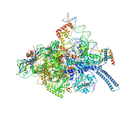 | | Mtb RNAP Holo/RbpA/double fork DNA -closed clamp | | 分子名称: | DNA (26-MER), DNA (31-MER), DNA-directed RNA polymerase subunit alpha, ... | | 著者 | Darst, S.A, Campbell, E.A, Boyaci Selcuk, H, Chen, J, Lilic, M. | | 登録日 | 2017-12-27 | | 公開日 | 2018-03-28 | | 最終更新日 | 2024-03-13 | | 実験手法 | ELECTRON MICROSCOPY (3.27 Å) | | 主引用文献 | Fidaxomicin jamsMycobacterium tuberculosisRNA polymerase motions needed for initiation via RbpA contacts.
Elife, 7, 2018
|
|
7D2O
 | |
3L1N
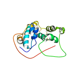
 | | Crystal structure of Mp1p ligand binding domain 2 complexd with palmitic acid | | 分子名称: | Cell wall antigen, PALMITIC ACID | | 著者 | Liao, S, Tung, E.T, Zheng, W, Chong, K, Xu, Y, Bartlam, M, Rao, Z, Yuen, K.Y. | | 登録日 | 2009-12-14 | | 公開日 | 2010-01-05 | | 最終更新日 | 2021-11-10 | | 実験手法 | X-RAY DIFFRACTION (1.3 Å) | | 主引用文献 | Crystal structure of the Mp1p ligand binding domain 2 reveals its function as a fatty acid-binding protein.
J.Biol.Chem., 285, 2010
|
|
3TXQ
 | |
1Y6U
 | |
1YFD
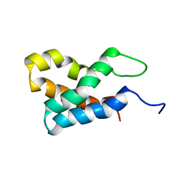
 | | Crystal structure of the Y122H mutant of ribonucleotide reductase R2 protein from E. coli | | 分子名称: | MERCURY (II) ION, MU-OXO-DIIRON, Ribonucleoside-diphosphate reductase 1 beta chain | | 著者 | Kolberg, M, Logan, D.T, Bleifuss, G, Poetsch, S, Sjoeberg, B.M, Graeslund, A, Lubitz, W, Lassmann, G, Lendzian, F. | | 登録日 | 2004-12-31 | | 公開日 | 2005-02-15 | | 最終更新日 | 2023-08-23 | | 実験手法 | X-RAY DIFFRACTION (1.9 Å) | | 主引用文献 | A new tyrosyl radical on Phe208 as ligand to the diiron center in Escherichia coli ribonucleotide reductase, mutant R2-Y122H. Combined x-ray diffraction and EPR/ENDOR studies
J.Biol.Chem., 280, 2005
|
|
4FLG
 | | HIV-1 protease mutant I47V complexed with reaction intermediate | | 分子名称: | CHLORIDE ION, GLUTAMIC ACID, GLYCEROL, ... | | 著者 | Yu, X, Shen, C.H, Weber, I.T. | | 登録日 | 2012-06-14 | | 公開日 | 2012-10-17 | | 最終更新日 | 2024-03-13 | | 実験手法 | X-RAY DIFFRACTION (1.31 Å) | | 主引用文献 | Capturing the Reaction Pathway in Near-Atomic-Resolution Crystal Structures of HIV-1 Protease.
Biochemistry, 51, 2012
|
|
4U6H
 | | Vaccinia L1/M12B9-Fab complex | | 分子名称: | Heavy chain of murine anti-vaccinia L1 IgG2a antibody M12B9, Light chain of murine anti-vaccinia L1 IgG2a antibody M12B9, Protein L1 | | 著者 | Matho, M.H, Schlossman, A, Zajonc, D.M. | | 登録日 | 2014-07-29 | | 公開日 | 2014-08-06 | | 最終更新日 | 2023-09-27 | | 実験手法 | X-RAY DIFFRACTION (3.1 Å) | | 主引用文献 | Potent neutralization of vaccinia virus by divergent murine antibodies targeting a common site of vulnerability in l1 protein.
J.Virol., 88, 2014
|
|
3GWH
 | | Crystallographic Ab Initio protein solution far below atomic resolution | | 分子名称: | PHOSPHATE ION, Transcriptional antiterminator (BglG family) | | 著者 | Rodriguez, D.D, Grosse, C, Himmel, S, Gonzalez, C, Becker, S, Sheldrick, G.M, Uson, I. | | 登録日 | 2009-04-01 | | 公開日 | 2010-04-07 | | 最終更新日 | 2024-03-20 | | 実験手法 | X-RAY DIFFRACTION (1.95 Å) | | 主引用文献 | Crystallographic ab initio protein structure solution below atomic resolution
Nat.Methods, 6, 2009
|
|
7DXJ
 | |
3GI4
 | | Crystal structure of protease inhibitor, KB60 in complex with wild type HIV-1 protease | | 分子名称: | 5S)-N-[(1S,2R)-3-[(1,3-Benzodioxol-5-ylsulfonyl)(2-methylpropyl)amino]-2-hydroxy-1-(phenylmethyl)propyl]-2-oxo-3-[3-(tr ifluoromethyl)phenyl]-5-oxazolidinecarboxamide, ACETATE ION, PHOSPHATE ION, ... | | 著者 | Nalam, M.N.L, Schiffer, C.A. | | 登録日 | 2009-03-05 | | 公開日 | 2010-03-09 | | 最終更新日 | 2024-02-21 | | 実験手法 | X-RAY DIFFRACTION (1.85 Å) | | 主引用文献 | Evaluating the substrate-envelope hypothesis: structural analysis of novel HIV-1 protease inhibitors designed to be robust against drug resistance.
J.Virol., 84, 2010
|
|
2PIN
 | | Thyroid receptor beta in complex with inhibitor | | 分子名称: | 1-(4-HEXYLPHENYL)PROP-2-EN-1-ONE, SULFATE ION, Thyroid hormone receptor beta-1, ... | | 著者 | Estebanez-Perpina, E, Jouravel, N, Baxter, J.D, Guy, L.R, Webb, P, Fletterick, R.J. | | 登録日 | 2007-04-13 | | 公開日 | 2008-02-26 | | 最終更新日 | 2024-02-21 | | 実験手法 | X-RAY DIFFRACTION (2.3 Å) | | 主引用文献 | Structural insight into the mode of action of a direct inhibitor of coregulator binding to the thyroid hormone receptor.
Mol.Endocrinol., 21, 2007
|
|
2A9N
 | | A Mutation Designed to Alter Crystal Packing Permits Structural Analysis of a Tight-binding Fluorescein-scFv complex | | 分子名称: | 4-(2,7-DIFLUORO-6-HYDROXY-3-OXO-3H-XANTHEN-9-YL)ISOPHTHALIC ACID, fluorescein-scfv | | 著者 | Cambillau, C, Spinelli, S, Honegger, A, Pluckthun, A. | | 登録日 | 2005-07-12 | | 公開日 | 2005-10-25 | | 最終更新日 | 2024-04-03 | | 実験手法 | X-RAY DIFFRACTION (3 Å) | | 主引用文献 | A mutation designed to alter crystal packing permits structural analysis of a tight-binding fluorescein-scFv complex.
Protein Sci., 14, 2005
|
|
7DXK
 | |
4F8O
 | | X-ray structure of PsaA from Yersinia pestis, in complex with lactose and AEBSF | | 分子名称: | 4-(2-AMINOETHYL)BENZENESULFONYL FLUORIDE, CHLORIDE ION, GUANIDINE, ... | | 著者 | Bao, R, Esser, L, Xia, D. | | 登録日 | 2012-05-17 | | 公開日 | 2013-05-22 | | 最終更新日 | 2020-07-29 | | 実験手法 | X-RAY DIFFRACTION (1.9 Å) | | 主引用文献 | Structural basis for the specific recognition of dual receptors by the homopolymeric pH 6 antigen (Psa) fimbriae of Yersinia pestis.
Proc.Natl.Acad.Sci.USA, 110, 2013
|
|
1MWA
 | | 2C/H-2KBM3/DEV8 ALLOGENEIC COMPLEX | | 分子名称: | 2-acetamido-2-deoxy-beta-D-glucopyranose, 2-acetamido-2-deoxy-beta-D-glucopyranose-(1-4)-2-acetamido-2-deoxy-beta-D-glucopyranose, 2C T CELL RECEPTOR ALPHA CHAIN, ... | | 著者 | Luz, J.G, Huang, M.D, Garcia, K.C, Rudolph, M.G, Teyton, L, Wilson, I.A. | | 登録日 | 2002-09-27 | | 公開日 | 2002-11-27 | | 最終更新日 | 2020-07-29 | | 実験手法 | X-RAY DIFFRACTION (2.4 Å) | | 主引用文献 | Structural comparison of allogeneic and syngeneic T cell receptor-peptide-major histocompatibility
complex complexes: a buried alloreactive mutation subtly alters peptide presentation substantially
increasing V(beta) Interactions.
J.EXP.MED., 195, 2002
|
|
2PK2
 | | Cyclin box structure of the P-TEFb subunit Cyclin T1 derived from a fusion complex with EIAV Tat | | 分子名称: | Cyclin-T1, Protein Tat | | 著者 | Anand, K, Schulte, A, Fujinaga, K, Scheffzek, K, Geyer, M. | | 登録日 | 2007-04-17 | | 公開日 | 2007-07-03 | | 最終更新日 | 2024-02-21 | | 実験手法 | X-RAY DIFFRACTION (2.67 Å) | | 主引用文献 | Cyclin Box Structure of the P-TEFb Subunit Cyclin T1 Derived from a Fusion Complex with EIAV Tat.
J.Mol.Biol., 370, 2007
|
|
4TX9
 | | Crystal structure of HisAp from Streptomyces sviceus with degraded ProFAR | | 分子名称: | AMINOIMIDAZOLE 4-CARBOXAMIDE RIBONUCLEOTIDE, Phosphoribosyl isomerase A, SULFATE ION | | 著者 | Michalska, K, Verduzco-Castro, E.A, Endres, M, Barona-Gomez, F, Joachimiak, A, Midwest Center for Structural Genomics (MCSG) | | 登録日 | 2014-07-02 | | 公開日 | 2014-08-06 | | 最終更新日 | 2023-12-27 | | 実験手法 | X-RAY DIFFRACTION (1.6 Å) | | 主引用文献 | Co-occurrence of analogous enzymes determines evolution of a novel ( beta alpha )8-isomerase sub-family after non-conserved mutations in flexible loop.
Biochem. J., 473, 2016
|
|
1MTN
 | | BOVINE ALPHA-CHYMOTRYPSIN:BPTI CRYSTALLIZATION | | 分子名称: | ALPHA-CHYMOTRYPSIN, BASIC PANCREATIC TRYPSIN INHIBITOR, SULFATE ION | | 著者 | Capasso, C, Rizzi, M, Menegatti, E, Ascenzi, P, Bolognesi, M. | | 登録日 | 1996-03-28 | | 公開日 | 1996-08-17 | | 最終更新日 | 2023-08-09 | | 実験手法 | X-RAY DIFFRACTION (2.8 Å) | | 主引用文献 | Crystal structure of the bovine alpha-chymotrypsin:Kunitz inhibitor complex. An example of multiple protein:protein recognition sites.
J.Mol.Recog., 10, 1997
|
|


