3H4K
 
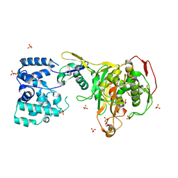
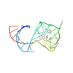
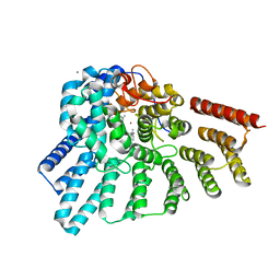
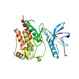
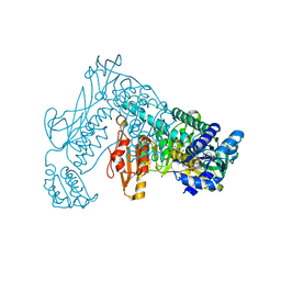 | | Crystal structure of the wild type Thioredoxin glutatione reductase from Schistosoma mansoni in complex with auranofin | | 分子名称: | FLAVIN-ADENINE DINUCLEOTIDE, GLUTATHIONE, GOLD ION, ... | | 著者 | Angelucci, F, Dimastrogiovanni, D, Miele, A.E, Boumis, G, Brunori, M, Bellelli, A. | | 登録日 | 2009-04-20 | | 公開日 | 2009-08-25 | | 最終更新日 | 2023-11-01 | | 実験手法 | X-RAY DIFFRACTION (2.55 Å) | | 主引用文献 | Inhibition of Schistosoma mansoni thioredoxin-glutathione reductase by auranofin: structural and kinetic aspects.
J.Biol.Chem., 284, 2009
|
|
2Y2A
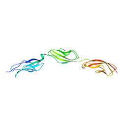
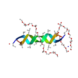
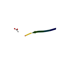 | | Structure of segment KLVFFA from the amyloid-beta peptide (Ab, residues 16-21), alternate polymorph I | | 分子名称: | ACETATE ION, AMYLOID BETA A4 PROTEIN | | 著者 | Colletier, J, Laganowsky, A, Sawaya, M.R, Eisenberg, D. | | 登録日 | 2010-12-14 | | 公開日 | 2011-10-26 | | 最終更新日 | 2024-05-08 | | 実験手法 | X-RAY DIFFRACTION (1.91 Å) | | 主引用文献 | Molecular Basis for Amyloid-{Beta} Polymorphism.
Proc.Natl.Acad.Sci.USA, 108, 2011
|
|
5BQQ
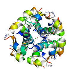 | | Human insulin with intra-chain chemical crosslink between modified B27 and B30 | | 分子名称: | CHLORIDE ION, Insulin, PHENOL, ... | | 著者 | Brzozowski, A.M, Turkenburg, J.P, Jiracek, J, Zakova, L. | | 登録日 | 2015-05-29 | | 公開日 | 2016-02-03 | | 最終更新日 | 2024-01-10 | | 実験手法 | X-RAY DIFFRACTION (1.54 Å) | | 主引用文献 | Rational steering of insulin binding specificity by intra-chain chemical crosslinking.
Sci Rep, 6, 2016
|
|
2FUB
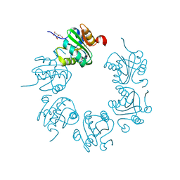
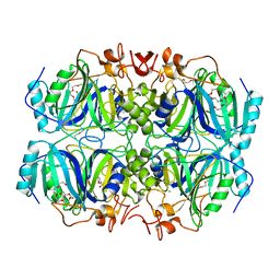
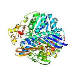
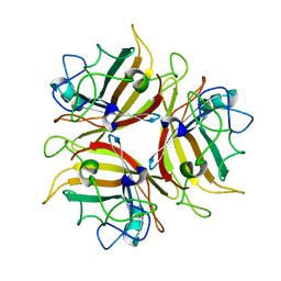
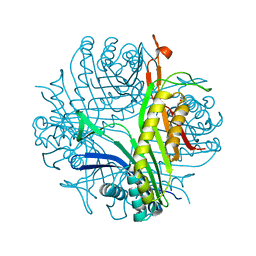 | | Crystal structure of urate oxidase at 140 MPa | | 分子名称: | 8-AZAXANTHINE, CYSTEINE, Uricase | | 著者 | Colloc'h, N, Girard, E, Fourme, R. | | 登録日 | 2006-01-26 | | 公開日 | 2006-02-14 | | 最終更新日 | 2023-11-15 | | 実験手法 | X-RAY DIFFRACTION (2.3 Å) | | 主引用文献 | High pressure macromolecular crystallography: The 140-MPa crystal structure at 2.3 A resolution of urate oxidase, a 135-kDa tetrameric assembly
Biochim.Biophys.Acta, 1764, 2006
|
|
2FUF
 | |
5BQZ
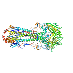 | | Crystal structure of hemagglutinin of A/Chicken/Guangdong/S1311/2010 (H6N6) in complex with human-like receptor LSTc | | 分子名称: | 2-acetamido-2-deoxy-beta-D-glucopyranose, 2-acetamido-2-deoxy-beta-D-glucopyranose-(1-4)-2-acetamido-2-deoxy-beta-D-glucopyranose, HEMAGGLUTININ HA1 CHAIN, ... | | 著者 | Ni, F, Kondrashkina, E, Wang, Q. | | 登録日 | 2015-05-29 | | 公開日 | 2015-10-14 | | 最終更新日 | 2023-09-27 | | 実験手法 | X-RAY DIFFRACTION (2.89 Å) | | 主引用文献 | Structural and Functional Studies of Influenza Virus A/H6 Hemagglutinin.
Plos One, 10, 2015
|
|
4KDL
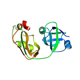 | | Crystal structure of p97/VCP N in complex with OTU1 UBXL | | 分子名称: | Transitional endoplasmic reticulum ATPase, Ubiquitin thioesterase OTU1 | | 著者 | Kim, S.J, Kim, E.E. | | 登録日 | 2013-04-25 | | 公開日 | 2014-03-19 | | 最終更新日 | 2023-09-20 | | 実験手法 | X-RAY DIFFRACTION (1.81 Å) | | 主引用文献 | Structural Basis for Ovarian Tumor Domain-containing Protein 1 (OTU1) Binding to p97/Valosin-containing Protein (VCP).
J.Biol.Chem., 289, 2014
|
|
5BR3
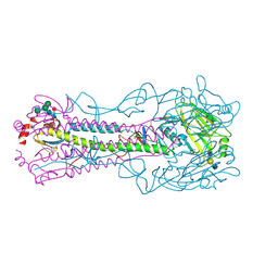 | | Crystal structure of hemagglutinin of A/Taiwan/2/2013 (H6N1) in complex with LSTa | | 分子名称: | 2-acetamido-2-deoxy-beta-D-glucopyranose, HEMAGGLUTININ HA1 CHAIN, HEMAGGLUTININ HA2 CHAIN, ... | | 著者 | Ni, F, Kondrashkina, E, Wang, Q. | | 登録日 | 2015-05-29 | | 公開日 | 2015-08-12 | | 最終更新日 | 2020-07-29 | | 実験手法 | X-RAY DIFFRACTION (2.55 Å) | | 主引用文献 | Structural and Functional Studies of Influenza Virus A/H6 Hemagglutinin.
Plos One, 7, 2015
|
|
3DPX
 | |
2XKM
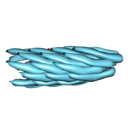 | | Consensus structure of Pf1 filamentous bacteriophage from X-ray fibre diffraction and solid-state NMR | | 分子名称: | CAPSID PROTEIN G8P | | 著者 | Straus, S.K, P Scott, W.R, Schwieters, C.D, Marvin, D.A. | | 登録日 | 2010-07-09 | | 公開日 | 2010-11-24 | | 最終更新日 | 2023-12-20 | | 実験手法 | FIBER DIFFRACTION (3.3 Å), SOLID-STATE NMR | | 主引用文献 | Consensus Structure of Pf1 Filamentous Bacteriophage from X-Ray Fibre Diffraction and Solid-State NMR.
Eur.Biophys.J., 40, 2011
|
|
6BXU
 | |
4Z6T
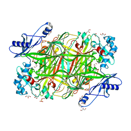
 | | Structure of H200N variant of Homoprotocatechuate 2,3-Dioxygenase from B.fuscum in complex with 4-sulfonyl catechol at 1.50 Ang resolution | | 分子名称: | 3,4-dihydroxybenzenesulfonic acid, CALCIUM ION, CHLORIDE ION, ... | | 著者 | Kovaleva, E.G, Lipscomb, J.D. | | 登録日 | 2015-04-06 | | 公開日 | 2015-08-26 | | 最終更新日 | 2023-09-27 | | 実験手法 | X-RAY DIFFRACTION (1.5 Å) | | 主引用文献 | Structural Basis for Substrate and Oxygen Activation in Homoprotocatechuate 2,3-Dioxygenase: Roles of Conserved Active Site Histidine 200.
Biochemistry, 54, 2015
|
|
3DWK
 | |
3DQ5
 | |
3DQE
 | |
6C63
 | |
2XDC
 | |
2FRV
 | | CRYSTAL STRUCTURE OF THE OXIDIZED FORM OF NI-FE HYDROGENASE | | 分子名称: | CARBONMONOXIDE-(DICYANO) IRON, FE3-S4 CLUSTER, IRON/SULFUR CLUSTER, ... | | 著者 | Volbeda, A, Frey, M, Fontecilla-Camps, J.C. | | 登録日 | 1997-06-10 | | 公開日 | 1998-06-17 | | 最終更新日 | 2023-08-09 | | 実験手法 | X-RAY DIFFRACTION (2.54 Å) | | 主引用文献 | Structure of the [Nife] Hydrogenase Active Site: Evidence for Biologically Uncommon Fe Ligands
J.Am.Chem.Soc., 118, 1996
|
|
2XKU
 | | Prion-like conversion during amyloid formation at atomic resolution | | 分子名称: | BETA-2-MICROGLOBULIN | | 著者 | Eichner, T, Kalverda, A.P, Thompson, G.S, Homans, S.W, Radford, S.E. | | 登録日 | 2010-07-12 | | 公開日 | 2011-02-09 | | 最終更新日 | 2023-06-14 | | 実験手法 | SOLUTION NMR | | 主引用文献 | Conformational Conversion During Amyloid Formation at Atomic Resolution.
Mol.Cell, 41, 2011
|
|
4KFD
 | | Crystal structure of Hansenula polymorpha copper amine oxidase-1 reduced by methylamine at pH 6.0 | | 分子名称: | COPPER (II) ION, GLYCEROL, HYDROGEN PEROXIDE, ... | | 著者 | Johnson, B.J, Yukl, E.T, Klema, V.J, Wilmot, C.M. | | 登録日 | 2013-04-26 | | 公開日 | 2013-08-21 | | 実験手法 | X-RAY DIFFRACTION (1.69 Å) | | 主引用文献 | Structural evidence for the semiquinone in a copper amine oxidase from Hansenula polymorpha: implications for the catalytic mechanism
J.Biol.Chem., 2013
|
|
3DSX
 | | Crystal structure of RabGGTase(DELTA LRR; DELTA IG)in complex with di-prenylated peptide Ser-Cys(GG)-Ser-Cys(GG) derivated from Rab7 | | 分子名称: | CALCIUM ION, GERAN-8-YL GERAN, Geranylgeranyl transferase type-2 subunit alpha, ... | | 著者 | Guo, Z, Yu, S, Goody, R.S, Alexandrov, K, Blankenfeldt, W. | | 登録日 | 2008-07-14 | | 公開日 | 2008-09-09 | | 最終更新日 | 2023-11-01 | | 実験手法 | X-RAY DIFFRACTION (2.1 Å) | | 主引用文献 | Structures of RabGGTase-substrate/product complexes provide insights into the evolution of protein prenylation
Embo J., 27, 2008
|
|
4ZDG
 | |
2FVT
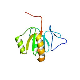 | | NMR Structure of the Rpa2829 protein from Rhodopseudomonas palustris: Northeast Structural Genomics Target RpR43 | | 分子名称: | conserved hypothetical protein | | 著者 | Cort, J.R, Ho, C.K, Cunningham, K, Ma, L.C, Conover, K, Xiao, R, Montelione, G.T, Kennedy, M.A, Northeast Structural Genomics Consortium (NESG) | | 登録日 | 2006-01-31 | | 公開日 | 2006-02-14 | | 最終更新日 | 2024-05-29 | | 実験手法 | SOLUTION NMR | | 主引用文献 | NMR Structure of the Rpa2829 protein from Rhodopseudomonas palustris
To be Published
|
|
1M5U
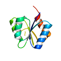 | | CRYSTAL STRUCTURE OF THE RESPONSE REGULATOR DIVK. STRUCTURE AT PH 8.0 IN THE APO-FORM | | 分子名称: | cell division response regulator DivK | | 著者 | Guillet, V, Ohta, N, Cabantous, S, Newton, A, Samama, J.-P, Structural Proteomics in Europe (SPINE) | | 登録日 | 2002-07-10 | | 公開日 | 2002-11-15 | | 最終更新日 | 2024-04-03 | | 実験手法 | X-RAY DIFFRACTION (1.87 Å) | | 主引用文献 | Crystallographic and biochemical studies of DivK reveal novel features of an essential response regulator in Caulobacter crescentus
J.Biol.Chem., 277, 2002
|
|
5AM7
 | | FGFR1 mutant with an inhibitor | | 分子名称: | 4-amino-5-fluoro-3-[5-(4-methylpiperazin-1-yl)-1H-benzimidazol-2-yl]quinolin-2(1H)-one, CHLORIDE ION, FIBROBLAST GROWTH FACTOR RECEPTOR 1 | | 著者 | Bunney, T.D, Wan, S, Thiyagarajan, N, Sutto, L, Williams, S.V, Ashford, P, Koss, H, Knowles, M.A, Gervasio, F.L, Coveney, P.V, Katan, M. | | 登録日 | 2015-03-10 | | 公開日 | 2015-03-18 | | 最終更新日 | 2024-01-10 | | 実験手法 | X-RAY DIFFRACTION (1.957 Å) | | 主引用文献 | The Effect of Mutations on Drug Sensitivity and Kinase Activity of Fibroblast Growth Factor Receptors: A Combined Experimental and Theoretical Study
Ebiomedicine, 2, 2015
|
|


