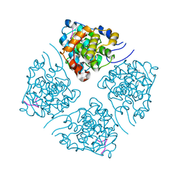
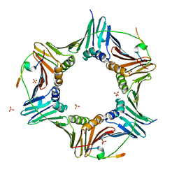
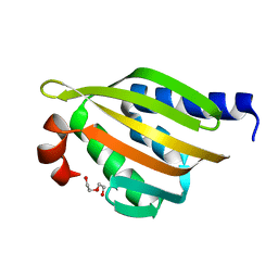
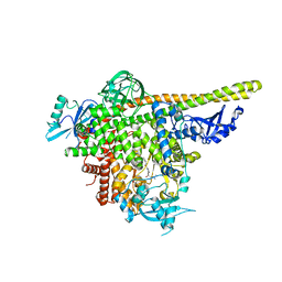
5I5M
 
 | | Shewanella denitrificans nitrous oxide reductase, Ca2+-reconstituted form | | 分子名称: | (R,R)-2,3-BUTANEDIOL, CALCIUM ION, Nitrous-oxide reductase, ... | | 著者 | Schneider, L.K, Einsle, O. | | 登録日 | 2016-02-15 | | 公開日 | 2016-03-02 | | 最終更新日 | 2024-01-10 | | 実験手法 | X-RAY DIFFRACTION (1.37 Å) | | 主引用文献 | Role of Calcium in Secondary Structure Stabilization during Maturation of Nitrous Oxide Reductase.
Biochemistry, 55, 2016
|
|
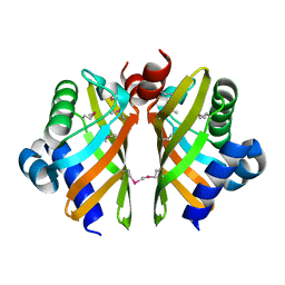
6HMQ
 | | STRUCTURE OF PROTEIN KINASE CK2 CATALYTIC SUBUNIT (ISOFORM CK2ALPHA'; CSNK2A2 GENE PRODUCT) IN COMPLEX WITH THE BENZOTRIAZOLE-TYPE INHIBITOR MB002 | | 分子名称: | 1,2-ETHANEDIOL, 2-AMINO-2-HYDROXYMETHYL-PROPANE-1,3-DIOL, 3-(4,5,6,7-tetrabromo-1H-benzotriazol-1-yl)propan-1-ol, ... | | 著者 | Niefind, K, Lindenblatt, D, Applegate, V.M, Jose, J, Le Borgne, M. | | 登録日 | 2018-09-12 | | 公開日 | 2019-03-27 | | 最終更新日 | 2024-05-15 | | 実験手法 | X-RAY DIFFRACTION (0.97 Å) | | 主引用文献 | Diacritic Binding of an Indenoindole Inhibitor by CK2 alpha Paralogs Explored by a Reliable Path to Atomic Resolution CK2 alpha ' Structures.
Acs Omega, 4, 2019
|
|
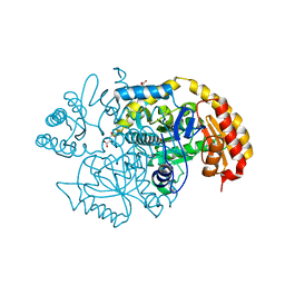
7LOX
 | | The structure of Agmatinase from E. Coli at 3.2 A displaying guanidine in the active site | | 分子名称: | Agmatinase, GUANIDINE, MANGANESE (II) ION | | 著者 | Maturana, P, Figueroa, M, Gonzalez-Ordenes, F, Villalobos, P, Martinez-Oyanedel, J, Uribe, E.A, Castro-Fernandez, V. | | 登録日 | 2021-02-11 | | 公開日 | 2021-05-12 | | 最終更新日 | 2023-10-18 | | 実験手法 | X-RAY DIFFRACTION (3.2 Å) | | 主引用文献 | Crystal Structure of Escherichia coli Agmatinase: Catalytic Mechanism and Residues Relevant for Substrate Specificity.
Int J Mol Sci, 22, 2021
|
|
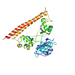
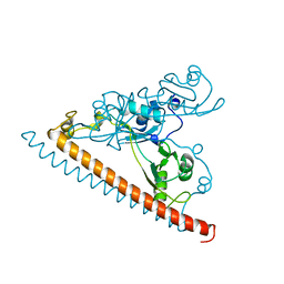
6D32
 | | Crystal structure of Xenopus Smoothened in complex with cyclopamine | | 分子名称: | Cyclopamine, Smoothened,Soluble cytochrome b562,Smoothened | | 著者 | Huang, P, Zheng, S, Kim, Y, Kruse, A.C, Salic, A. | | 登録日 | 2018-04-14 | | 公開日 | 2018-05-23 | | 最終更新日 | 2023-10-04 | | 実験手法 | X-RAY DIFFRACTION (3.751 Å) | | 主引用文献 | Structural Basis of Smoothened Activation in Hedgehog Signaling.
Cell, 174, 2018
|
|
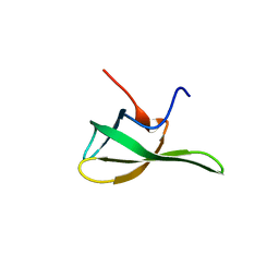
5IEH
 | |
7LUO
 | | N-terminus of Skp2 bound to Cyclin A | | 分子名称: | S-phase kinase-associated protein 2,Cyclin-A2, Skp2 Motif 1 uncharacterized fragment 1, Skp2 Motif 1 uncharacterized fragment 2 | | 著者 | Kelso, S, Ceccarelli, D.F, Sicheri, F. | | 登録日 | 2021-02-22 | | 公開日 | 2021-05-12 | | 最終更新日 | 2023-10-18 | | 実験手法 | X-RAY DIFFRACTION (3.17 Å) | | 主引用文献 | Bipartite binding of the N terminus of Skp2 to cyclin A.
Structure, 29, 2021
|
|
6HNL
 | | Selenomethionine derivative of IdmH 96-104 loop truncation variant | | 分子名称: | Putative polyketide cyclase IdmH | | 著者 | Drulyte, I, Obajdin, J, Trinh, C, Hemsworth, G.R, Berry, A. | | 登録日 | 2018-09-16 | | 公開日 | 2019-11-06 | | 最終更新日 | 2019-11-20 | | 実験手法 | X-RAY DIFFRACTION (2.2 Å) | | 主引用文献 | Crystal structure of the putative cyclase IdmH from the indanomycin nonribosomal peptide synthase/polyketide synthase.
Iucrj, 6, 2019
|
|
5UC7
 | | Crystal structure of BioA / 7,8-diaminopelargonic acid aminotransferase / DAPA synthase from Citrobacter rodentium, PLP complex | | 分子名称: | Adenosylmethionine-8-amino-7-oxononanoate aminotransferase, D(-)-TARTARIC ACID | | 著者 | Stogios, P.J, Evdokimova, E, Di Leo, R, Savchenko, A, Anderson, W.F, Center for Structural Genomics of Infectious Diseases (CSGID) | | 登録日 | 2016-12-21 | | 公開日 | 2017-01-25 | | 最終更新日 | 2023-11-15 | | 実験手法 | X-RAY DIFFRACTION (1.835 Å) | | 主引用文献 | Crystal structure of BioA / 7,8-diaminopelargonic acid aminotransferase / DAPA synthase from Citrobacter rodentium, PLP complex
To Be Published
|
|
7LRQ
 | | Crystal structure of human SFPQ/NONO heterodimer, conserved DBHS region | | 分子名称: | CHLORIDE ION, Non-POU domain-containing octamer-binding protein, Splicing factor, ... | | 著者 | Marshall, A.C, Bond, C.S, Mohnen, I. | | 登録日 | 2021-02-17 | | 公開日 | 2021-05-12 | | 最終更新日 | 2023-10-25 | | 実験手法 | X-RAY DIFFRACTION (2.3 Å) | | 主引用文献 | Paraspeckle subnuclear bodies depend on dynamic heterodimerisation of DBHS RNA-binding proteins via their structured domains.
J.Biol.Chem., 298, 2022
|
|
6HVO
 | | Crystal structure of human PCNA in complex with three peptides of p12 subunit of human polymerase delta | | 分子名称: | DNA polymerase delta subunit 4, Proliferating cell nuclear antigen, SULFATE ION | | 著者 | Gonzalez-Magana, A, Romano-Moreno, M, Rojas, A.L, Blanco, F.J, De Biasio, A. | | 登録日 | 2018-10-11 | | 公開日 | 2019-01-23 | | 最終更新日 | 2019-03-27 | | 実験手法 | X-RAY DIFFRACTION (2.1 Å) | | 主引用文献 | The p12 subunit of human polymerase delta uses an atypical PIP box for molecular recognition of proliferating cell nuclear antigen (PCNA).
J.Biol.Chem., 294, 2019
|
|
7LRU
 | | Crystal structure of SFPQ-NONO-SFPQ chimeric protein homodimer | | 分子名称: | Splicing factor, proline- and glutamine-rich,Isoform 2 of Non-POU domain-containing octamer-binding protein,Isoform Short of Splicing factor, proline- and glutamine-rich | | 著者 | Marshall, A.C, Bond, C.S, Mohnen, I, Knott, G.J. | | 登録日 | 2021-02-17 | | 公開日 | 2021-05-12 | | 最終更新日 | 2023-10-18 | | 実験手法 | X-RAY DIFFRACTION (1.6 Å) | | 主引用文献 | Crystal structure of SFPQ-NONO-SFPQ chimeric protein homodimer
To Be Published
|
|
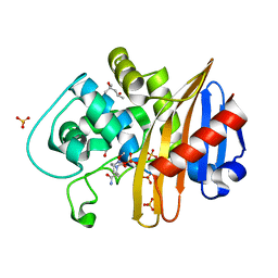
5I0S
 | |
7LOL
 | | The structure of Agmatinase from E. Coli at 1.8 A displaying urea and agmatine | | 分子名称: | 2-AMINO-2-HYDROXYMETHYL-PROPANE-1,3-DIOL, AGMATINE, Agmatinase, ... | | 著者 | Maturana, P, Figueroa, M, Gonzalez-Ordenes, F, Villalobos, P, Martinez-Oyanedel, J, Uribe, E.A, Castro-Fernandez, V. | | 登録日 | 2021-02-10 | | 公開日 | 2021-05-12 | | 最終更新日 | 2023-10-18 | | 実験手法 | X-RAY DIFFRACTION (1.8 Å) | | 主引用文献 | Crystal Structure of Escherichia coli Agmatinase: Catalytic Mechanism and Residues Relevant for Substrate Specificity.
Int J Mol Sci, 22, 2021
|
|
5TRV
 | | Crystal structure of a de novo designed protein with curved beta-sheet | | 分子名称: | DI(HYDROXYETHYL)ETHER, denovo NTF2 | | 著者 | Basanta, B, Oberdorfer, G, Marcos, E, Chidyausiku, T.M, Sankaran, B, Baker, D. | | 登録日 | 2016-10-27 | | 公開日 | 2017-01-25 | | 最終更新日 | 2024-03-06 | | 実験手法 | X-RAY DIFFRACTION (2.91 Å) | | 主引用文献 | Principles for designing proteins with cavities formed by curved beta sheets.
Science, 355, 2017
|
|
6HXQ
 | |
5I9S
 | | MicroED structure of proteinase K at 1.75 A resolution | | 分子名称: | Proteinase K, SULFATE ION | | 著者 | Hattne, J, Shi, D, de la Cruz, M.J, Reyes, F.E, Gonen, T. | | 登録日 | 2016-02-20 | | 公開日 | 2016-06-08 | | 最終更新日 | 2023-08-30 | | 実験手法 | ELECTRON CRYSTALLOGRAPHY (1.75 Å) | | 主引用文献 | Modeling truncated pixel values of faint reflections in MicroED images.
J.Appl.Crystallogr., 49, 2016
|
|
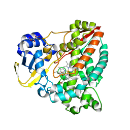
6HYC
 | |
7LNO
 | | Structure of apo-CDD-1 beta-lactamase in imidazole and MPD | | 分子名称: | (4S)-2-METHYL-2,4-PENTANEDIOL, Beta-lactamase, SULFATE ION | | 著者 | Smith, C.A, Vakulenko, S.B, Stewart, N.K. | | 登録日 | 2021-02-08 | | 公開日 | 2021-05-12 | | 最終更新日 | 2023-11-15 | | 実験手法 | X-RAY DIFFRACTION (1.58 Å) | | 主引用文献 | In Crystallo Time-Resolved Interaction of the Clostridioides difficile CDD-1 enzyme with Avibactam Provides New Insights into the Catalytic Mechanism of Class D beta-lactamases.
Acs Infect Dis., 7, 2021
|
|
6HQ9
 | | Crystal structure of the Tudor domain of human ERCC6-L2 | | 分子名称: | DNA excision repair protein ERCC-6-like 2 | | 著者 | Newman, J.A, Gavard, A.E, Nathan, W.J, Pinkas, D.M, von Delft, F, Arrowsmith, C.H, Edwards, A, Bountra, C, Gileadi, O. | | 登録日 | 2018-09-24 | | 公開日 | 2018-10-17 | | 最終更新日 | 2024-01-24 | | 実験手法 | X-RAY DIFFRACTION (1.982 Å) | | 主引用文献 | Crystal structure of the Tudor domain of human ERCC6-L2
To Be Published
|
|
7LNQ
 | | Structure of the avibactam-CDD-1 3 minute complex in imidazole and MPD | | 分子名称: | (2S,5R)-1-formyl-5-[(sulfooxy)amino]piperidine-2-carboxamide, (4S)-2-METHYL-2,4-PENTANEDIOL, Beta-lactamase, ... | | 著者 | Smith, C.A, Vakulenko, S.B, Stewart, N.K. | | 登録日 | 2021-02-08 | | 公開日 | 2021-05-12 | | 最終更新日 | 2023-11-15 | | 実験手法 | X-RAY DIFFRACTION (1.73 Å) | | 主引用文献 | In Crystallo Time-Resolved Interaction of the Clostridioides difficile CDD-1 enzyme with Avibactam Provides New Insights into the Catalytic Mechanism of Class D beta-lactamases.
Acs Infect Dis., 7, 2021
|
|
6HQD
 | |
6CMC
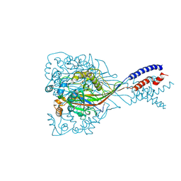 | |
5UL1
 | | The co-structure of 3-amino-6-(4-((1-(dimethylamino)propan-2-yl)sulfonyl)phenyl)-N-phenylpyrazine-2-carboxamide and a rationally designed PI3K-alpha mutant that mimics ATR | | 分子名称: | 3-amino-6-(4-{[(2S)-1-(dimethylamino)propan-2-yl]sulfonyl}phenyl)-N-phenylpyrazine-2-carboxamide, Phosphatidylinositol 3-kinase regulatory subunit alpha, Phosphatidylinositol 4,5-bisphosphate 3-kinase catalytic subunit alpha isoform | | 著者 | Knapp, M.S, Elling, R.A, Mamo, M. | | 登録日 | 2017-01-23 | | 公開日 | 2017-05-10 | | 最終更新日 | 2024-04-03 | | 実験手法 | X-RAY DIFFRACTION (3 Å) | | 主引用文献 | Rationally Designed PI3K alpha Mutants to Mimic ATR and Their Use to Understand Binding Specificity of ATR Inhibitors.
J. Mol. Biol., 429, 2017
|
|
5I2U
 | | Crystal structure of a novel Halo-Tolerant Cellulase from Soil Metagenome | | 分子名称: | Cellulase, GLYCEROL, MAGNESIUM ION, ... | | 著者 | Garg, R, Brahma, V, Srivastava, R, Verma, L, Karthikeyan, S, Sahni, G. | | 登録日 | 2016-02-09 | | 公開日 | 2017-01-18 | | 最終更新日 | 2023-11-08 | | 実験手法 | X-RAY DIFFRACTION (2.2 Å) | | 主引用文献 | Biochemical and structural characterization of a novel halotolerant cellulase from soil metagenome
Sci Rep, 6, 2016
|
|
6CMQ
 | | Structure of human SHP2 without N-SH2 domain | | 分子名称: | Tyrosine-protein phosphatase non-receptor type 11 | | 著者 | Padua, R.A.P, Sun, Y, Marko, I, Pitsawong, W, Kern, D. | | 登録日 | 2018-03-06 | | 公開日 | 2018-11-14 | | 最終更新日 | 2023-10-04 | | 実験手法 | X-RAY DIFFRACTION (2.9 Å) | | 主引用文献 | Mechanism of activating mutations and allosteric drug inhibition of the phosphatase SHP2.
Nat Commun, 9, 2018
|
|


