7V9D
 
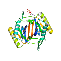
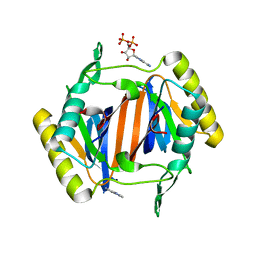
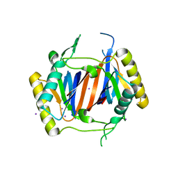
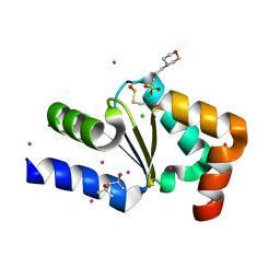
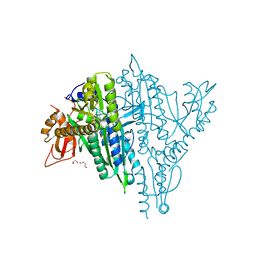 | | Plasmodium falciparum Prolyl-tRNA Synthetase (PfPRS) in Complex with inhibitor L95 and azetidine | | 分子名称: | (2S)-azetidine-2-carboxylic acid, 1,2-ETHANEDIOL, 1,4-BUTANEDIOL, ... | | 著者 | Manickam, Y, Malhotra, N, Sharma, A. | | 登録日 | 2021-08-24 | | 公開日 | 2022-09-07 | | 最終更新日 | 2023-11-29 | | 実験手法 | X-RAY DIFFRACTION (1.937 Å) | | 主引用文献 | Plasmodium falciparum Prolyl-tRNA Synthetase (PfPRS) in Complex with inhibitor L95 and azetidine
To Be Published
|
|
5C5V
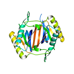
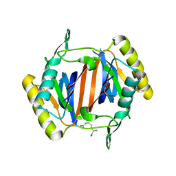
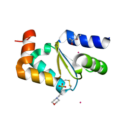
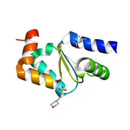
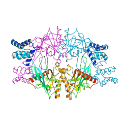 | | Recombinant Inorganic Pyrophosphatase from T brucei brucei | | 分子名称: | 1,2-ETHANEDIOL, Acidocalcisomal pyrophosphatase, BROMIDE ION, ... | | 著者 | Jamwal, A, Yogavel, M, Sharma, A. | | 登録日 | 2015-06-22 | | 公開日 | 2015-11-04 | | 最終更新日 | 2024-03-20 | | 実験手法 | X-RAY DIFFRACTION (2.35 Å) | | 主引用文献 | Structural and Functional Highlights of Vacuolar Soluble Protein 1 from Pathogen Trypanosoma brucei brucei
J.Biol.Chem., 290, 2015
|
|
3KNP
 | |
3KOB
 | |
3KO4
 | |
3KOC
 | |
3KNF
 | |
3KO7
 | |
3KO3
 | |
3KOD
 | |
3KO9
 | |
3KO5
 | |
3LMU
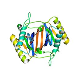
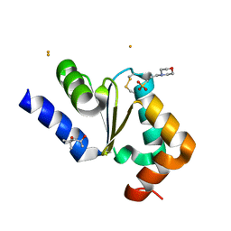
 | | Crystal structure of DTD from Plasmodium falciparum | | 分子名称: | D-tyrosyl-tRNA(Tyr) deacylase, IODIDE ION | | 著者 | Manickam, Y, Bhatt, T.K, Khan, S, Sharma, A. | | 登録日 | 2010-02-01 | | 公開日 | 2010-03-02 | | 最終更新日 | 2024-03-20 | | 実験手法 | X-RAY DIFFRACTION (3.3 Å) | | 主引用文献 | Structure of D-tyrosyl-tRNATyr deacylase using home-source Cu Kalpha and moderate-quality iodide-SAD data: structural polymorphism and HEPES-bound enzyme states
Acta Crystallogr.,Sect.D, 66, 2010
|
|
3AFQ
 | | Crystal structure of the single-stranded DNA binding protein from Mycobacterium leprae (Form II) | | 分子名称: | Single-stranded DNA-binding protein | | 著者 | Kaushal, P.S, Singh, P, Sharma, A, Muniyappa, K, Vijayan, M. | | 登録日 | 2010-03-10 | | 公開日 | 2010-10-06 | | 最終更新日 | 2023-11-01 | | 実験手法 | X-RAY DIFFRACTION (2.8 Å) | | 主引用文献 | X-ray and molecular-dynamics studies on Mycobacterium leprae single-stranded DNA-binding protein and comparison with other eubacterial SSB structures
Acta Crystallogr.,Sect.D, 66, 2010
|
|
3AFP
 | | Crystal structure of the single-stranded DNA binding protein from Mycobacterium leprae (Form I) | | 分子名称: | CADMIUM ION, GLYCEROL, Single-stranded DNA-binding protein | | 著者 | Kaushal, P.S, Singh, P, Sharma, A, Muniyappa, K, Vijayan, M. | | 登録日 | 2010-03-10 | | 公開日 | 2010-10-06 | | 最終更新日 | 2023-11-01 | | 実験手法 | X-RAY DIFFRACTION (2.05 Å) | | 主引用文献 | X-ray and molecular-dynamics studies on Mycobacterium leprae single-stranded DNA-binding protein and comparison with other eubacterial SSB structures
Acta Crystallogr.,Sect.D, 66, 2010
|
|
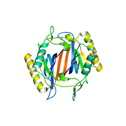
7DIN
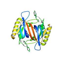
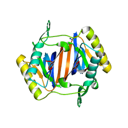
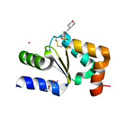
 | | Structure of PfGrx1 with cobalt | | 分子名称: | (4S)-2-METHYL-2,4-PENTANEDIOL, 3[N-MORPHOLINO]PROPANE SULFONIC ACID, COBALT (II) ION, ... | | 著者 | Manickam, Y, Sharma, A. | | 登録日 | 2020-11-19 | | 公開日 | 2021-11-24 | | 実験手法 | X-RAY DIFFRACTION (1.799 Å) | | 主引用文献 | Interaction of metals with PfGrx1
To Be Published
|
|
7DIP
 | |
7DIS
 | |
7DIZ
 | |
7DIR
 | |
7DIT
 | |
7DJ0
 | |
7DIK
 | |
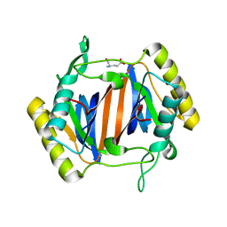
7DIQ
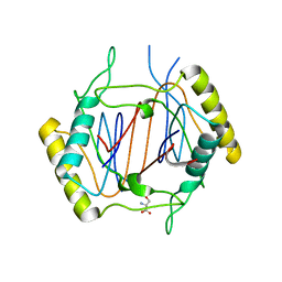
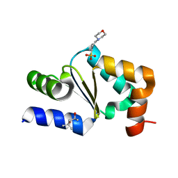
 | | Structure of PfGrx1 with zinc | | 分子名称: | (4S)-2-METHYL-2,4-PENTANEDIOL, 3[N-MORPHOLINO]PROPANE SULFONIC ACID, Glutaredoxin, ... | | 著者 | Manickam, Y, Sharma, A. | | 登録日 | 2020-11-19 | | 公開日 | 2021-11-24 | | 最終更新日 | 2023-11-29 | | 実験手法 | X-RAY DIFFRACTION (1.87 Å) | | 主引用文献 | Interaction of metals with PfGrx1
To Be Published
|
|
7DIU
 | |


