Loading
PDBj
メニューPDBj@FacebookPDBj@TwitterPDBj@YouTubewwPDB FoundationwwPDB
RCSB PDBPDBeBMRBAdv. SearchSearch help
PDB: 43 件

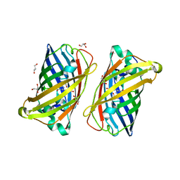
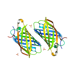
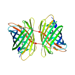
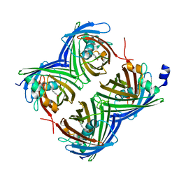
7SF9
DownloadVisualize
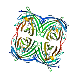
BU of 7sf9 by Molmil
Branchiostoma Floridae Violet Fluorescent Protein
分子名称: Violet Fluorescent Protein
著者Pletnev, S, Pletneva, N, Pletnev, V.Z, Muslinkina, L.
登録日2021-10-03
公開日2022-03-30
最終更新日2023-10-18
実験手法X-RAY DIFFRACTION (1.8 Å)
主引用文献A novel violet fluorescent protein contains a unique oxidized tyrosine as the simplest chromophore ever reported in fluorescent proteins.
Protein Sci., 31, 2022
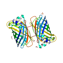
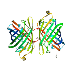
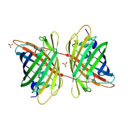
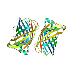
3LVD
DownloadVisualize
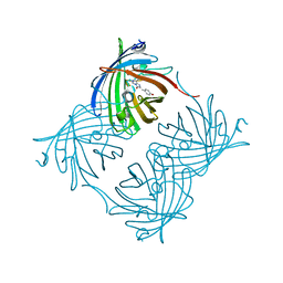
BU of 3lvd by Molmil
Crystal structure of GFP-like protein aceGFP_G222E (A. coerulescens). UV-photoconverted green form.
分子名称: GLYCEROL, Green fluorescent protein
著者Pletneva, N.V, Pletnev, V.Z, Pletnev, S.V.
登録日2010-02-19
公開日2010-03-09
最終更新日2023-11-15
実験手法X-RAY DIFFRACTION (1.751 Å)
主引用文献Structural evidence for a dehydrated intermediate in green fluorescent protein chromophore biosynthesis.
J.Biol.Chem., 285, 2010
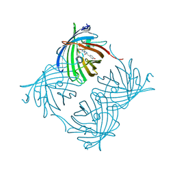
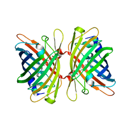
3LVC
DownloadVisualize
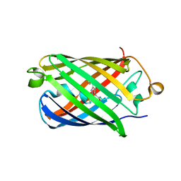
BU of 3lvc by Molmil
Crystal structure of GFP-like protein aceGFP_G222E (A. coerulescens). Colorless form.
分子名称: GLYCEROL, Green fluorescent protein
著者Pletneva, N.V, Pletnev, V.Z, Pletnev, S.V.
登録日2010-02-19
公開日2010-03-09
最終更新日2023-11-15
実験手法X-RAY DIFFRACTION (1.14 Å)
主引用文献Structural evidence for a dehydrated intermediate in green fluorescent protein chromophore biosynthesis.
J.Biol.Chem., 285, 2010
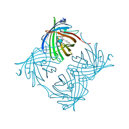
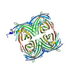
3LVA
DownloadVisualize
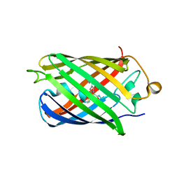
BU of 3lva by Molmil
Crystal structure of colorless GFP-like protein from Aequorea coerulescens
分子名称: GLYCEROL, Green fluorescent protein, SULFATE ION
著者Pletneva, N.V, Pletnev, V.Z, Pletnev, S.V.
登録日2010-02-19
公開日2010-03-09
最終更新日2023-11-15
実験手法X-RAY DIFFRACTION (1.5 Å)
主引用文献Structural evidence for a dehydrated intermediate in green fluorescent protein chromophore biosynthesis.
J.Biol.Chem., 285, 2010
2PXW
DownloadVisualize
BU of 2pxw by Molmil
Crystal Structure of N66D Mutant of Green Fluorescent Protein from Zoanthus sp. at 2.4 A Resolution (Transition State)
分子名称: GFP-like fluorescent chromoprotein FP506
著者Pletnev, S.V, Pletneva, N.V, Tikhonova, T.V, Pletnev, V.Z.
登録日2007-05-14
公開日2007-09-25
最終更新日2024-04-03
実験手法X-RAY DIFFRACTION (2.4 Å)
主引用文献Refined crystal structures of red and green fluorescent proteins from the button polyp Zoanthus.
Acta Crystallogr.,Sect.D, 63, 2007
2PXS
DownloadVisualize
BU of 2pxs by Molmil
Crystal Structure of N66D Mutant of Green Fluorescent Protein from Zoanthus sp. at 2.2 A Resolution (Mature State)
分子名称: GFP-like fluorescent chromoprotein FP506
著者Pletnev, S.V, Pletneva, N.V, Tikhonova, T.V, Pletnev, V.Z.
登録日2007-05-14
公開日2007-09-25
最終更新日2024-04-03
実験手法X-RAY DIFFRACTION (2.2 Å)
主引用文献Refined crystal structures of red and green fluorescent proteins from the button polyp Zoanthus.
Acta Crystallogr.,Sect.D, 63, 2007
3PJ5
DownloadVisualize
BU of 3pj5 by Molmil
Crystal structure of far-red fluorescent protein Katushka crystallized at pH 5.0
分子名称: Red fluorescent protein eqFP578, SULFATE ION
著者Pletnev, S, Pletneva, N.V, Pletnev, V.Z.
登録日2010-11-08
公開日2011-05-25
最終更新日2012-02-22
実験手法X-RAY DIFFRACTION (1.6 Å)
主引用文献Crystallographic study of red fluorescent protein eqFP578 and its far-red variant Katushka reveals opposite pH-induced isomerization of chromophore.
Protein Sci., 20, 2011
3PJB
DownloadVisualize
BU of 3pjb by Molmil
Crystal structure of red fluorescent protein eqFP578 crystallized at pH 4.0
分子名称: GLYCEROL, Red fluorescent protein eqFP578
著者Pletnev, S, Pletneva, N.V, Pletnev, V.Z.
登録日2010-11-09
公開日2011-05-25
最終更新日2018-01-24
実験手法X-RAY DIFFRACTION (1.75 Å)
主引用文献Crystallographic study of red fluorescent protein eqFP578 and its far-red variant Katushka reveals opposite pH-induced isomerization of chromophore.
Protein Sci., 20, 2011
4HE4
DownloadVisualize
BU of 4he4 by Molmil
Crystal structure of the yellow fluorescent protein phiYFP (Phialidium sp.)
分子名称: Yellow fluorescent protein
著者Pletneva, N, Pletnev, S, Pletnev, V.Z.
登録日2012-10-03
公開日2013-05-29
最終更新日2023-12-06
実験手法X-RAY DIFFRACTION (2.05 Å)
主引用文献Yellow fluorescent protein phiYFPv (Phialidium): structure and structure-based mutagenesis.
Acta Crystallogr.,Sect.D, 69, 2013
3PIB
DownloadVisualize
BU of 3pib by Molmil
Crystal structure of red fluorescent protein eqFP578 crystallized at pH 5.5
分子名称: GLYCEROL, eqFP578 fluorescent protein
著者Pletnev, S, Pletneva, N.V, Pletnev, V.Z.
登録日2010-11-05
公開日2011-05-25
最終更新日2012-02-22
実験手法X-RAY DIFFRACTION (1.154 Å)
主引用文献Crystallographic study of red fluorescent protein eqFP578 and its far-red variant Katushka reveals opposite pH-induced isomerization of chromophore.
Protein Sci., 20, 2011
3PJ7
DownloadVisualize
BU of 3pj7 by Molmil
Crystal structure of far-red fluorescent protein Katushka crystallized at pH 8.5
分子名称: Red fluorescent protein eqFP578
著者Pletnev, S, Pletneva, N.V, Pletnev, V.Z.
登録日2010-11-08
公開日2011-05-25
最終更新日2012-02-22
実験手法X-RAY DIFFRACTION (1.85 Å)
主引用文献Crystallographic study of red fluorescent protein eqFP578 and its far-red variant Katushka reveals opposite pH-induced isomerization of chromophore.
Protein Sci., 20, 2011
2OJK
DownloadVisualize
BU of 2ojk by Molmil
Crystal Structure of Green Fluorescent Protein from Zoanthus sp at 2.2 A Resolution
分子名称: GFP-like fluorescent chromoprotein FP506
著者Pletneva, N.V, Pletnev, S.V, Tikhonova, T.V, Pletnev, V.Z.
登録日2007-01-12
公開日2007-09-25
最終更新日2023-11-15
実験手法X-RAY DIFFRACTION (2.2 Å)
主引用文献Refined crystal structures of red and green fluorescent proteins from the button polyp Zoanthus.
Acta Crystallogr.,Sect.D, 63, 2007
4EDS
DownloadVisualize
BU of 4eds by Molmil
Crystal structure of far-red fluorescent protein eqFP670
分子名称: far-red fluorescent protein eqFP650
著者Pletnev, S, Pletnev, V.Z, Pletneva, N.V.
登録日2012-03-27
公開日2012-09-05
最終更新日2013-03-27
実験手法X-RAY DIFFRACTION (1.6 Å)
主引用文献Structural basis for bathochromic shift of fluorescence in far-red fluorescent proteins eqFP650 and eqFP670.
Acta Crystallogr.,Sect.D, 68, 2012
4EDO
DownloadVisualize
BU of 4edo by Molmil
Crystal structure of far-red fluorescent protein eqFP650
分子名称: far-red fluorescent protein eqFP650
著者Pletnev, S, Pletnev, V.Z, Pletneva, N.V.
登録日2012-03-27
公開日2012-09-05
最終更新日2013-03-27
実験手法X-RAY DIFFRACTION (1.8 Å)
主引用文献Structural basis for bathochromic shift of fluorescence in far-red fluorescent proteins eqFP650 and eqFP670.
Acta Crystallogr.,Sect.D, 68, 2012
3GL4
DownloadVisualize
BU of 3gl4 by Molmil
X-ray structure of photobleached killerred
分子名称: KillerRed
著者Pletnev, S, Pletneva, N.V, Pletnev, V.Z.
登録日2009-03-11
公開日2009-09-08
最終更新日2023-11-15
実験手法X-RAY DIFFRACTION (2.15 Å)
主引用文献Structural basis for phototoxicity of the genetically encoded photosensitizer KillerRed.
J.Biol.Chem., 284, 2009
3GB3
DownloadVisualize
BU of 3gb3 by Molmil
X-ray structure of genetically encoded photosensitizer KillerRed in native form
分子名称: KillerRed, SULFATE ION
著者Pletnev, S, Pletneva, N.V, Pletnev, V.Z.
登録日2009-02-18
公開日2009-09-08
最終更新日2023-11-15
実験手法X-RAY DIFFRACTION (1.75 Å)
主引用文献Structural basis for phototoxicity of the genetically encoded photosensitizer KillerRed.
J.Biol.Chem., 284, 2009
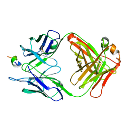
1F90
DownloadVisualize
BU of 1f90 by Molmil
FAB FRAGMENT OF MONOCLONAL ANTIBODY (LNKB-2) AGAINST HUMAN INTERLEUKIN-2 IN COMPLEX WITH ANTIGENIC PEPTIDE
分子名称: ANTIGENIC NONAPEPTIDE, FAB FRAGMENT OF MONOCLONAL ANTIBODY
著者Afonin, P.V, Fokin, A.V, Tsigannik, I.N, Mikhailova, I.Y, Onoprienko, L.V, Mikhaleva, I.I, Ivanov, V.T, Mareeva, T.Y, Nesmeyanov, V.A, Li, N, Duax, W.L, Pletnev, V.Z.
登録日2000-07-06
公開日2001-07-11
最終更新日2018-04-04
実験手法X-RAY DIFFRACTION (2.6 Å)
主引用文献Crystal structure of an anti-interleukin-2 monoclonal antibody Fab complexed with an antigenic nonapeptide.
Protein Sci., 10, 2001
1BQP
DownloadVisualize
BU of 1bqp by Molmil
THE STRUCTURE OF THE PEA LECTIN-D-MANNOPYRANOSE COMPLEX
分子名称: CALCIUM ION, MANGANESE (II) ION, PROTEIN (LECTIN), ...
著者Ruzeinikov, S.N, Mikhailova, I.Y, Tsygannik, I.N, Pangborn, W, Duax, W, Pletnev, V.Z.
登録日1998-08-17
公開日1998-08-26
最終更新日2023-08-09
実験手法X-RAY DIFFRACTION (2.1 Å)
主引用文献The Structure of the Pea Lectin-D-Mannopyranose Complex at a 2.1 A Resolution
RUSS.J.BIOORGANIC CHEM., 24, 1998
<12


224201

件を2024-08-28に公開中

PDB statisticsPDBj update infoContact PDBjnumon