4OVK
 
 | |
3SSR
 | | CcmK2 dodecamer - form 2 | | 分子名称: | Carbon dioxide concentrating mechanism protein, SULFATE ION | | 著者 | Kimber, M.S, Samborska, B. | | 登録日 | 2011-07-08 | | 公開日 | 2012-07-11 | | 最終更新日 | 2023-09-13 | | 実験手法 | X-RAY DIFFRACTION (1.6 Å) | | 主引用文献 | A CcmK2 double layer is the dominant architectural feature of the beta-carboxysomal shell facet
Structure, 2012
|
|
2WOI
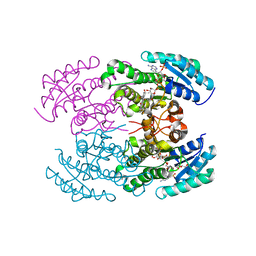
 | | Trypanothione reductase from Trypanosoma brucei | | 分子名称: | CHLORIDE ION, FLAVIN-ADENINE DINUCLEOTIDE, SODIUM ION, ... | | 著者 | Alphey, M.S, Fairlamb, A.H. | | 登録日 | 2009-07-24 | | 公開日 | 2010-08-04 | | 最終更新日 | 2023-12-20 | | 実験手法 | X-RAY DIFFRACTION (2.1 Å) | | 主引用文献 | Dihydroquinazolines as a Novel Class of Trypanosoma Brucei Trypanothione Reductase Inhibitors: Discovery, Synthesis, and Characterization of Their Binding Mode by Protein Crystallography.
J.Med.Chem., 54, 2011
|
|
4OZL
 | | GlnK2 from Haloferax mediterranei complexed with AMP | | 分子名称: | ADENOSINE MONOPHOSPHATE, Nitrogen regulatory protein P-II, SULFATE ION | | 著者 | Palanca, C, Pedro-Roig, L, Llacer, J.L, Camacho, M, Bonete, M.J, Rubio, V. | | 登録日 | 2014-02-17 | | 公開日 | 2014-07-02 | | 最終更新日 | 2023-12-27 | | 実験手法 | X-RAY DIFFRACTION (1.4942 Å) | | 主引用文献 | The structure of a PII signaling protein from a halophilic archaeon reveals novel traits and high-salt adaptations.
Febs J., 281, 2014
|
|
4OIH
 | | Importin Alpha in Complex with the Bipartite NLS of Prp20 | | 分子名称: | Guanine nucleotide exchange factor SRM1, Importin subunit alpha-1 | | 著者 | Roman, N, Christie, M, Swarbrick, C.M.D, Kobe, B, Forwood, J.K. | | 登録日 | 2014-01-19 | | 公開日 | 2014-02-19 | | 最終更新日 | 2023-11-08 | | 実験手法 | X-RAY DIFFRACTION (2.1 Å) | | 主引用文献 | Structural Characterisation of the Nuclear Import Receptor Importin Alpha in Complex with the Bipartite NLS of Prp20
Plos One, 8, 2013
|
|
4OPR
 | | Crystal structure of stabilized TEM-1 beta-lactamase variant v.13 carrying G238S mutation | | 分子名称: | CALCIUM ION, SULFATE ION, TEM-94 ES-beta-lactamase, ... | | 著者 | Dellus-Gur, E, Elias, M, Fraser, J.S, Tawfik, D.S. | | 登録日 | 2014-02-06 | | 公開日 | 2015-05-20 | | 最終更新日 | 2018-01-17 | | 実験手法 | X-RAY DIFFRACTION (1.45 Å) | | 主引用文献 | Negative Epistasis and Evolvability in TEM-1 beta-Lactamase--The Thin Line between an Enzyme's Conformational Freedom and Disorder.
J. Mol. Biol., 427, 2015
|
|
4OQ0
 | | Crystal structure of stabilized TEM-1 beta-lactamase variant v.13 carrying R164S/G238S mutation in complex with boron-based inhibitor EC25 | | 分子名称: | 1,2-ETHANEDIOL, 3-[(2R)-2-{[(2R)-2-amino-2-phenylacetyl]amino}-2-(dihydroxyboranyl)ethyl]benzoic acid, CALCIUM ION, ... | | 著者 | Dellus-Gur, E, Elias, M, Fraser, J.S, Tawfik, D.S. | | 登録日 | 2014-02-07 | | 公開日 | 2015-05-20 | | 最終更新日 | 2018-01-17 | | 実験手法 | X-RAY DIFFRACTION (1.42 Å) | | 主引用文献 | Negative Epistasis and Evolvability in TEM-1 beta-Lactamase--The Thin Line between an Enzyme's Conformational Freedom and Disorder.
J. Mol. Biol., 427, 2015
|
|
4OQG
 | | Crystal structure of TEM-1 beta-lactamase in complex with boron-based inhibitor EC25 | | 分子名称: | 3-[(2R)-2-{[(2R)-2-amino-2-phenylacetyl]amino}-2-(dihydroxyboranyl)ethyl]benzoic acid, Ampicillin resistance protein, ZINC ION | | 著者 | Dellus-Gur, E, Elias, M, Fraser, J.S, Tawfik, D.S. | | 登録日 | 2014-02-09 | | 公開日 | 2015-05-20 | | 最終更新日 | 2023-09-20 | | 実験手法 | X-RAY DIFFRACTION (2.4 Å) | | 主引用文献 | Negative Epistasis and Evolvability in TEM-1 beta-Lactamase--The Thin Line between an Enzyme's Conformational Freedom and Disorder.
J. Mol. Biol., 427, 2015
|
|
4OSO
 | | The crystal structure of landomycin C-6 ketoreductase LanV with bound NADP and rabelomycin | | 分子名称: | DI(HYDROXYETHYL)ETHER, NADP NICOTINAMIDE-ADENINE-DINUCLEOTIDE PHOSPHATE, Reductase homolog, ... | | 著者 | Paananen, P, Niiranen, L, Patrikainen, P, Metsa-Ketela, M. | | 登録日 | 2014-02-13 | | 公開日 | 2014-10-01 | | 最終更新日 | 2023-09-20 | | 実験手法 | X-RAY DIFFRACTION (2.5 Å) | | 主引用文献 | Structure-based engineering of angucyclinone 6-ketoreductases.
Chem.Biol., 21, 2014
|
|
4OUG
 | | Crystal structure of the ternary complex of camel peptidoglycan recognition protein, PGRP-S with lipopolysaccharide and palmitic acid at 2.46 A resolution | | 分子名称: | (R)-((2R,3S,4R,5R,6R)-3-HYDROXY-2-(HYDROXYMETHYL)-5-((R)-3-HYDROXYTETRADECANAMIDO)-6-(PHOSPHONOOXY)TETRAHYDRO-2H-PYRAN-4-YL) 3-HYDROXYTETRADECANOATE, GLYCEROL, L(+)-TARTARIC ACID, ... | | 著者 | Yamini, S, Sharma, P, Yadav, S.P, Sinha, M, Kaur, P, Sharma, S, Singh, T.P. | | 登録日 | 2014-02-17 | | 公開日 | 2014-03-05 | | 最終更新日 | 2023-11-08 | | 実験手法 | X-RAY DIFFRACTION (2.46 Å) | | 主引用文献 | Crystal structure of the ternary complex of camel peptidoglycan recognition protein, PGRP-S with lipopolysaccharide and palmitic acid at 2.46 A resolution
To be Published
|
|
4P1O
 | | Crystal structure of the Bateman domain of murine magnesium transporter CNNM2 bound to ATP-Mg | | 分子名称: | ADENOSINE-5'-TRIPHOSPHATE, MAGNESIUM ION, Metal transporter CNNM2 | | 著者 | Corral-Rodriguez, M.A, Stuiver, M, Abascal-Palacios, G, Diercks, T, Oyenarte, I, Ereno-Orbea, J, Encinar, J.A, Spiwok, V, Terashima, H, Accardi, A, Muller, D, Martinez-Cruz, L.A. | | 登録日 | 2014-02-27 | | 公開日 | 2015-04-15 | | 最終更新日 | 2023-12-27 | | 実験手法 | X-RAY DIFFRACTION (3.06 Å) | | 主引用文献 | Structural and ligand binding properties of the Bateman domain of human magnesium transporters CNNM2 and CNNM4
To Be Published
|
|
4P1Q
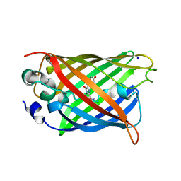 | | GREEN FLUORESCENT PROTEIN E222H VARIANT | | 分子名称: | Green fluorescent protein, SODIUM ION | | 著者 | Klein, M, Carius, Y, Auerbach, D, Franz, S, Jung, G, Lancaster, C.R.D. | | 登録日 | 2014-02-27 | | 公開日 | 2014-07-16 | | 最終更新日 | 2023-11-15 | | 実験手法 | X-RAY DIFFRACTION (1.5 Å) | | 主引用文献 | Replacement of Highly Conserved E222 by the Photostable Non-photoconvertible Histidine in GFP.
Chembiochem, 15, 2014
|
|
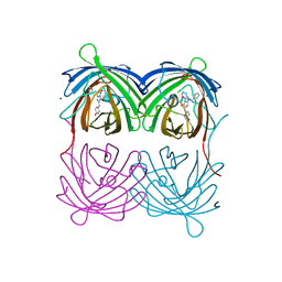
4P24
 | | pore forming toxin | | 分子名称: | (4S)-2-METHYL-2,4-PENTANEDIOL, Alpha-hemolysin | | 著者 | Sugawara, T, Yamashita, D, Tanaka, Y, Tanaka, I, Yao, M. | | 登録日 | 2014-03-01 | | 公開日 | 2015-03-11 | | 最終更新日 | 2023-12-27 | | 実験手法 | X-RAY DIFFRACTION (3.1 Å) | | 主引用文献 | Structural basis for pore-forming mechanism of staphylococcal alpha-hemolysin.
Toxicon, 108, 2015
|
|
4P76
 | | Cellular response to a crystal-forming protein | | 分子名称: | Photoconvertible fluorescent protein, SODIUM ION | | 著者 | Tsutsui, H, Jinno, Y, Shoda, K, Tomita, A, Matsuda, M, Yamashita, E, Katayama, H, Nakagawa, A, Miyawaki, A. | | 登録日 | 2014-03-26 | | 公開日 | 2015-04-29 | | 最終更新日 | 2023-11-15 | | 実験手法 | X-RAY DIFFRACTION (2.9 Å) | | 主引用文献 | A diffraction-quality protein crystal processed as an autophagic cargo
Mol.Cell, 58, 2015
|
|
4P3Z
 | | Chlamydia pneumoniae CopN (D29 construct) | | 分子名称: | CopN, GLYCEROL, SULFATE ION | | 著者 | Nawrotek, A, Guimaraes, B.G, Knossow, M, Gigant, B. | | 登録日 | 2014-03-10 | | 公開日 | 2014-07-30 | | 最終更新日 | 2023-12-27 | | 実験手法 | X-RAY DIFFRACTION (1.77 Å) | | 主引用文献 | Biochemical and Structural Insights into Microtubule Perturbation by CopN from Chlamydia pneumoniae.
J.Biol.Chem., 289, 2014
|
|
4P3N
 | | Structural Basis for Full-Spectrum Inhibition of Threonyl-tRNA Synthetase by Borrelidin 1 | | 分子名称: | (1R,2R)-2-[(2S,4E,6E,8R,9S,11R,13S,15S,16S)-7-cyano-8,16-dihydroxy-9,11,13,15-tetramethyl-18-oxooxacyclooctadeca-4,6-dien-2-yl]cyclopentanecarboxylic acid, Threonine--tRNA ligase, cytoplasmic, ... | | 著者 | Fang, P, Yu, X, Chen, K, Chen, X, Guo, M. | | 登録日 | 2014-03-09 | | 公開日 | 2015-03-11 | | 最終更新日 | 2023-12-27 | | 実験手法 | X-RAY DIFFRACTION (2.6 Å) | | 主引用文献 | Structural basis for full-spectrum inhibition of translational functions on a tRNA synthetase.
Nat Commun, 6, 2015
|
|
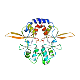
4P58
 | | Crystal structure of mouse comt bound to an inhibitor | | 分子名称: | 1',3'-dimethyl-1H,1'H-3,4'-bipyrazole, Catechol O-methyltransferase | | 著者 | Lanier, M. | | 登録日 | 2014-03-15 | | 公開日 | 2014-06-25 | | 最終更新日 | 2023-09-27 | | 実験手法 | X-RAY DIFFRACTION (2.06 Å) | | 主引用文献 | A fragment-based approach to identifying S-adenosyl-l-methionine -competitive inhibitors of catechol O-methyl transferase (COMT).
J.Med.Chem., 57, 2014
|
|
2VUP
 | |
4PUF
 | | Complex between the Salmonella T3SS effector SlrP and its human target thioredoxin-1 | | 分子名称: | E3 ubiquitin-protein ligase SlrP, Thioredoxin | | 著者 | Zouhir, S, Bernal-Bayard, J, Cordero-Alba, M, Cardenal-Munoz, E, Guimaraes, B, Lazar, N, Ramos-Morales, F, Nessler, S. | | 登録日 | 2014-03-13 | | 公開日 | 2014-09-17 | | 最終更新日 | 2024-02-28 | | 実験手法 | X-RAY DIFFRACTION (3.296 Å) | | 主引用文献 | The structure of the Slrp-Trx1 complex sheds light on the autoinhibition mechanism of the type III secretion system effectors of the NEL family.
Biochem.J., 464, 2014
|
|
4P3U
 | |
4P40
 | | Chlamydia pneumoniae CopN | | 分子名称: | CHLORIDE ION, CopN, DIMETHYLAMINE, ... | | 著者 | Nawrotek, A, Guimaraes, B.G, Knossow, M, Gigant, B. | | 登録日 | 2014-03-10 | | 公開日 | 2014-07-30 | | 最終更新日 | 2023-12-27 | | 実験手法 | X-RAY DIFFRACTION (1.2 Å) | | 主引用文献 | Biochemical and Structural Insights into Microtubule Perturbation by CopN from Chlamydia pneumoniae.
J.Biol.Chem., 289, 2014
|
|
4P4N
 | |
4PEO
 | | Crystal structure of a hypothetical protein from Staphylococcus aureus. | | 分子名称: | Hypothetical protein | | 著者 | McGrath, T.E, Kisselman, G, Romanov, V, Wu-Brown, J, Soloveychik, M, Chan, T.S.Y, Gordon, R.D, Thambipillai, D, Dharamsi, A, Mansoury, K, Battaile, K.P, Edwards, A.M, Pai, E.F, Chirgadze, N.Y. | | 登録日 | 2014-04-24 | | 公開日 | 2015-05-06 | | 最終更新日 | 2024-02-28 | | 実験手法 | X-RAY DIFFRACTION (1.73 Å) | | 主引用文献 | Crystal structure of a hypothetical protein from Staphylococcus aureus.
To Be Published
|
|
3SSS
 | | CcmK1 with residues 103-113 deleted | | 分子名称: | CHLORIDE ION, Carbon dioxide concentrating mechanism protein | | 著者 | Kimber, M.S, Samborska, B. | | 登録日 | 2011-07-08 | | 公開日 | 2012-07-11 | | 最終更新日 | 2023-09-13 | | 実験手法 | X-RAY DIFFRACTION (2.05 Å) | | 主引用文献 | A CcmK2 double layer is the dominant architectural feature of the beta-carboxysomal shell facet
Structure, 2012
|
|
4P7E
 | | Triazolopyridine compounds as selective JAK1 inhibitors: from hit identification to GLPG0634 | | 分子名称: | N-(5-{4-[(1,1-dioxidothiomorpholin-4-yl)methyl]phenyl}[1,2,4]triazolo[1,5-a]pyridin-2-yl)cyclopropanecarboxamide, Tyrosine-protein kinase JAK2 | | 著者 | Menet, C.C.J, Fletcher, S, Van Lommen, G, Geney, R, Blanc, J, Smits, K, Jouannigot, N, van der Aar, E.M, Clement-Lacroix, P, Lepescheux, L, Galien, R, Vayssiere, B, Nelles, L, Christophe, T, Brys, R, Uhring, M, Ciesielski, F, Van Rompaey, L. | | 登録日 | 2014-03-27 | | 公開日 | 2014-11-19 | | 最終更新日 | 2023-12-20 | | 実験手法 | X-RAY DIFFRACTION (2.4 Å) | | 主引用文献 | Triazolopyridines as Selective JAK1 Inhibitors: From Hit Identification to GLPG0634.
J.Med.Chem., 57, 2014
|
|


