Loading
PDBj
MenuPDBj@FacebookPDBj@TwitterPDBj@YouTubewwPDB FoundationwwPDB
RCSB PDBPDBeBMRBAdv. SearchSearch help
Search by PDB author
3HBZ
DownloadVisualize
BU of 3hbz by Molmil
Crystal structure of a putative glycoside hydrolase (bt_2081) from bacteroides thetaiotaomicron vpi-5482 at 2.05 A resolution
Descriptor: ACETATE ION, CACODYLATE ION, CALCIUM ION, ...
Authors:Joint Center for Structural Genomics (JCSG)
Deposit date:2009-05-05
Release date:2009-05-26
Last modified:2023-02-01
Method:X-RAY DIFFRACTION (2.05 Å)
Cite:Structure of Bacteroides thetaiotaomicron BT2081 at 2.05 A resolution: the first structural representative of a new protein family that may play a role in carbohydrate metabolism.
Acta Crystallogr.,Sect.F, 66, 2010
3D00
DownloadVisualize
BU of 3d00 by Molmil
Crystal structure of a tungsten formylmethanofuran dehydrogenase subunit e (fmde)-like protein (syn_00638) from syntrophus aciditrophicus at 1.90 A resolution
Descriptor: CHLORIDE ION, Tungsten formylmethanofuran dehydrogenase subunit E, ZINC ION
Authors:Joint Center for Structural Genomics (JCSG)
Deposit date:2008-04-30
Release date:2008-05-20
Last modified:2023-02-01
Method:X-RAY DIFFRACTION (1.9 Å)
Cite:Structures of three members of Pfam PF02663 (FmdE) implicated in microbial methanogenesis reveal a conserved alpha+beta core domain and an auxiliary C-terminal treble-clef zinc finger.
Acta Crystallogr.,Sect.F, 66, 2010
2GLZ
DownloadVisualize
BU of 2glz by Molmil
Crystal structure of a formylmethanofuran dehydrogenase subunit e-like protein (dhaf_2992) from desulfitobacterium hafniense dcb-2 at 1.45 A resolution
Descriptor: 1,2-ETHANEDIOL, NICKEL (II) ION, ZINC ION, ...
Authors:Joint Center for Structural Genomics (JCSG)
Deposit date:2006-04-05
Release date:2006-04-18
Last modified:2023-01-25
Method:X-RAY DIFFRACTION (1.45 Å)
Cite:Structures of three members of Pfam PF02663 (FmdE) implicated in microbial methanogenesis reveal a conserved alpha+beta core domain and an auxiliary C-terminal treble-clef zinc finger.
Acta Crystallogr.,Sect.F, 66, 2010
3CGH
DownloadVisualize
BU of 3cgh by Molmil
Crystal structure of a susd homolog (bt_3984) from bacteroides thetaiotaomicron vpi-5482 at 1.70 A resolution
Descriptor: ACETATE ION, SusD homolog, ZINC ION
Authors:Joint Center for Structural Genomics (JCSG)
Deposit date:2008-03-05
Release date:2008-03-18
Last modified:2023-02-01
Method:X-RAY DIFFRACTION (1.7 Å)
Cite:Structure of BT_3984, a member of the SusD/RagB family of nutrient-binding molecules.
Acta Crystallogr.,Sect.F, 66, 2010
2GVK
DownloadVisualize
BU of 2gvk by Molmil
Crystal structure of a dye-decolorizing peroxidase (DyP) from Bacteroides thetaiotaomicron VPI-5482 at 1.6 A resolution
Descriptor: 1,2-ETHANEDIOL, ACETIC ACID, CHLORIDE ION, ...
Authors:Joint Center for Structural Genomics (JCSG)
Deposit date:2006-05-02
Release date:2006-05-16
Last modified:2023-01-25
Method:X-RAY DIFFRACTION (1.6 Å)
Cite:Crystal structures of two novel dye-decolorizing peroxidases reveal a beta-barrel fold with a conserved heme-binding motif.
Proteins, 69, 2007
1J6U
DownloadVisualize
BU of 1j6u by Molmil
Crystal structure of UDP-N-acetylmuramate-alanine ligase MurC (TM0231) from Thermotoga maritima at 2.3 A resolution
Descriptor: UDP-N-acetylmuramate-alanine ligase MurC
Authors:Joint Center for Structural Genomics (JCSG)
Deposit date:2002-08-29
Release date:2002-11-06
Last modified:2023-01-25
Method:X-RAY DIFFRACTION (2.3 Å)
Cite:Crystal structure of an Udp-n-acetylmuramate-alanine ligase MurC (TM0231) from Thermotoga maritima at 2.3 A resolution.
Proteins, 55, 2004
3T2L
DownloadVisualize
BU of 3t2l by Molmil
Crystal structure of a Putative cell adhesion protein (BF1858) from Bacteroides fragilis NCTC 9343 at 2.33 A resolution
Descriptor: CHLORIDE ION, GLYCEROL, Putative cell adhesion protein
Authors:Joint Center for Structural Genomics (JCSG)
Deposit date:2011-07-22
Release date:2011-08-10
Last modified:2023-02-01
Method:X-RAY DIFFRACTION (2.33 Å)
Cite:A Distinct Type of Pilus from the Human Microbiome.
Cell, 165, 2016
3G23
DownloadVisualize
BU of 3g23 by Molmil
Crystal structure of a ld-carboxypeptidase a (saro_1426) from novosphingobium aromaticivorans dsm at 1.89 A resolution
Descriptor: GLYCEROL, LD-carboxypeptidase A, SULFATE ION, ...
Authors:Joint Center for Structural Genomics (JCSG)
Deposit date:2009-01-30
Release date:2009-02-10
Last modified:2023-02-01
Method:X-RAY DIFFRACTION (1.89 Å)
Cite:Structure and function of a novel LD-carboxypeptidase a involved in peptidoglycan recycling.
J.Bacteriol., 195, 2013
3G0T
DownloadVisualize
BU of 3g0t by Molmil
Crystal structure of putative aspartate aminotransferase (NP_905498.1) from Porphyromonas gingivalis W83 at 1.75 A resolution
Descriptor: 1,2-ETHANEDIOL, 2-{2-[2-(2-{2-[2-(2-ETHOXY-ETHOXY)-ETHOXY]-ETHOXY}-ETHOXY)-ETHOXY]-ETHOXY}-ETHANOL, CHLORIDE ION, ...
Authors:Joint Center for Structural Genomics (JCSG)
Deposit date:2009-01-28
Release date:2009-02-10
Last modified:2023-02-01
Method:X-RAY DIFFRACTION (1.75 Å)
Cite:Molecular characterization of novel pyridoxal-5'-phosphate-dependent enzymes from the human microbiome.
Protein Sci., 23, 2014
3E0F
DownloadVisualize
BU of 3e0f by Molmil
Crystal structure of a putative metal-dependent phosphoesterase (bad_1165) from bifidobacterium adolescentis atcc 15703 at 2.40 A resolution
Descriptor: ACETATE ION, FE (III) ION, PHOSPHATE ION, ...
Authors:Joint Center for Structural Genomics (JCSG)
Deposit date:2008-07-31
Release date:2008-09-02
Last modified:2023-02-01
Method:X-RAY DIFFRACTION (2.4 Å)
Cite:Crystal structure of a metal-dependent phosphoesterase (YP_910028.1) from Bifidobacterium adolescentis: Computational prediction and experimental validation of phosphoesterase activity.
Proteins, 79, 2011
3R4R
DownloadVisualize
BU of 3r4r by Molmil
Crystal structure of a fimbrial assembly protein (BDI_3522) from Parabacteroides distasonis ATCC 8503 at 2.38 A resolution
Descriptor: 1,2-ETHANEDIOL, SULFATE ION, hypothetical fimbrial assembly protein
Authors:Joint Center for Structural Genomics (JCSG)
Deposit date:2011-03-17
Release date:2011-03-30
Last modified:2023-02-01
Method:X-RAY DIFFRACTION (2.38 Å)
Cite:A Distinct Type of Pilus from the Human Microbiome.
Cell, 165, 2016
1VPZ
DownloadVisualize
BU of 1vpz by Molmil
Crystal structure of a putative carbon storage regulator protein (csra, pa0905) from pseudomonas aeruginosa at 2.05 A resolution
Descriptor: Carbon storage regulator homolog
Authors:Joint Center for Structural Genomics (JCSG)
Deposit date:2004-11-24
Release date:2004-12-14
Last modified:2023-01-25
Method:X-RAY DIFFRACTION (2.05 Å)
Cite:Crystal structure of the global regulatory protein CsrA from Pseudomonas putida at 2.05 A resolution reveals a new fold.
Proteins, 61, 2005
3ETN
DownloadVisualize
BU of 3etn by Molmil
Crystal structure of putative phosphosugar isomerase involved in capsule formation (YP_209877.1) from Bacteroides fragilis NCTC 9343 at 1.70 A resolution
Descriptor: 1,2-ETHANEDIOL, CYTIDINE 5'-MONOPHOSPHATE 3-DEOXY-BETA-D-GULO-OCT-2-ULO-PYRANOSONIC ACID, putative phosphosugar isomerase involved in capsule formation
Authors:Joint Center for Structural Genomics (JCSG)
Deposit date:2008-10-08
Release date:2008-10-21
Last modified:2023-02-01
Method:X-RAY DIFFRACTION (1.7 Å)
Cite:Structural analysis of arabinose-5-phosphate isomerase from Bacteroides fragilis and functional implications.
Acta Crystallogr.,Sect.D, 70, 2014
3BY7
DownloadVisualize
BU of 3by7 by Molmil
CRYSTAL STRUCTURE OF A PROTEIN STRUCTURALLY SIMILAR TO SM/LSM-LIKE RNA-BINDING PROTEINS (JCVI_PEP_1096686650277) FROM UNCULTURED MARINE ORGANISM AT 2.60 A RESOLUTION
Descriptor: uncharacterized protein
Authors:Joint Center for Structural Genomics (JCSG)
Deposit date:2008-01-15
Release date:2008-01-29
Last modified:2023-02-01
Method:X-RAY DIFFRACTION (2.6 Å)
Cite:Crystal structure of a novel Sm-like protein of putative cyanophage origin at 2.60 A resolution.
Proteins, 75, 2009
2FNA
DownloadVisualize
BU of 2fna by Molmil
Crystal structure of an archaeal aaa+ atpase (sso1545) from sulfolobus solfataricus p2 at 2.00 A resolution
Descriptor: 1,2-ETHANEDIOL, ADENOSINE-5'-DIPHOSPHATE, Conserved hypothetical protein, ...
Authors:Joint Center for Structural Genomics (JCSG)
Deposit date:2006-01-10
Release date:2006-02-07
Last modified:2023-01-25
Method:X-RAY DIFFRACTION (2 Å)
Cite:Crystal structure of a novel archaeal AAA+ ATPase SSO1545 from Sulfolobus solfataricus.
Proteins, 74, 2009
3H41
DownloadVisualize
BU of 3h41 by Molmil
CRYSTAL STRUCTURE OF A NLPC/P60 FAMILY PROTEIN (BCE_2878) FROM BACILLUS CEREUS ATCC 10987 AT 1.79 A RESOLUTION
Descriptor: ALANINE, D-GLUTAMIC ACID, NLP/P60 family protein, ...
Authors:Joint Center for Structural Genomics (JCSG)
Deposit date:2009-04-17
Release date:2009-05-05
Last modified:2023-02-01
Method:X-RAY DIFFRACTION (1.79 Å)
Cite:Structure of the gamma-D-glutamyl-L-diamino acid endopeptidase YkfC from Bacillus cereus in complex with L-Ala-[gamma]-D-Glu: insights into substrate recognition by NlpC/P60 cysteine peptidases
Acta Crystallogr.,Sect.F, 66, 2010
3H0N
DownloadVisualize
BU of 3h0n by Molmil
Crystal structure of a duf1470 family protein (jann_2411) from jannaschia sp. ccs1 at 1.45 A resolution
Descriptor: ACETATE ION, GLYCEROL, NICKEL (II) ION, ...
Authors:Joint Center for Structural Genomics (JCSG)
Deposit date:2009-04-09
Release date:2009-04-21
Last modified:2023-02-01
Method:X-RAY DIFFRACTION (1.45 Å)
Cite:The structure of Jann_2411 (DUF1470) from Jannaschia sp. at 1.45  resolution reveals a new fold (the ABATE domain) and suggests its possible role as a transcription regulator.
Acta Crystallogr.,Sect.F, 66, 2010
3GO5
DownloadVisualize
BU of 3go5 by Molmil
Crystal structure of a multidomain protein with nucleic acid binding domains (sp_0946) from streptococcus pneumoniae tigr4 at 1.40 A resolution
Descriptor: 1,2-ETHANEDIOL, CHLORIDE ION, Multidomain protein with S1 RNA-binding domains
Authors:Joint Center for Structural Genomics (JCSG)
Deposit date:2009-03-18
Release date:2009-04-07
Last modified:2023-02-01
Method:X-RAY DIFFRACTION (1.4 Å)
Cite:Structure of a virulence regulatory factor CvfB reveals a novel winged helix RNA binding module.
Structure, 18, 2010
2F46
DownloadVisualize
BU of 2f46 by Molmil
Crystal structure of a putative phosphatase (nma1982) from neisseria meningitidis z2491 at 1.41 A resolution
Descriptor: CHLORIDE ION, UNKNOWN LIGAND, hypothetical protein
Authors:Joint Center for Structural Genomics (JCSG)
Deposit date:2005-11-22
Release date:2006-02-07
Last modified:2023-01-25
Method:X-RAY DIFFRACTION (1.41 Å)
Cite:Crystal structure of NMA1982 from Neisseria meningitidis at 1.5 A resolution provides a structural scaffold for nonclassical, eukaryotic-like phosphatases.
Proteins, 69, 2007
2FG0
DownloadVisualize
BU of 2fg0 by Molmil
Crystal structure of a putative gamma-d-glutamyl-l-diamino acid endopeptidase (npun_r0659) from nostoc punctiforme pcc 73102 at 1.79 A resolution
Descriptor: COG0791: Cell wall-associated hydrolases (invasion-associated proteins), GLYCEROL
Authors:Joint Center for Structural Genomics (JCSG)
Deposit date:2005-12-20
Release date:2006-01-10
Last modified:2023-01-25
Method:X-RAY DIFFRACTION (1.79 Å)
Cite:Structural Basis of Murein Peptide Specificity of a gamma-D-Glutamyl-L-Diamino Acid Endopeptidase.
Structure, 17, 2009
3UFI
DownloadVisualize
BU of 3ufi by Molmil
Crystal structure of a putative cell adhesion protein (BACOVA_04980) from Bacteroides ovatus ATCC 8483 at 2.18 A resolution
Descriptor: CHLORIDE ION, SULFATE ION, hypothetical protein BACOVA_04980
Authors:Joint Center for Structural Genomics (JCSG)
Deposit date:2011-11-01
Release date:2011-12-14
Last modified:2023-02-01
Method:X-RAY DIFFRACTION (2.18 Å)
Cite:A Distinct Type of Pilus from the Human Microbiome.
Cell, 165, 2016
2HBW
DownloadVisualize
BU of 2hbw by Molmil
Crystal structure of a putative endopeptidase (ava_3396) from anabaena variabilis atcc 29413 at 1.05 A resolution
Descriptor: ACETATE ION, NLP/P60 protein, UNKNOWN LIGAND
Authors:Joint Center for Structural Genomics (JCSG)
Deposit date:2006-06-14
Release date:2006-08-08
Last modified:2023-01-25
Method:X-RAY DIFFRACTION (1.05 Å)
Cite:Structural Basis of Murein Peptide Specificity of a gamma-D-Glutamyl-L-Diamino Acid Endopeptidase.
Structure, 17, 2009
3SY6
DownloadVisualize
BU of 3sy6 by Molmil
Crystal structure of a fimbrial protein BF1861 [Bacteroides fragilis NCTC 9343] (BF1861) from Bacteroides fragilis NCTC 9343 at 1.90 A resolution
Descriptor: 2-[N-CYCLOHEXYLAMINO]ETHANE SULFONIC ACID, Fimbrial protein BF1861
Authors:Joint Center for Structural Genomics (JCSG)
Deposit date:2011-07-15
Release date:2011-08-24
Last modified:2023-02-01
Method:X-RAY DIFFRACTION (1.9 Å)
Cite:A Distinct Type of Pilus from the Human Microbiome.
Cell, 165, 2016
3TX8
DownloadVisualize
BU of 3tx8 by Molmil
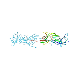
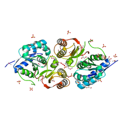
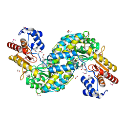
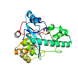
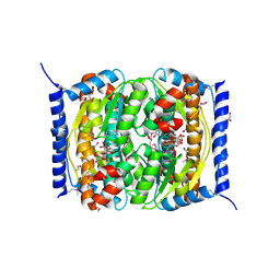
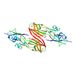
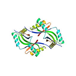
Crystal structure of a succinyl-diaminopimelate desuccinylase (ArgE) from Corynebacterium glutamicum ATCC 13032 at 2.97 A resolution
Descriptor: CHLORIDE ION, PHOSPHATE ION, Succinyl-diaminopimelate desuccinylase
Authors:Joint Center for Structural Genomics (JCSG), Brunger, A.T, Terwilliger, T.C, Read, R.J, Adams, P.D, Levitt, M, Schroder, G.F.
Deposit date:2011-09-22
Release date:2011-10-26
Last modified:2023-12-06
Method:X-RAY DIFFRACTION (2.972 Å)
Cite:Application of DEN refinement and automated model building to a difficult case of molecular-replacement phasing: the structure of a putative succinyl-diaminopimelate desuccinylase from Corynebacterium glutamicum.
Acta Crystallogr.,Sect.D, 68, 2012
1VQ3
DownloadVisualize
BU of 1vq3 by Molmil
Crystal structure of Phosphoribosylformylglycinamidine synthase, purS subunit (EC 6.3.5.3) (TM1244) from Thermotoga maritima at 1.90 A resolution
Descriptor: Phosphoribosylformylglycinamidine synthase, purS subunit
Authors:Joint Center for Structural Genomics (JCSG)
Deposit date:2004-12-15
Release date:2004-12-28
Last modified:2023-09-20
Method:X-RAY DIFFRACTION (1.9 Å)
Cite:Crystal structure of phosphoribosylformyl-glycinamidine synthase II, PurS subunit (TM1244) from Thermotoga maritima at 1.90 A resolution.
Proteins, 65, 2006

225399

PDB entries from 2024-09-25

PDB statisticsPDBj update infoContact PDBjnumon