Loading
PDBj
MenuPDBj@FacebookPDBj@TwitterPDBj@YouTubewwPDB FoundationwwPDB
RCSB PDBPDBeBMRBAdv. SearchSearch help
Search by PDB author
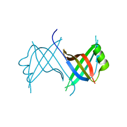
2MOA
DownloadVisualize
BU of 2moa by Molmil
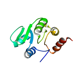
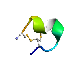
Solution NMR structure of peptide ImI1 (peak 2)
Descriptor: Alpha-conotoxin ImI
Authors:Heinis, C, Chen, S.
Deposit date:2014-04-24
Release date:2014-09-24
Last modified:2023-11-15
Method:SOLUTION NMR
Cite:Dithiol amino acids can structurally shape and enhance the ligand-binding properties of polypeptides.
Nat Chem, 6, 2014
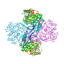
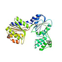
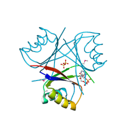
1AT1
DownloadVisualize
BU of 1at1 by Molmil
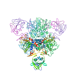
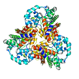
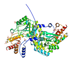
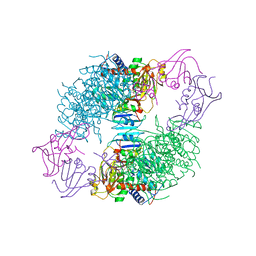
CRYSTAL STRUCTURES OF PHOSPHONOACETAMIDE LIGATED T AND PHOSPHONOACETAMIDE AND MALONATE LIGATED R STATES OF ASPARTATE CARBAMOYLTRANSFERASE AT 2.8-ANGSTROMS RESOLUTION AND NEUTRAL P*H
Descriptor: ASPARTATE CARBAMOYLTRANSFERASE REGULATORY CHAIN, ASPARTATE CARBAMOYLTRANSFERASE, CATALYTIC CHAIN, ...
Authors:Gouaux, J.E, Lipscomb, W.N.
Deposit date:1989-09-22
Release date:1990-10-15
Last modified:2024-02-07
Method:X-RAY DIFFRACTION (2.8 Å)
Cite:Crystal structures of phosphonoacetamide ligated T and phosphonoacetamide and malonate ligated R states of aspartate carbamoyltransferase at 2.8-A resolution and neutral pH.
Biochemistry, 29, 1990
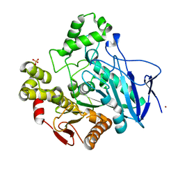
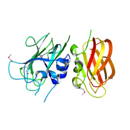
1C7I
DownloadVisualize
BU of 1c7i by Molmil
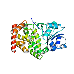
THERMOPHYLIC PNB ESTERASE
Descriptor: CALCIUM ION, PROTEIN (PARA-NITROBENZYL ESTERASE)
Authors:Spiller, B, Gershenson, A, Arnold, F, Stevens, R.
Deposit date:2000-02-21
Release date:2000-03-29
Last modified:2023-12-27
Method:X-RAY DIFFRACTION (2 Å)
Cite:A structural view of evolutionary divergence.
Proc.Natl.Acad.Sci.USA, 96, 1999
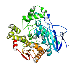
1QE3
DownloadVisualize
BU of 1qe3 by Molmil
PNB ESTERASE
Descriptor: PARA-NITROBENZYL ESTERASE, SULFATE ION, ZINC ION
Authors:Spiller, B, Gershenson, A, Arnold, F, Stevens, R.
Deposit date:1999-07-12
Release date:1999-07-21
Last modified:2024-02-14
Method:X-RAY DIFFRACTION (1.5 Å)
Cite:A structural view of evolutionary divergence.
Proc.Natl.Acad.Sci.USA, 96, 1999
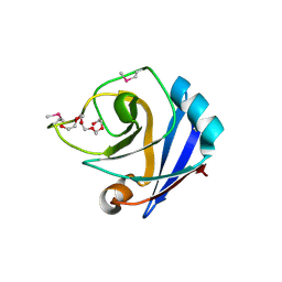
1ZX8
DownloadVisualize
BU of 1zx8 by Molmil
CRYSTAL STRUCTURE OF an atypical cyclophilin (peptidylprolyl cis-trans isomerase) (TM1367) FROM THERMOTOGA MARITIMA AT 1.90 A RESOLUTION
Descriptor: NICKEL (II) ION, PENTAETHYLENE GLYCOL, hypothetical protein TM1367
Authors:Joint Center for Structural Genomics (JCSG)
Deposit date:2005-06-07
Release date:2005-07-26
Last modified:2023-01-25
Method:X-RAY DIFFRACTION (1.9 Å)
Cite:Crystal structure of TM1367 from Thermotoga maritima at 1.90 A resolution reveals an atypical member of the cyclophilin (peptidylprolyl isomerase) fold.
Proteins, 63, 2006
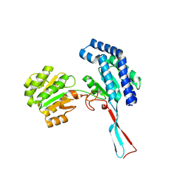
2AFB
DownloadVisualize
BU of 2afb by Molmil
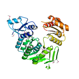
Crystal structure of 2-dehydro-3- deoxygluconokinase (EC 2.7.1.45) (tm0067) from THERMOTOGA MARITIMA at 2.05 A resolution
Descriptor: 2-keto-3-deoxygluconate kinase, CALCIUM ION, NICKEL (II) ION
Authors:Joint Center for Structural Genomics (JCSG)
Deposit date:2005-07-25
Release date:2006-02-14
Last modified:2023-01-25
Method:X-RAY DIFFRACTION (2.05 Å)
Cite:Crystal structure of 2-keto-3-deoxygluconate kinase (TM0067) from Thermotoga maritima at 2.05 A resolution.
Proteins, 70, 2007
1Z9F
DownloadVisualize
BU of 1z9f by Molmil
Crystal structure of single stranded DNA-binding protein (TM0604) from Thermotoga maritima at 2.60 A resolution
Descriptor: Single-strand binding protein
Authors:Joint Center for Structural Genomics (JCSG)
Deposit date:2005-04-01
Release date:2005-04-12
Last modified:2023-09-20
Method:X-RAY DIFFRACTION (2.3 Å)
Cite:Crystal structure of a single-stranded DNA-binding protein (TM0604) from Thermotoga maritima at 2.60 A resolution.
Proteins, 63, 2006
2B8N
DownloadVisualize
BU of 2b8n by Molmil
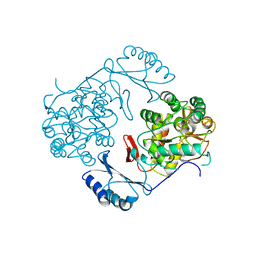
Crystal structure of Glycerate kinase (EC 2.7.1.31) (tm1585) from THERMOTOGA MARITIMA at 2.70 A resolution
Descriptor: glycerate kinase, putative
Authors:Joint Center for Structural Genomics (JCSG)
Deposit date:2005-10-07
Release date:2005-11-08
Last modified:2023-01-25
Method:X-RAY DIFFRACTION (2.53 Å)
Cite:Crystal structure of a glycerate kinase (TM1585) from Thermotoga maritima at 2.70 A resolution reveals a new fold
Proteins, 65, 2006
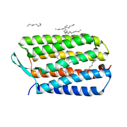
4HYX
DownloadVisualize
BU of 4hyx by Molmil
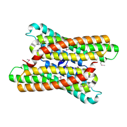
Crystal Structure Analysis of the Bacteriorhodopsin in Facial Amphiphile-4 DMPC Bicelle
Descriptor: Bacteriorhodopsin, DECANE, GLYCEROL, ...
Authors:Lee, S, Stout, C.D, Zhang, Q.
Deposit date:2012-11-14
Release date:2013-03-20
Last modified:2013-05-22
Method:X-RAY DIFFRACTION (1.99 Å)
Cite:Steroid-based facial amphiphiles for stabilization and crystallization of membrane proteins.
Proc.Natl.Acad.Sci.USA, 110, 2013
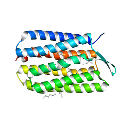
4HWL
DownloadVisualize
BU of 4hwl by Molmil
Crystal Structure Analysis of the Bacteriorhodopsin in Facial Amphiphile-7 DMPC Bicelle
Descriptor: Bacteriorhodopsin, GLYCEROL, HEPTANE, ...
Authors:Lee, S, Stout, C.D, Zhang, Q.
Deposit date:2012-11-08
Release date:2013-03-20
Last modified:2023-09-20
Method:X-RAY DIFFRACTION (2 Å)
Cite:Steroid-based facial amphiphiles for stabilization and crystallization of membrane proteins.
Proc.Natl.Acad.Sci.USA, 110, 2013
1NBE
DownloadVisualize
BU of 1nbe by Molmil
ASPARTATE TRANSCARBAMOYLASE REGULATORY CHAIN MUTANT (T82A)
Descriptor: ASPARTATE TRANSCARBAMOYLASE, D-MALATE, ZINC ION
Authors:Williams, M.K, Stec, B, Kantrowitz, E.R.
Deposit date:1998-04-25
Release date:1998-10-14
Last modified:2024-05-22
Method:X-RAY DIFFRACTION (2.6 Å)
Cite:A single mutation in the regulatory chain of Escherichia coli aspartate transcarbamoylase results in an extreme T-state structure.
J.Mol.Biol., 281, 1998
2JZD
DownloadVisualize
BU of 2jzd by Molmil
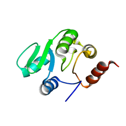
NMR structure of the domain 527-651 of the SARS-CoV nonstructural protein nsp3
Descriptor: Replicase polyprotein 1ab
Authors:Chatterjee, A, Johnson, M.A, Serrano, P, Pedrini, B, Joseph, J, Saikatendu, K, Neuman, B, Stevens, R.C, Wilson, I.A, Buchmeier, M.J, Kuhn, P, Wuthrich, K, Joint Center for Structural Genomics (JCSG)
Deposit date:2008-01-04
Release date:2008-02-05
Last modified:2024-05-29
Method:SOLUTION NMR
Cite:Nuclear magnetic resonance structure shows that the severe acute respiratory syndrome coronavirus-unique domain contains a macrodomain fold.
J.Virol., 83, 2009
2JZE
DownloadVisualize
BU of 2jze by Molmil
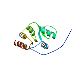
NMR structure of the domain 527-651 of the SARS-CoV nonstructural protein nsp3, single conformer closest to the mean coordinates of an ensemble of twenty energy minimized conformers
Descriptor: Replicase polyprotein 1ab
Authors:Chatterjee, A, Johnson, M.A, Serrano, P, Pedrini, B, Joseph, J, Saikatendu, K, Neuman, B, Stevens, R.C, Wilson, I.A, Buchmeier, M.J, Kuhn, P, Wuthrich, K, Joint Center for Structural Genomics (JCSG)
Deposit date:2008-01-04
Release date:2008-02-05
Last modified:2024-05-29
Method:SOLUTION NMR
Cite:Nuclear magnetic resonance structure shows that the severe acute respiratory syndrome coronavirus-unique domain contains a macrodomain fold.
J.Virol., 83, 2009
2JZF
DownloadVisualize
BU of 2jzf by Molmil
NMR Conformer closest to the mean coordinates of the domain 513-651 of the SARS-CoV nonstructural protein nsp3
Descriptor: Replicase polyprotein 1ab
Authors:Chatterjee, A, Johnson, M.A, Serrano, P, Pedrini, B, Joseph, J, Saikatendu, K, Neuman, B, Stevens, R.C, Wilson, I.A, Buchmeier, M.J, Kuhn, P, Wuthrich, K, Joint Center for Structural Genomics (JCSG)
Deposit date:2008-01-04
Release date:2008-02-05
Last modified:2024-05-29
Method:SOLUTION NMR
Cite:Nuclear magnetic resonance structure shows that the severe acute respiratory syndrome coronavirus-unique domain contains a macrodomain fold.
J.Virol., 83, 2009
1J5S
DownloadVisualize
BU of 1j5s by Molmil
Crystal structure of uronate isomerase (TM0064) from Thermotoga maritima at 2.85 A resolution
Descriptor: URONATE ISOMERASE
Authors:Joint Center for Structural Genomics (JCSG)
Deposit date:2002-07-02
Release date:2002-07-17
Last modified:2024-05-22
Method:X-RAY DIFFRACTION (2.85 Å)
Cite:Crystal structure of uronate isomerase (TM0064) from Thermotoga maritima at 2.85 A resolution.
Proteins, 53, 2003
1J6U
DownloadVisualize
BU of 1j6u by Molmil
Crystal structure of UDP-N-acetylmuramate-alanine ligase MurC (TM0231) from Thermotoga maritima at 2.3 A resolution
Descriptor: UDP-N-acetylmuramate-alanine ligase MurC
Authors:Joint Center for Structural Genomics (JCSG)
Deposit date:2002-08-29
Release date:2002-11-06
Last modified:2023-01-25
Method:X-RAY DIFFRACTION (2.3 Å)
Cite:Crystal structure of an Udp-n-acetylmuramate-alanine ligase MurC (TM0231) from Thermotoga maritima at 2.3 A resolution.
Proteins, 55, 2004
1PHZ
DownloadVisualize
BU of 1phz by Molmil
STRUCTURE OF PHOSPHORYLATED PHENYLALANINE HYDROXYLASE
Descriptor: FE (III) ION, PROTEIN (PHENYLALANINE HYDROXYLASE)
Authors:Kobe, B, Jennings, I.G, House, C.M, Michell, B.J, Cotton, R.G, Kemp, B.E.
Deposit date:1998-11-11
Release date:1999-04-30
Last modified:2024-04-03
Method:X-RAY DIFFRACTION (2.2 Å)
Cite:Structural basis of autoregulation of phenylalanine hydroxylase.
Nat.Struct.Biol., 6, 1999
1O0X
DownloadVisualize
BU of 1o0x by Molmil
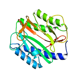
Crystal structure of Methionine aminopeptidase (TM1478) from Thermotoga maritima at 1.90 A resolution
Descriptor: Methionine aminopeptidase
Authors:Joint Center for Structural Genomics (JCSG)
Deposit date:2002-09-13
Release date:2002-11-06
Last modified:2023-09-20
Method:X-RAY DIFFRACTION (1.9 Å)
Cite:Crystal structure of a methionine aminopeptidase (TM1478) from Thermotoga maritima at 1.9 A resolution.
Proteins, 56, 2004
1O51
DownloadVisualize
BU of 1o51 by Molmil
Crystal structure of a putative PII-like signaling protein (TM0021) from Thermotoga maritima at 2.50 A resolution
Descriptor: ADENOSINE-5'-DIPHOSPHATE, Hypothetical protein TM0021, SULFATE ION
Authors:Joint Center for Structural Genomics (JCSG)
Deposit date:2003-08-01
Release date:2003-08-19
Last modified:2023-01-25
Method:X-RAY DIFFRACTION (2.5 Å)
Cite:Crystal structure of a putative PII-like signaling protein (TM0021) from Thermotoga maritima at 2.5 A resolution
Proteins, 54, 2004
1O59
DownloadVisualize
BU of 1o59 by Molmil
Crystal structure of Allantoicase (yir029w) from Saccharomyces cerevisiae at 2.40 A resolution
Descriptor: Allantoicase
Authors:Joint Center for Structural Genomics (JCSG)
Deposit date:2003-08-22
Release date:2003-09-02
Last modified:2023-01-25
Method:X-RAY DIFFRACTION (2.4 Å)
Cite:Crystal structure of an allantoicase (YIR029W) from Saccharomyces cerevisiae at 2.4 A resolution
Proteins, 56, 2004
1O20
DownloadVisualize
BU of 1o20 by Molmil
Crystal structure of Gamma-glutamyl phosphate reductase (TM0293) from Thermotoga maritima at 2.00 A resolution
Descriptor: Gamma-glutamyl phosphate reductase
Authors:Joint Center for Structural Genomics (JCSG)
Deposit date:2003-02-12
Release date:2003-04-01
Last modified:2023-01-25
Method:X-RAY DIFFRACTION (2 Å)
Cite:Crystal structure of gamma-glutamyl phosphate reductase (TM0293) from Thermotoga maritima at 2.0 A resolution.
Proteins, 54, 2004
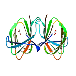
1O4T
DownloadVisualize
BU of 1o4t by Molmil
Crystal structure of a predicted oxalate decarboxylase (tm1287) from thermotoga maritima at 1.95 A resolution
Descriptor: MANGANESE (II) ION, OXALATE ION, putative oxalate decarboxylase
Authors:Joint Center for Structural Genomics (JCSG)
Deposit date:2003-07-03
Release date:2003-07-15
Last modified:2023-09-20
Method:X-RAY DIFFRACTION (1.95 Å)
Cite:Crystal structure of a putative oxalate decarboxylase (TM1287) from Thermotoga maritima at 1.95 A resolution
Proteins, 56, 2004
1O4S
DownloadVisualize
BU of 1o4s by Molmil
Crystal structure of Aspartate aminotransferase (TM1255) from Thermotoga maritima at 1.90 A resolution
Descriptor: Aspartate aminotransferase, PYRIDOXAL-5'-PHOSPHATE, SULFATE ION
Authors:Joint Center for Structural Genomics (JCSG)
Deposit date:2003-06-26
Release date:2003-07-08
Last modified:2023-09-20
Method:X-RAY DIFFRACTION (1.9 Å)
Cite:Crystal structure of an aspartate aminotransferase (TM1255) from Thermotoga maritima at 1.90 A resolution
Proteins, 55, 2004
1KQ3
DownloadVisualize
BU of 1kq3 by Molmil
CRYSTAL STRUCTURE OF A GLYCEROL DEHYDROGENASE (TM0423) FROM THERMOTOGA MARITIMA AT 1.5 A RESOLUTION
Descriptor: 2-AMINO-2-HYDROXYMETHYL-PROPANE-1,3-DIOL, CHLORIDE ION, ZINC ION, ...
Authors:Wilson, I.A, Miller, M.D, Joint Center for Structural Genomics (JCSG)
Deposit date:2002-01-03
Release date:2002-02-27
Last modified:2024-02-14
Method:X-RAY DIFFRACTION (1.5 Å)
Cite:Structural genomics of the Thermotoga maritima proteome implemented in a high-throughput structure determination pipeline
Proc.Natl.Acad.Sci.USA, 99, 2002
1O5H
DownloadVisualize
BU of 1o5h by Molmil
Crystal structure of formiminotetrahydrofolate cyclodeaminase (TM1560) from Thermotoga maritima at 2.80 A resolution
Descriptor: Formiminotetrahydrofolate cyclodeaminase
Authors:Joint Center for Structural Genomics (JCSG)
Deposit date:2003-09-17
Release date:2003-09-30
Last modified:2023-01-25
Method:X-RAY DIFFRACTION (2.8 Å)
Cite:Crystal structure of a formiminotetrahydrofolate cyclodeaminase (TM1560) from Thermotoga maritima at 2.80 A resolution reveals a new fold
Proteins, 58, 2005

225399

PDB entries from 2024-09-25

PDB statisticsPDBj update infoContact PDBjnumon