Loading
PDBj
MenuPDBj@FacebookPDBj@TwitterPDBj@YouTubewwPDB FoundationwwPDB
RCSB PDBPDBeBMRBAdv. SearchSearch help
Search by PDB author
3R4Q
DownloadVisualize
BU of 3r4q by Molmil
Crystal structure of Lactoylglutathione lyase from Agrobacterium tumefaciens
Descriptor: COBALT (II) ION, ISOPROPYL ALCOHOL, Lactoylglutathione lyase
Authors:Agarwal, R, Almo, S.C, Swaminathan, S, New York Structural Genomics Research Consortium (NYSGRC)
Deposit date:2011-03-17
Release date:2011-04-06
Last modified:2024-02-21
Method:X-RAY DIFFRACTION (2.51 Å)
Cite:Crystal structure of Lactoylglutathione lyase from Agrobacterium tumefaciens
To be Published
2PGW
DownloadVisualize
BU of 2pgw by Molmil
Crystal structure of a putative muconate cycloisomerase from Sinorhizobium meliloti 1021
Descriptor: GLYCEROL, Muconate cycloisomerase
Authors:Kumaran, D, Burley, S.K, Swaminathan, S, New York SGX Research Center for Structural Genomics (NYSGXRC)
Deposit date:2007-04-10
Release date:2007-04-24
Last modified:2021-02-03
Method:X-RAY DIFFRACTION (1.95 Å)
Cite:Crystal structure of a putative muconate cycloisomerase from Sinorhizobium meliloti 1021
To be Published
2Q07
DownloadVisualize
BU of 2q07 by Molmil
Crystal structure of AF0587, a protein of unknown function
Descriptor: Uncharacterized protein AF0587
Authors:Sugadev, R, Burley, S.K, Swaminathan, S, New York SGX Research Center for Structural Genomics (NYSGXRC)
Deposit date:2007-05-21
Release date:2007-06-05
Last modified:2021-02-03
Method:X-RAY DIFFRACTION (2.04 Å)
Cite:Crystal structure of AF0587, a protein of unknown function.
To be Published
2PWT
DownloadVisualize
BU of 2pwt by Molmil
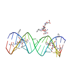
Crystal structure of the bacterial ribosomal decoding site complexed with aminoglycoside containing the L-HABA group
Descriptor: 22-mer of the ribosomal decoding site, DOUBLY FUNCTIONALIZED PAROMOMYCIN PM-II-162
Authors:Kondo, J, Pachamuthu, K, Francois, B, Szychowski, J, Hanessian, S, Westhof, E.
Deposit date:2007-05-13
Release date:2007-09-18
Last modified:2024-03-13
Method:X-RAY DIFFRACTION (1.8 Å)
Cite:Crystal Structure of the Bacterial Ribosomal Decoding Site Complexed with a Synthetic Doubly Functionalized Paromomycin Derivative: a New Specific Binding Mode to an A-Minor Motif Enhances in vitro Antibacterial Activity
Chemmedchem, 2, 2007
3R3H
DownloadVisualize
BU of 3r3h by Molmil
Crystal structure of O-methyltransferase from Legionella pneumophila
Descriptor: O-methyltransferase, SAM-dependent
Authors:Agarwal, R, Almo, S.C, Swaminathan, S, New York Structural Genomics Research Consortium (NYSGRC)
Deposit date:2011-03-15
Release date:2011-04-06
Last modified:2024-02-21
Method:X-RAY DIFFRACTION (2.65 Å)
Cite:Crystal structure of O-methyltransferase from Legionella pneumophila
To be Published
2QGO
DownloadVisualize
BU of 2qgo by Molmil
Crystal structure of a putative Fe-S biosynthesis protein from Lactobacillus acidophilus
Descriptor: Putative Fe-S biosynthesis protein
Authors:Agarwal, R, Burley, S.K, Swaminathan, S, New York SGX Research Center for Structural Genomics (NYSGXRC)
Deposit date:2007-06-29
Release date:2007-07-31
Last modified:2024-02-21
Method:X-RAY DIFFRACTION (2.04 Å)
Cite:Structural analysis of Fe-S cluster proteins.
To be Published
2PZZ
DownloadVisualize
BU of 2pzz by Molmil
2.2 A resolution crystal structure of UPF0201 protein from Methanococcus jannaschii
Descriptor: UPF0201 protein MJ1564
Authors:Rao, K.N, Burley, S.K, Swaminathan, S, New York SGX Research Center for Structural Genomics (NYSGXRC)
Deposit date:2007-05-18
Release date:2007-05-29
Last modified:2021-02-03
Method:X-RAY DIFFRACTION (2.2 Å)
Cite:UPF201 archaeal specific family members reveal structural similarity to RNA-binding proteins but low likelihood for RNA-binding function.
Plos One, 3, 2008
2PAJ
DownloadVisualize
BU of 2paj by Molmil
Crystal structure of an amidohydrolase from an environmental sample of Sargasso sea
Descriptor: ZINC ION, putative cytosine/guanine deaminase
Authors:Agarwal, R, Burley, S.K, Swaminathan, S, New York SGX Research Center for Structural Genomics (NYSGXRC)
Deposit date:2007-03-27
Release date:2007-04-03
Last modified:2024-02-21
Method:X-RAY DIFFRACTION (2.7 Å)
Cite:Discovery and structure determination of the orphan enzyme isoxanthopterin deaminase.
Biochemistry, 49, 2010
2PLT
DownloadVisualize
BU of 2plt by Molmil
STRUCTURE DETERMINATION OF PLASTOCYANIN FROM A CRYSTAL SPECIMEN WITH HEMIHEDRAL TWINNING FRACTION OF ONE-HALF
Descriptor: CALCIUM ION, COPPER (II) ION, PLASTOCYANIN
Authors:Redinbo, M.R, Merchant, S, Yeates, T.O.
Deposit date:1993-05-06
Release date:1993-10-31
Last modified:2024-02-21
Method:X-RAY DIFFRACTION (1.5 Å)
Cite:The 1.5-A crystal structure of plastocyanin from the green alga Chlamydomonas reinhardtii.
Biochemistry, 32, 1993
2QVH
DownloadVisualize
BU of 2qvh by Molmil
Crystal structure of O-succinylbenzoate synthase complexed with O-succinyl benzoate (OSB)
Descriptor: 2-SUCCINYLBENZOATE, MAGNESIUM ION, O-succinylbenzoate-CoA synthase
Authors:Sugadev, R, Burley, S.K, Swaminathan, S, New York SGX Research Center for Structural Genomics (NYSGXRC)
Deposit date:2007-08-08
Release date:2007-08-28
Last modified:2023-11-15
Method:X-RAY DIFFRACTION (1.76 Å)
Cite:Crystal structure of O-succinylbenzoate synthase complexed with O-succinyl benzoate.
To be Published
3QW7
DownloadVisualize
BU of 3qw7 by Molmil
Crystal structure of the protease domain of Botulinum Neurotoxin Serotype A with a peptide inhibitor RRFC
Descriptor: Botulinum neurotoxin type A, SODIUM ION, SULFATE ION, ...
Authors:Kumaran, D, Swaminathan, S.
Deposit date:2011-02-27
Release date:2012-02-08
Last modified:2023-09-13
Method:X-RAY DIFFRACTION (1.5 Å)
Cite:Peptide inhibitors of botulinum neurotoxin serotype A: design, inhibition, cocrystal structures, structure-activity relationship and pharmacophore modeling.
Acta Crystallogr.,Sect.D, 68, 2012
3QW6
DownloadVisualize
BU of 3qw6 by Molmil
Crystal structure of the protease domain of Botulinum Neurotoxin Serotype A with a peptide inhibitor RYGC
Descriptor: Botulinum neurotoxin type A, SODIUM ION, SULFATE ION, ...
Authors:Kumaran, D, Swaminathan, S.
Deposit date:2011-02-26
Release date:2012-02-08
Last modified:2023-09-13
Method:X-RAY DIFFRACTION (1.6 Å)
Cite:Peptide inhibitors of botulinum neurotoxin serotype A: design, inhibition, cocrystal structures, structure-activity relationship and pharmacophore modeling.
Acta Crystallogr.,Sect.D, 68, 2012
2QS8
DownloadVisualize
BU of 2qs8 by Molmil
Crystal structure of a Xaa-Pro dipeptidase with bound methionine in the active site
Descriptor: MAGNESIUM ION, METHIONINE, Xaa-Pro Dipeptidase
Authors:Kumaran, D, Burley, S.K, Swaminathan, S, New York SGX Research Center for Structural Genomics (NYSGXRC)
Deposit date:2007-07-30
Release date:2007-08-21
Last modified:2021-02-03
Method:X-RAY DIFFRACTION (2.33 Å)
Cite:Functional annotation of two new carboxypeptidases from the amidohydrolase superfamily of enzymes.
Biochemistry, 48, 2009
2QVC
DownloadVisualize
BU of 2qvc by Molmil
Crystal structure of a periplasmic sugar ABC transporter from Thermotoga maritima
Descriptor: Sugar ABC transporter, periplasmic sugar-binding protein, beta-D-glucopyranose
Authors:Palani, K, Kumaran, D, Burley, S.K, Swaminathan, S, New York SGX Research Center for Structural Genomics (NYSGXRC)
Deposit date:2007-08-08
Release date:2007-08-28
Last modified:2021-02-03
Method:X-RAY DIFFRACTION (2.4 Å)
Cite:Structure of a periplasmic glucose-binding protein from Thermotoga maritima.
Acta Crystallogr.,Sect.F, 68, 2012
2QZJ
DownloadVisualize
BU of 2qzj by Molmil
Crystal structure of a two-component response regulator from Clostridium difficile
Descriptor: Two-component response regulator
Authors:Sugadev, R, Burley, S.K, Swaminathan, S, New York SGX Research Center for Structural Genomics (NYSGXRC)
Deposit date:2007-08-16
Release date:2007-09-04
Last modified:2021-02-03
Method:X-RAY DIFFRACTION (2.89 Å)
Cite:Crystal structure of a two-component response regulator from Clostridium difficile.
To be Published
2QXY
DownloadVisualize
BU of 2qxy by Molmil
Crystal structure of a response regulator from Thermotoga maritima
Descriptor: Response regulator, SULFATE ION
Authors:Palani, K, Kumaran, D, Burley, S.K, Swaminathan, S, New York SGX Research Center for Structural Genomics (NYSGXRC)
Deposit date:2007-08-13
Release date:2007-08-28
Last modified:2021-02-03
Method:X-RAY DIFFRACTION (1.95 Å)
Cite:Crystal structure of a response regulator from Thermotoga maritima.
To be Published
3T81
DownloadVisualize
BU of 3t81 by Molmil
Crystal Structure of diiron adenine deaminase
Descriptor: Adenine deaminase 2, FE (III) ION
Authors:Bagaria, A, Kumaran, D, Burley, S.K, Swaminathan, S, New York SGX Research Center for Structural Genomics (NYSGXRC)
Deposit date:2011-08-01
Release date:2011-11-02
Last modified:2023-12-06
Method:X-RAY DIFFRACTION (2.63 Å)
Cite:Crystal Structure of diiron adenine deaminase
TO BE PUBLISHED
3T4Z
DownloadVisualize
BU of 3t4z by Molmil
Crystal Structure of NaK2K Channel Y55W Mutant
Descriptor: POTASSIUM ION, Potassium channel protein
Authors:Sauer, D.B, Zeng, W, Raghunathan, S, Jiang, Y.
Deposit date:2011-07-26
Release date:2011-10-05
Last modified:2024-02-28
Method:X-RAY DIFFRACTION (1.901 Å)
Cite:Protein interactions central to stabilizing the K+ channel selectivity filter in a four-sited configuration for selective K+ permeation.
Proc.Natl.Acad.Sci.USA, 108, 2011
2OY9
DownloadVisualize
BU of 2oy9 by Molmil
Crystal structure of an unknown conserved protein- Pfam: UPF0223
Descriptor: MAGNESIUM ION, UPF0223 protein BH2638
Authors:Sugadev, R, Burley, S.K, Swaminathan, S, New York SGX Research Center for Structural Genomics (NYSGXRC)
Deposit date:2007-02-21
Release date:2007-03-20
Last modified:2021-02-03
Method:X-RAY DIFFRACTION (1.6 Å)
Cite:Crystal structure of an unknown conserved protein - Pfam: UPF0223
To be Published
2RJO
DownloadVisualize
BU of 2rjo by Molmil
Crystal structure of Twin-arginine translocation pathway signal protein from Burkholderia phytofirmans
Descriptor: SULFATE ION, Twin-arginine translocation pathway signal protein, beta-D-galactopyranose
Authors:Agarwal, R, Burley, S.K, Swaminathan, S, New York SGX Research Center for Structural Genomics (NYSGXRC)
Deposit date:2007-10-15
Release date:2007-10-23
Last modified:2021-02-03
Method:X-RAY DIFFRACTION (2.05 Å)
Cite:Crystal structure of twin-arginine translocation pathway signal protein from Burkholderia phytofirmans.
To be Published
2RDY
DownloadVisualize
BU of 2rdy by Molmil
Crystal structure of a putative glycoside hydrolase family protein from Bacillus halodurans
Descriptor: BH0842 protein
Authors:Sugadev, R, Burley, S.K, Swaminathan, S, New York SGX Research Center for Structural Genomics (NYSGXRC)
Deposit date:2007-09-25
Release date:2007-10-09
Last modified:2021-02-03
Method:X-RAY DIFFRACTION (2.03 Å)
Cite:Crystal structure of a putative glycoside hydrolase family protein from Bacillus halodurans.
To be Published
4M1A
DownloadVisualize
BU of 4m1a by Molmil
Crystal structure of a Domain of unknown function (DUF1904) from Sebaldella termitidis ATCC 33386
Descriptor: Hypothetical protein
Authors:Kumaran, D, Chamala, S, Evans, B, Foti, R, Gizzi, A, Hillerich, B, Kar, A, Lafleur, J, Seidel, R, Villigas, G, Zencheck, W, Al Obaidi, N, Almo, S.C, Swaminathan, S, New York Structural Genomics Research Consortium (NYSGRC)
Deposit date:2013-08-02
Release date:2013-08-14
Method:X-RAY DIFFRACTION (1.9 Å)
Cite:Crystal structure of a Domain of unknown function (DUF1904) from Sebaldellatermitidis ATCC 33386
To be Published
4LKB
DownloadVisualize
BU of 4lkb by Molmil
Crystal structure of a putative 4-Oxalocrotonate Tautomerase from Nostoc sp. PCC 7120
Descriptor: GLYCEROL, SULFATE ION, hypothetical protein alr4568/putative 4-Oxalocrotonate Tautomerase
Authors:Kumaran, D, Chamala, S, Evans, B, Foti, R, Gizzi, A, Hillerich, B, Kar, A, Lafleur, J, Seidel, R, Villigas, G, Zencheck, W, Al Obaidi, N, Almo, S.C, Swaminathan, S, New York Structural Genomics Research Consortium (NYSGRC)
Deposit date:2013-07-07
Release date:2013-07-24
Method:X-RAY DIFFRACTION (2.16 Å)
Cite:Crystal structure of a putative 4-Oxalocrotonate Tautomerase from Nostoc sp. PCC 7120
To be Published
4LK5
DownloadVisualize
BU of 4lk5 by Molmil
Crystal structure of a enoyl-CoA hydratase from Mycobacterium avium subsp. paratuberculosis K-10
Descriptor: enoyl-CoA hydratase
Authors:Kumaran, D, Chamala, S, Evans, B, Foti, R, Gizzi, A, Hillerich, B, Kar, A, Lafleur, J, Seidel, R, Villigas, G, Zencheck, W, Al Obaidi, N, Almo, S.C, Swaminathan, S, New York Structural Genomics Research Consortium (NYSGRC)
Deposit date:2013-07-06
Release date:2013-07-24
Method:X-RAY DIFFRACTION (2.5 Å)
Cite:Crystal structure of a enoyl-CoA hydratase from Mycobacterium avium subsp. paratuberculosis K-10
To be Published
4NEC
DownloadVisualize
BU of 4nec by Molmil
Conversion of a Disulfide Bond into a Thioacetal Group during Echinomycin Biosynthesis
Descriptor: 2-CARBOXYQUINOXALINE, ACETATE ION, Echinomycin, ...
Authors:Hotta, K, Keegan, R.M, Ranganathan, S, Fang, M, Bibby, J, Winn, M.D, Sato, M, Lian, M, Watanabe, K, Rigden, D.J, Kim, C.-Y.
Deposit date:2013-10-29
Release date:2014-01-15
Last modified:2024-04-03
Method:X-RAY DIFFRACTION (1.5 Å)
Cite:Conversion of a disulfide bond into a thioacetal group during echinomycin biosynthesis.
Angew.Chem.Int.Ed.Engl., 53, 2014

225399

數據於2024-09-25公開中

PDB statisticsPDBj update infoContact PDBjnumon