Loading
PDBj
MenuPDBj@FacebookPDBj@TwitterPDBj@YouTubewwPDB FoundationwwPDB
RCSB PDBPDBeBMRBAdv. SearchSearch help
Search by PDB author
5J92
DownloadVisualize
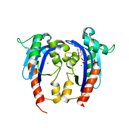
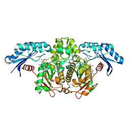
BU of 5j92 by Molmil
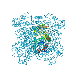
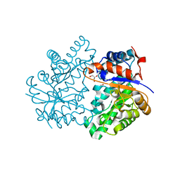
Crystal structure of a putative alpha-ketoglutarate dependent 2,4-D dioxygenase from Burkholderia xenovorans
Descriptor: ACETATE ION, FE (II) ION, Putative alpha KG dependent 2,4-D dioxygenase
Authors:Seattle Structural Genomics Center for Infectious Disease (SSGCID)
Deposit date:2016-04-08
Release date:2016-05-11
Last modified:2023-09-27
Method:X-RAY DIFFRACTION (1.95 Å)
Cite:Crystal structure of a putative alpha-ketoglutarate dependent 2,4-D dioxygenase from Burkholderia xenovorans
to be published
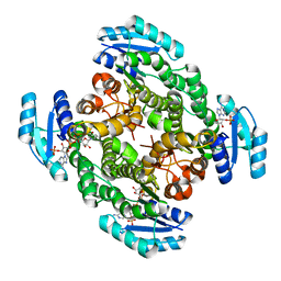
5JC8
DownloadVisualize
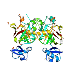
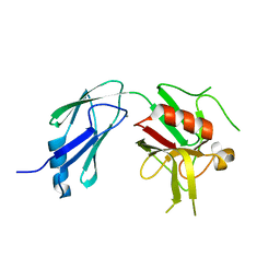
BU of 5jc8 by Molmil
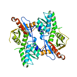
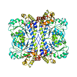
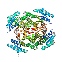
Crystal structure of a putative short-chain dehydrogenase/reductase from Burkholderia xenovorans
Descriptor: 1,2-ETHANEDIOL, 2-[BIS-(2-HYDROXY-ETHYL)-AMINO]-2-HYDROXYMETHYL-PROPANE-1,3-DIOL, MAGNESIUM ION, ...
Authors:Seattle Structural Genomics Center for Infectious Disease (SSGCID)
Deposit date:2016-04-14
Release date:2016-05-11
Last modified:2023-09-27
Method:X-RAY DIFFRACTION (1.45 Å)
Cite:Crystal structure of a putative short-chain dehydrogenase/reductase from Burkholderia xenovorans
to be published
6UWW
DownloadVisualize
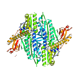
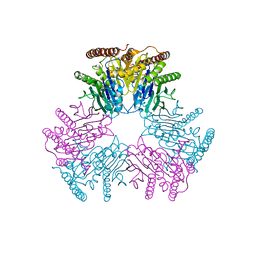
BU of 6uww by Molmil
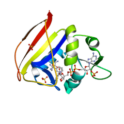
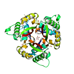
Crystal structure of dihydrofolate reductase from Mycobacterium ulcerans with P218 inhibitor
Descriptor: 3-(2-{3-[(2,4-diamino-6-ethylpyrimidin-5-yl)oxy]propoxy}phenyl)propanoic acid, Dihydrofolate reductase, NADP NICOTINAMIDE-ADENINE-DINUCLEOTIDE PHOSPHATE, ...
Authors:Seattle Structural Genomics Center for Infectious Disease, Seattle Structural Genomics Center for Infectious Disease (SSGCID)
Deposit date:2019-11-05
Release date:2020-11-11
Last modified:2023-10-11
Method:X-RAY DIFFRACTION (0.92 Å)
Cite:Crystal structure of dihydrofolate reductase from Mycobacterium ulcerans with SDDC-0001565 inhibitor
to be published
6V45
DownloadVisualize
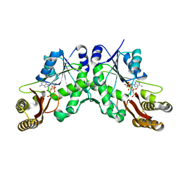
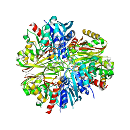
BU of 6v45 by Molmil
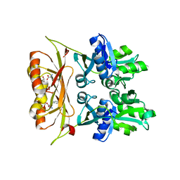
Crystal structure of a Probable carnitine operon oxidoreductase caia from Brucella melitensis
Descriptor: Probable carnitine operon oxidoreductase caia, SULFATE ION
Authors:Seattle Structural Genomics Center for Infectious Disease (SSGCID)
Deposit date:2019-11-27
Release date:2019-12-11
Last modified:2024-04-03
Method:X-RAY DIFFRACTION (2.6 Å)
Cite:Crystal structure of a Probable carnitine operon oxidoreductase caia from Brucella melitensis
to be published
6UZI
DownloadVisualize
BU of 6uzi by Molmil
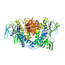
Crystal structure of Dihydrolipoyl dehydrogenase from Elizabethkingia anophelis NUHP1
Descriptor: 1,2-ETHANEDIOL, CHLORIDE ION, Dihydrolipoyl dehydrogenase, ...
Authors:Seattle Structural Genomics Center for Infectious Disease (SSGCID)
Deposit date:2019-11-15
Release date:2020-01-15
Last modified:2023-10-11
Method:X-RAY DIFFRACTION (2.8 Å)
Cite:Crystal structure of Dihydrolipoyl dehydrogenase from Elizabethkingia anophelis NUHP1
TO BE PUBLISHED
5J3B
DownloadVisualize
BU of 5j3b by Molmil
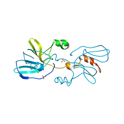
Structure of translation elongation factor P from Acinetobacter baumannii
Descriptor: CHLORIDE ION, Elongation factor P, POTASSIUM ION
Authors:Seattle Structural Genomics Center for Infectious Disease (SSGCID)
Deposit date:2016-03-30
Release date:2016-05-04
Last modified:2023-09-27
Method:X-RAY DIFFRACTION (2.3 Å)
Cite:Structure of translation elongation factor P from Acinetobacter baumannii
to be published
4HVJ
DownloadVisualize
BU of 4hvj by Molmil
Crystal structure of a putative uncharacterized protein from Mycobacterium tuberculosis in complex with AMP
Descriptor: ADENOSINE MONOPHOSPHATE, MAGNESIUM ION, Putative uncharacterized protein
Authors:Seattle Structural Genomics Center for Infectious Disease (SSGCID)
Deposit date:2012-11-06
Release date:2012-11-28
Last modified:2023-09-20
Method:X-RAY DIFFRACTION (2.1 Å)
Cite:Mycobacterium tuberculosis Rv2179c Protein Establishes a New Exoribonuclease Family with Broad Phylogenetic Distribution.
J.Biol.Chem., 289, 2014
6V77
DownloadVisualize
BU of 6v77 by Molmil
Crystal structure of a putative HpcE protein from Mycobacterium smegmatis
Descriptor: 1,2-ETHANEDIOL, Putative HpcE protein, SULFATE ION, ...
Authors:Seattle Structural Genomics Center for Infectious Disease (SSGCID)
Deposit date:2019-12-07
Release date:2020-01-15
Last modified:2023-10-11
Method:X-RAY DIFFRACTION (1.75 Å)
Cite:Crystal structure of a putative HpcE protein from Mycobacterium smegmatis
To be Published
6UYH
DownloadVisualize
BU of 6uyh by Molmil
Crystal structure of prolyl-tRNA synthetase from Naegleria fowleri in complex with halofuginone and AMPPNP
Descriptor: 1,2-ETHANEDIOL, 7-bromo-6-chloro-3-{3-[(2R,3S)-3-hydroxypiperidin-2-yl]-2-oxopropyl}quinazolin-4(3H)-one, MAGNESIUM ION, ...
Authors:Seattle Structural Genomics Center for Infectious Disease (SSGCID)
Deposit date:2019-11-13
Release date:2019-11-27
Last modified:2023-10-11
Method:X-RAY DIFFRACTION (1.75 Å)
Cite:Crystal structure of prolyl-tRNA synthetase from Naegleria fowleri in complex with halofuginone and AMPPNP
TO BE PUBLISHED
5KWV
DownloadVisualize
BU of 5kwv by Molmil
Crystal Structure of a Pantoate-beta-alanine Ligase from Neisseria gonorrhoeae with bound AMPPNP
Descriptor: PHOSPHOAMINOPHOSPHONIC ACID-ADENYLATE ESTER, Pantothenate synthetase
Authors:Seattle Structural Genomics Center for Infectious Disease (SSGCID)
Deposit date:2016-07-19
Release date:2016-07-27
Last modified:2023-10-04
Method:X-RAY DIFFRACTION (2.25 Å)
Cite:Crystal Structure of a Pantoate-beta-alanine Ligase from Neisseria gonorrhoeae with bound AMPPNP
to be published
4HWG
DownloadVisualize
BU of 4hwg by Molmil
Structure of UDP-N-acetylglucosamine 2-epimerase from Rickettsia bellii
Descriptor: PHOSPHATE ION, UDP-N-acetylglucosamine 2-epimerase
Authors:Seattle Structural Genomics Center for Infectious Disease (SSGCID)
Deposit date:2012-11-07
Release date:2012-11-28
Last modified:2023-09-20
Method:X-RAY DIFFRACTION (2 Å)
Cite:Structure of UDP-N-acetylglucosamine 2-epimerase from Rickettsia bellii
TO BE PUBLISHED
4I1Y
DownloadVisualize
BU of 4i1y by Molmil
The structure of Cysteine synthase from Mycobacterium ulcerans Agy99
Descriptor: CHLORIDE ION, Cysteine synthase, SULFATE ION
Authors:Seattle Structural Genomics Center for Infectious Disease (SSGCID)
Deposit date:2012-11-21
Release date:2012-12-26
Last modified:2023-09-20
Method:X-RAY DIFFRACTION (2.6 Å)
Cite:Increasing the structural coverage of tuberculosis drug targets.
Tuberculosis (Edinb), 95, 2015
6V3M
DownloadVisualize
BU of 6v3m by Molmil
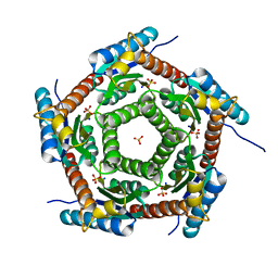
Crystal structure of 2C-methyl-D-erythritol 2,4-cyclodiphosphate synthase (IspF) Burkholderia pseudomallei in compomplex with ligand HGN-0961 (BSI110840)
Descriptor: 1,2-ETHANEDIOL, 2-C-methyl-D-erythritol 2,4-cyclodiphosphate synthase, 5-{[(propan-2-yl)carbamoyl]amino}-1,3,4-thiadiazole-2-sulfonamide, ...
Authors:Seattle Structural Genomics Center for Infectious Disease (SSGCID)
Deposit date:2019-11-26
Release date:2020-12-09
Last modified:2023-10-11
Method:X-RAY DIFFRACTION (1.55 Å)
Cite:Crystal structure of 2C-methyl-D-erythritol 2,4-cyclodiphosphate synthase (IspF) Burkholderia pseudomallei in compomplex with ligand HGN-0961 (BSI110840)
to be published
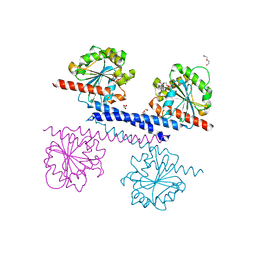
6VH5
DownloadVisualize
BU of 6vh5 by Molmil
Crystal structure of prephenate dehydratase from brucella melitensis biovar abortus 2308 in complex with phenylalanine
Descriptor: 1,2-ETHANEDIOL, PHENYLALANINE, Prephenate dehydratase:Amino acid-binding ACT
Authors:Seattle Structural Genomics Center for Infectious Disease (SSGCID)
Deposit date:2020-01-09
Release date:2020-01-29
Last modified:2024-04-03
Method:X-RAY DIFFRACTION (2.4 Å)
Cite:Crystal structure of prephenate dehydratase from Brucella melitensis biovar abortus 2308 in complex with phenylalanine
to be published
4IJN
DownloadVisualize
BU of 4ijn by Molmil
Crystal structure of an acetate kinase from Mycobacterium smegmatis bound to AMP and sulfate
Descriptor: 1,2-ETHANEDIOL, ADENOSINE MONOPHOSPHATE, Acetate kinase, ...
Authors:Seattle Structural Genomics Center for Infectious Disease (SSGCID)
Deposit date:2012-12-21
Release date:2013-01-16
Last modified:2023-09-20
Method:X-RAY DIFFRACTION (1.7 Å)
Cite:Increasing the structural coverage of tuberculosis drug targets.
Tuberculosis (Edinb), 95, 2015
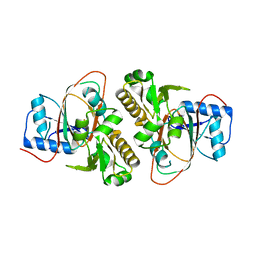
4K73
DownloadVisualize
BU of 4k73 by Molmil
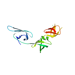
X-ray crystal structure of an L,D-transpeptidase from Mycobacterium tuberculosis H37Rv
Descriptor: CALCIUM ION, L,D-transpeptidase
Authors:Seattle Structural Genomics Center for Infectious Disease (SSGCID)
Deposit date:2013-04-16
Release date:2013-06-12
Last modified:2024-02-28
Method:X-RAY DIFFRACTION (1.65 Å)
Cite:X-ray crystal structure of an L,D-transpeptidase from Mycobacterium tuberculosis H37Rv (CASP Target)
TO BE PUBLISHED
6VIN
DownloadVisualize
BU of 6vin by Molmil
Crystallographic structure of the circularly permuted human Taspase1 protein
Descriptor: Threonine aspartase 1
Authors:Martin-Garcia, J.M, Fromme, P.
Deposit date:2020-01-13
Release date:2021-01-13
Last modified:2023-10-11
Method:X-RAY DIFFRACTION (3.04 Å)
Cite:Structural insights into the function of the catalytically active human Taspase1.
Structure, 29, 2021
4K9D
DownloadVisualize
BU of 4k9d by Molmil
X-ray crystal structure of a Glyceraldehyde 3-phosphate dehydrogenase from Brugia malayi bound to the co-factor NAD
Descriptor: 1,2-ETHANEDIOL, Glyceraldehyde-3-phosphate dehydrogenase, NICOTINAMIDE-ADENINE-DINUCLEOTIDE
Authors:Seattle Structural Genomics Center for Infectious Disease (SSGCID)
Deposit date:2013-04-19
Release date:2013-05-01
Last modified:2023-09-20
Method:X-RAY DIFFRACTION (2.1 Å)
Cite:X-ray crystal structure of a Glyceraldehyde 3-phosphate dehydrogenase from Brugia malayi bound to the co-factor NAD
TO BE PUBLISHED
4K3Z
DownloadVisualize
BU of 4k3z by Molmil
Crystal structure of D-erythrulose 4-phosphate dehydrogenase from Brucella melitensis, solved by iodide SAD
Descriptor: 1,2-ETHANEDIOL, D-erythrulose 4-phosphate dehydrogenase, GLYCEROL
Authors:Seattle Structural Genomics Center for Infectious Disease (SSGCID)
Deposit date:2013-04-11
Release date:2013-05-15
Last modified:2024-03-13
Method:X-RAY DIFFRACTION (1.95 Å)
Cite:Crystal structure of D-erythrulose 4-phosphate dehydrogenase from Brucella melitensis, solved by iodide SAD
To be Published
4KAM
DownloadVisualize
BU of 4kam by Molmil
X-ray crystal structure of O-acetylhomoserine sulfhydrylase MetC from Mycobacterium marinum ATCC BAA-535 / M
Descriptor: O-acetylhomoserine sulfhydrylase MetC, SULFATE ION
Authors:Seattle Structural Genomics Center for Infectious Disease (SSGCID)
Deposit date:2013-04-22
Release date:2013-05-01
Last modified:2023-09-20
Method:X-RAY DIFFRACTION (2 Å)
Cite:Increasing the structural coverage of tuberculosis drug targets.
Tuberculosis (Edinb), 95, 2015
4IYQ
DownloadVisualize
BU of 4iyq by Molmil
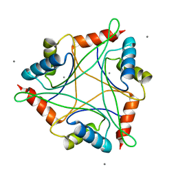
Crystal structure of divalent ion tolerance protein CutA1 from Ehrlichia chaffeensis
Descriptor: CALCIUM ION, Divalent ion tolerance protein CutA1
Authors:Seattle Structural Genomics Center for Infectious Disease (SSGCID)
Deposit date:2013-01-29
Release date:2013-02-13
Last modified:2023-09-20
Method:X-RAY DIFFRACTION (2.55 Å)
Cite:Crystal structure of divalent ion tolerance protein CutA1 from Ehrlichia chaffeensis
To be Published
4J07
DownloadVisualize
BU of 4j07 by Molmil
Crystal structure of a PROBABLE RIBOFLAVIN SYNTHASE, BETA CHAIN RIBH (6,7-dimethyl-8-ribityllumazine synthase, DMRL synthase, Lumazine synthase) from Mycobacterium leprae
Descriptor: 6,7-dimethyl-8-ribityllumazine synthase, SODIUM ION, SULFATE ION
Authors:Seattle Structural Genomics Center for Infectious Disease (SSGCID)
Deposit date:2013-01-30
Release date:2013-03-06
Last modified:2023-09-20
Method:X-RAY DIFFRACTION (1.95 Å)
Cite:Crystal structure of a Probable Riboflavin Synthase, Beta chain RIBH (6,7-dimethyl-8-ribityllumazine synthase, DMRL synthase, Lumazine synthase) from Mycobacterium leprae
TO BE PUBLISHED
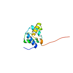
2LKY
DownloadVisualize
BU of 2lky by Molmil
Solution structure of MSMEG_1053, the second DUF3349 annotated protein in the genome of Mycobacterium smegmatis, Seattle Structural Genomics Center for Infectious Disease target MysmA.17112.b
Descriptor: Uncharacterized protein
Authors:Buchko, G.W, Seattle Structural Genomics Center for Infectious Disease (SSGCID)
Deposit date:2011-10-22
Release date:2011-11-16
Last modified:2024-05-15
Method:SOLUTION NMR
Cite:Structural diversity in the Mycobacteria DUF3349 superfamily.
Protein Sci., 29, 2020
4K6F
DownloadVisualize
BU of 4k6f by Molmil
X-ray crystal structure of a putative Acetoacetyl-CoA reductase from Burkholderia cenocepacia bound to the co-factor NADP
Descriptor: NADP NICOTINAMIDE-ADENINE-DINUCLEOTIDE PHOSPHATE, Putative Acetoacetyl-CoA reductase
Authors:Seattle Structural Genomics Center for Infectious Disease (SSGCID)
Deposit date:2013-04-15
Release date:2013-05-15
Last modified:2023-09-20
Method:X-RAY DIFFRACTION (1.5 Å)
Cite:X-ray crystal structure of a putative Acetoacetyl-CoA reductase from Burkholderia cenocepacia bound to the co-factor NADP
TO BE PUBLISHED
6VS4
DownloadVisualize
BU of 6vs4 by Molmil
Crystal structure of ADP RIBOSYLATION FACTOR-LIKE GTP BINDING PROTEIN /Small COPII coat GTPase SAR1 from Encephalitozoon cuniculi in complex with GDP
Descriptor: DI(HYDROXYETHYL)ETHER, GUANOSINE-5'-DIPHOSPHATE, MAGNESIUM ION, ...
Authors:Seattle Structural Genomics Center for Infectious Disease (SSGCID)
Deposit date:2020-02-10
Release date:2020-03-11
Last modified:2024-04-03
Method:X-RAY DIFFRACTION (2.4 Å)
Cite:Crystal structure of ADP RIBOSYLATION FACTOR-LIKE GTP BINDING PROTEIN /Small COPII coat GTPase SAR1 from Encephalitozoon cuniculi in complex with GDP
to be published

225399

건을2024-09-25부터공개중

PDB statisticsPDBj update infoContact PDBjnumon