1MA9
 
 | | Crystal structure of the complex of human vitamin D binding protein and rabbit muscle actin | | 分子名称: | ADENOSINE-5'-TRIPHOSPHATE, Actin, Alpha Skeletal Muscle, ... | | 著者 | Verboven, C, Bogaerts, I, Waelkens, E, Rabijns, A, Van Baelen, H, Bouillon, R, De Ranter, C. | | 登録日 | 2002-08-02 | | 公開日 | 2003-02-04 | | 最終更新日 | 2011-07-13 | | 実験手法 | X-RAY DIFFRACTION (2.4 Å) | | 主引用文献 | Actin-DBP: the perfect structural fit?
Acta Crystallogr.,Sect.D, 59, 2003
|
|
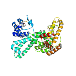
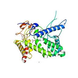
1NNL
 | | Crystal structure of Human Phosphoserine Phosphatase | | 分子名称: | CALCIUM ION, CHLORIDE ION, L-3-phosphoserine phosphatase | | 著者 | Peeraer, Y, Rabijns, A, Verboven, C, Collet, J.F, Van Schaftingen, E, De Ranter, C. | | 登録日 | 2003-01-14 | | 公開日 | 2003-06-03 | | 最終更新日 | 2024-03-13 | | 実験手法 | X-RAY DIFFRACTION (1.53 Å) | | 主引用文献 | High-resolution structure of human phosphoserine phosphatase in open conformation.
Acta Crystallogr.,Sect.D, 59, 2003
|
|
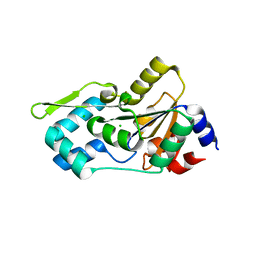
1J78
 | | Crystallographic analysis of the human vitamin D binding protein | | 分子名称: | 3-{2-[1-(5-HYDROXY-1,5-DIMETHYL-HEXYL)-7A-METHYL-OCTAHYDRO-INDEN-4-YLIDENE]-ETHYLIDENE}-4-METHYLENE-CYCLOHEXANOL, OLEIC ACID, vitamin D binding protein | | 著者 | Verboven, C, Rabijns, A, De Maeyer, M, Van Baelen, H, Bouillon, R, De Ranter, C. | | 登録日 | 2001-05-16 | | 公開日 | 2002-02-06 | | 最終更新日 | 2018-01-31 | | 実験手法 | X-RAY DIFFRACTION (2.31 Å) | | 主引用文献 | A structural basis for the unique binding features of the human vitamin D-binding protein.
Nat.Struct.Biol., 9, 2002
|
|
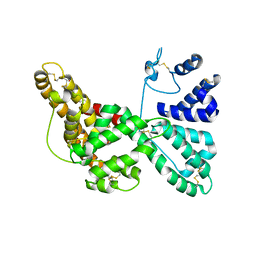
1J7E
 | | A Structural Basis for the Unique Binding Features of the Human Vitamin D-binding Protein | | 分子名称: | 3-(2-{4-[2-(5-HYDROXY-2-METHYLENE-CYCLOHEXYLIDENE)-ETHYLIDENE]-7A-METHYL-OCTAHYDRO-INDEN-1-YL}-PROPYL)-PHENOL, OLEIC ACID, vitamin D binding protein | | 著者 | Verboven, C, Rabijns, A, De Maeyer, M, Van Baelen, H, Bouillon, R, De Ranter, C. | | 登録日 | 2001-05-16 | | 公開日 | 2002-02-06 | | 最終更新日 | 2023-08-16 | | 実験手法 | X-RAY DIFFRACTION (2.55 Å) | | 主引用文献 | A structural basis for the unique binding features of the human vitamin D-binding protein.
Nat.Struct.Biol., 9, 2002
|
|
2SAK
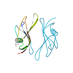 | | STAPHYLOKINASE (SAKSTAR VARIANT) | | 分子名称: | 2-AMINO-2-HYDROXYMETHYL-PROPANE-1,3-DIOL, STAPHYLOKINASE | | 著者 | Rabijns, A, De Bondt, H.L, De Maeyer, M, Lasters, I, De Ranter, C. | | 登録日 | 1997-02-20 | | 公開日 | 1998-02-25 | | 最終更新日 | 2024-02-21 | | 実験手法 | X-RAY DIFFRACTION (1.8 Å) | | 主引用文献 | Three-dimensional structure of staphylokinase, a plasminogen activator with therapeutic potential.
Nat.Struct.Biol., 4, 1997
|
|
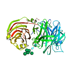
1R76
 | | Structure of a pectate lyase from Azospirillum irakense | | 分子名称: | CHLORIDE ION, GLYCEROL, ISOPROPYL ALCOHOL, ... | | 著者 | Novoa de Armas, H, Verboven, C, De Ranter, C, Desair, J, Vande Broek, A, Vanderleyden, J, Rabijns, A. | | 登録日 | 2003-10-20 | | 公開日 | 2004-06-01 | | 最終更新日 | 2024-02-14 | | 実験手法 | X-RAY DIFFRACTION (2.65 Å) | | 主引用文献 | Azospirillum irakense pectate lyase displays a toroidal fold.
Acta Crystallogr.,Sect.D, 60, 2004
|
|
2AC1
 | | Crystal structure of a cell-wall invertase from Arabidopsis thaliana | | 分子名称: | 2-acetamido-2-deoxy-beta-D-glucopyranose, 2-acetamido-2-deoxy-beta-D-glucopyranose-(1-4)-2-acetamido-2-deoxy-beta-D-glucopyranose, GLYCEROL, ... | | 著者 | Verhaest, M, Le Roy, K, De Ranter, C, Van Laere, A, Van den Ende, W, Rabijns, A. | | 登録日 | 2005-07-18 | | 公開日 | 2006-08-29 | | 最終更新日 | 2023-08-23 | | 実験手法 | X-RAY DIFFRACTION (2.15 Å) | | 主引用文献 | X-ray diffraction structure of a cell-wall invertase from Arabidopsis thaliana.
Acta Crystallogr.,Sect.D, 62, 2006
|
|


