6X2Y
 
 | |
6X2R
 | |
6BC1
 | |
6X2S
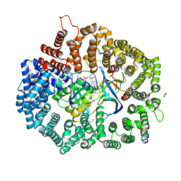
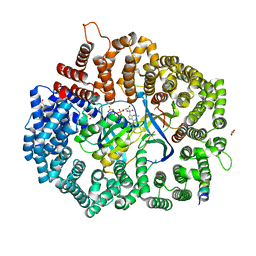
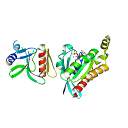
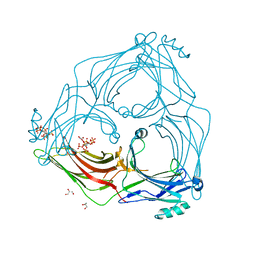
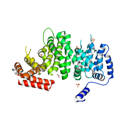
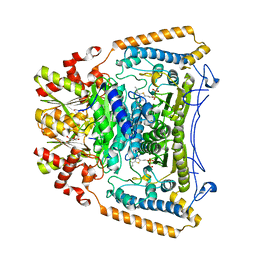
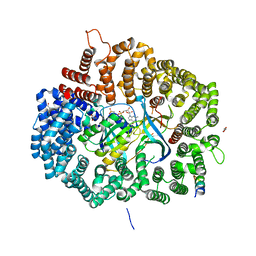 | | Crystal Structure of Mek1(NQ)NES peptide bound to CRM | | 分子名称: | Dual specificity mitogen-activated protein kinase kinase 1, Exportin-1, GLYCEROL, ... | | 著者 | Baumhardt, J.M. | | 登録日 | 2020-05-20 | | 公開日 | 2020-07-01 | | 最終更新日 | 2023-10-18 | | 実験手法 | X-RAY DIFFRACTION (2.496 Å) | | 主引用文献 | Recognition of nuclear export signals by CRM1 carrying the oncogenic E571K mutation.
Mol.Biol.Cell, 31, 2020
|
|
6BC0
 | |
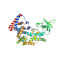
6AR0
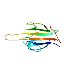 | | Structure of human SLMAP FHA domain | | 分子名称: | Sarcolemmal membrane-associated protein | | 著者 | Ni, L, Luo, X. | | 登録日 | 2017-08-21 | | 公開日 | 2018-07-04 | | 最終更新日 | 2023-10-04 | | 実験手法 | X-RAY DIFFRACTION (1.08 Å) | | 主引用文献 | SAV1 promotes Hippo kinase activation through antagonizing the PP2A phosphatase STRIPAK.
Elife, 6, 2017
|
|
6AR2
 | |
6COK
 | |
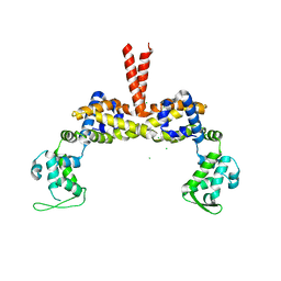
5TV1
 | | active arrestin-3 with inositol hexakisphosphate | | 分子名称: | Beta-arrestin-2, GLYCEROL, INOSITOL HEXAKISPHOSPHATE | | 著者 | Chen, Q, Gilbert, N.C, Perry, N.A, Vishniveteskiy, S, Gurevich, V.V, Iverson, T.M. | | 登録日 | 2016-11-07 | | 公開日 | 2017-11-22 | | 最終更新日 | 2023-10-04 | | 実験手法 | X-RAY DIFFRACTION (2.4 Å) | | 主引用文献 | Structural basis of arrestin-3 activation and signaling.
Nat Commun, 8, 2017
|
|
5V6R
 | |
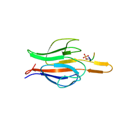
5V6B
 | | Crystal structure of GIPC1 | | 分子名称: | PDZ domain-containing protein GIPC1 | | 著者 | Shang, G, Zhang, X. | | 登録日 | 2017-03-16 | | 公開日 | 2017-05-31 | | 最終更新日 | 2023-10-04 | | 実験手法 | X-RAY DIFFRACTION (1.9 Å) | | 主引用文献 | Structure analyses reveal a regulated oligomerization mechanism of the PlexinD1/GIPC/myosin VI complex.
Elife, 6, 2017
|
|
5V6H
 | |
5V6E
 | |
5V6T
 | |
4K6J
 | | Human cohesin inhibitor WapL | | 分子名称: | ACETATE ION, SULFATE ION, Wings apart-like protein homolog | | 著者 | Tomchick, D.R, Yu, H, Ouyang, Z. | | 登録日 | 2013-04-16 | | 公開日 | 2013-06-19 | | 最終更新日 | 2024-02-28 | | 実験手法 | X-RAY DIFFRACTION (2.6205 Å) | | 主引用文献 | Structure of the human cohesin inhibitor Wapl.
Proc.Natl.Acad.Sci.USA, 110, 2013
|
|
3SEO
 | |
2QJO
 | | crystal structure of a bifunctional NMN adenylyltransferase/ADP ribose pyrophosphatase (NadM) complexed with ADPRP and NAD from Synechocystis sp. | | 分子名称: | ADENOSINE-5-DIPHOSPHORIBOSE, Bifunctional NMN adenylyltransferase/Nudix hydrolase, NICOTINAMIDE-ADENINE-DINUCLEOTIDE, ... | | 著者 | Huang, N, Sorci, L, Zhang, X, Brautigan, C, Raffaelli, N, Magni, G, Grishin, N.V, Osterman, A, Zhang, H. | | 登録日 | 2007-07-08 | | 公開日 | 2008-03-11 | | 最終更新日 | 2024-02-21 | | 実験手法 | X-RAY DIFFRACTION (2.6 Å) | | 主引用文献 | Bifunctional NMN Adenylyltransferase/ADP-Ribose Pyrophosphatase: Structure and Function in Bacterial NAD Metabolism.
Structure, 16, 2008
|
|
2QJT
 | | Crystal structure of a bifunctional NMN adenylyltransferase/ADP ribose pyrophosphatase complexed with AMP and MN ion from Francisella tularensis | | 分子名称: | ADENOSINE MONOPHOSPHATE, MANGANESE (II) ION, Nicotinamide-nucleotide adenylyltransferase | | 著者 | Huang, N, Sorci, L, Zhang, X, Brautigan, C, Raffaelli, N, Magni, G, Grishin, N.V, Osterman, A, Zhang, H. | | 登録日 | 2007-07-09 | | 公開日 | 2008-03-04 | | 最終更新日 | 2017-10-18 | | 実験手法 | X-RAY DIFFRACTION (2.3 Å) | | 主引用文献 | Bifunctional NMN Adenylyltransferase/ADP-Ribose Pyrophosphatase: Structure and Function in Bacterial NAD Metabolism.
Structure, 16, 2008
|
|
2R5W
 | | Crystal structure of a bifunctional NMN adenylyltransferase/ADP ribose pyrophosphatase from Francisella tularensis | | 分子名称: | CHLORIDE ION, MAGNESIUM ION, Nicotinamide-nucleotide adenylyltransferase | | 著者 | Huang, N, Sorci, L, Zhang, X, Brautigan, C, Li, X, Raffaelli, N, Grishin, N, Osterman, A, Zhang, H. | | 登録日 | 2007-09-04 | | 公開日 | 2008-03-04 | | 最終更新日 | 2011-07-13 | | 実験手法 | X-RAY DIFFRACTION (2.3 Å) | | 主引用文献 | Bifunctional NMN Adenylyltransferase/ADP-Ribose Pyrophosphatase: Structure and Function in Bacterial NAD Metabolism.
Structure, 16, 2008
|
|
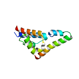
6ANJ
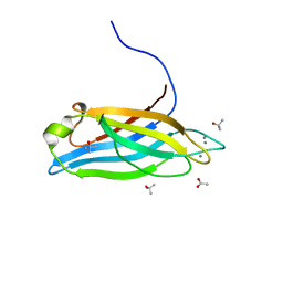 | | Synaptotagmin-7, C2A domain | | 分子名称: | ACETATE ION, CALCIUM ION, Synaptotagmin-7, ... | | 著者 | Tomchick, D.R, Rizo, J, Voleti, R. | | 登録日 | 2017-08-13 | | 公開日 | 2017-09-20 | | 最終更新日 | 2023-10-04 | | 実験手法 | X-RAY DIFFRACTION (1.698 Å) | | 主引用文献 | Exceptionally tight membrane-binding may explain the key role of the synaptotagmin-7 C2A domain in asynchronous neurotransmitter release.
Proc. Natl. Acad. Sci. U.S.A., 114, 2017
|
|
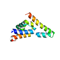
6ANK
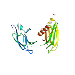 | | Synaptotagmin-7, C2A- and C2B-domains | | 分子名称: | 1,2-ETHANEDIOL, CALCIUM ION, Synaptotagmin-7 | | 著者 | Tomchick, D.R, Rizo, J, Voleti, R. | | 登録日 | 2017-08-13 | | 公開日 | 2017-09-20 | | 最終更新日 | 2023-10-04 | | 実験手法 | X-RAY DIFFRACTION (2.254 Å) | | 主引用文献 | Exceptionally tight membrane-binding may explain the key role of the synaptotagmin-7 C2A domain in asynchronous neurotransmitter release.
Proc. Natl. Acad. Sci. U.S.A., 114, 2017
|
|
2J9F
 | | Human branched-chain alpha-ketoacid dehydrogenase-decarboxylase E1b | | 分子名称: | 2-OXOISOVALERATE DEHYDROGENASE ALPHA SUBUNIT, 2-OXOISOVALERATE DEHYDROGENASE BETA SUBUNIT, C2-1-HYDROXY-3-METHYL-PROPYL-THIAMIN DIPHOSPHATE, ... | | 著者 | Jun, L, Machius, M, Chuang, J.L, Wynn, R.M, Chuang, D.T. | | 登録日 | 2006-11-07 | | 公開日 | 2007-02-27 | | 最終更新日 | 2023-12-13 | | 実験手法 | X-RAY DIFFRACTION (1.88 Å) | | 主引用文献 | The Two Active Sites in Human Branched-Chain Alpha- Keto Acid Dehydrogenase Operate Independently without an Obligatory Alternating-Site Mechanism.
J.Biol.Chem., 282, 2007
|
|


