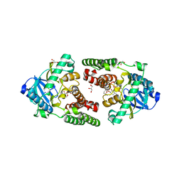
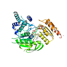
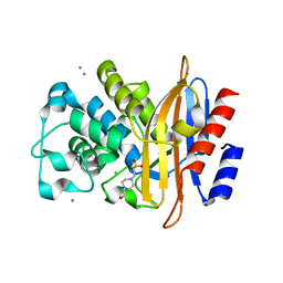
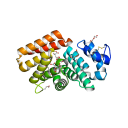
1A8E
 
 | | HUMAN SERUM TRANSFERRIN, RECOMBINANT N-TERMINAL LOBE | | 分子名称: | CARBONATE ION, FE (III) ION, SERUM TRANSFERRIN | | 著者 | Macgillivray, R.T.A, Moore, S.A, Chen, J, Anderson, B.F, Baker, H, Luo, Y, Bewley, M, Smith, C.A, Murphy, M.E.P, Wang, Y, Mason, A.B, Woodworth, R.C, Brayer, G.D, Baker, E.N. | | 登録日 | 1998-03-24 | | 公開日 | 1998-06-17 | | 最終更新日 | 2024-04-03 | | 実験手法 | X-RAY DIFFRACTION (1.6 Å) | | 主引用文献 | Two high-resolution crystal structures of the recombinant N-lobe of human transferrin reveal a structural change implicated in iron release.
Biochemistry, 37, 1998
|
|
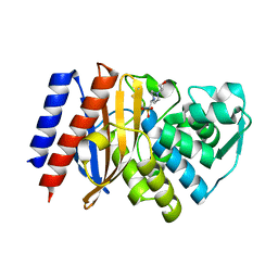
1DTG
 | | HUMAN TRANSFERRIN N-LOBE MUTANT H249E | | 分子名称: | CARBONATE ION, FE (III) ION, TRANSFERRIN | | 著者 | MacGillivray, R.T, Bewley, M.C, Smith, C.A, He, Q.Y, Mason, A.B. | | 登録日 | 2000-01-12 | | 公開日 | 2000-01-21 | | 最終更新日 | 2021-11-03 | | 実験手法 | X-RAY DIFFRACTION (2.4 Å) | | 主引用文献 | Mutation of the iron ligand His 249 to Glu in the N-lobe of human transferrin abolishes the dilysine "trigger" but does not significantly affect iron release.
Biochemistry, 39, 2000
|
|
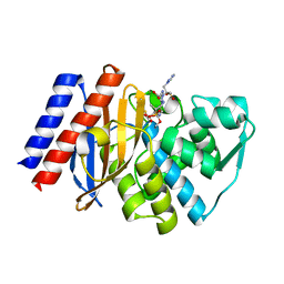
1CQ3
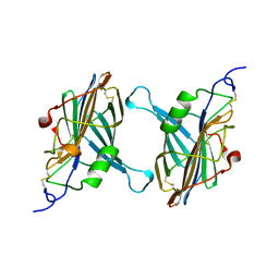 | | STRUCTURE OF A SOLUBLE SECRETED CHEMOKINE INHIBITOR, VCCI, FROM COWPOX VIRUS | | 分子名称: | VIRAL CHEMOKINE INHIBITOR | | 著者 | Carfi, A, Smith, C.A, Smolak, P.J, McGrew, J, Wiley, D.C. | | 登録日 | 1999-08-05 | | 公開日 | 1999-11-12 | | 最終更新日 | 2011-07-13 | | 実験手法 | X-RAY DIFFRACTION (1.85 Å) | | 主引用文献 | Structure of a soluble secreted chemokine inhibitor vCCI (p35) from cowpox virus.
Proc.Natl.Acad.Sci.USA, 96, 1999
|
|
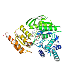
1E3J
 | | Ketose reductase (sorbitol dehydrogenase) from silverleaf whitefly | | 分子名称: | BORIC ACID, NADP(H)-DEPENDENT KETOSE REDUCTASE, PHOSPHATE ION, ... | | 著者 | Banfield, M.J, Salvucci, M.E, Baker, E.N, Smith, C.A. | | 登録日 | 2000-06-19 | | 公開日 | 2001-02-04 | | 最終更新日 | 2024-05-08 | | 実験手法 | X-RAY DIFFRACTION (2.3 Å) | | 主引用文献 | Crystal Structure of Nadp(H)-Dependent Ketose Reductase from Besimia Argentifolii at 2.3 Angstrom Resolution
J.Mol.Biol., 306, 2001
|
|
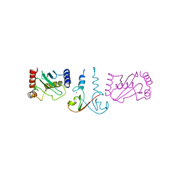
3EB5
 | | Structure of the cIAP2 RING domain | | 分子名称: | Baculoviral IAP repeat-containing protein 3, SODIUM ION, ZINC ION | | 著者 | Mace, P.D, Linke, K, Smith, C.A, Day, C.L. | | 登録日 | 2008-08-27 | | 公開日 | 2008-09-09 | | 最終更新日 | 2024-02-21 | | 実験手法 | X-RAY DIFFRACTION (2 Å) | | 主引用文献 | Structures of the cIAP2 RING domain reveal conformational changes associated with ubiquitin-conjugating enzyme (E2) recruitment.
J.Biol.Chem., 283, 2008
|
|
3EB6
 | | Structure of the cIAP2 RING domain bound to UbcH5b | | 分子名称: | Baculoviral IAP repeat-containing protein 3, Ubiquitin-conjugating enzyme E2 D2, ZINC ION | | 著者 | Mace, P.D, Linke, K, Schumacher, F.-R, Smith, C.A, Day, C.L. | | 登録日 | 2008-08-27 | | 公開日 | 2008-09-09 | | 最終更新日 | 2024-02-21 | | 実験手法 | X-RAY DIFFRACTION (3.4 Å) | | 主引用文献 | Structures of the cIAP2 RING Domain Reveal Conformational Changes Associated with Ubiquitin-conjugating Enzyme (E2) Recruitment.
J.Biol.Chem., 283, 2008
|
|
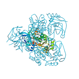
3HAV
 | | Structure of the streptomycin-ATP-APH(2")-IIa ternary complex | | 分子名称: | ADENOSINE-5'-TRIPHOSPHATE, Aminoglycoside phosphotransferase, MAGNESIUM ION, ... | | 著者 | Young, P.G, Baker, E.N, Vakulenko, S.B, Smith, C.A. | | 登録日 | 2009-05-02 | | 公開日 | 2009-11-17 | | 最終更新日 | 2023-09-06 | | 実験手法 | X-RAY DIFFRACTION (2.45 Å) | | 主引用文献 | The crystal structures of substrate and nucleotide complexes of Enterococcus faecium aminoglycoside-2''-phosphotransferase-IIa [APH(2'')-IIa] provide insights into substrate selectivity in the APH(2'') subfamily.
J.Bacteriol., 191, 2009
|
|
3HAM
 | | Structure of the gentamicin-APH(2")-IIa complex | | 分子名称: | (2R,3R,4R,5R)-2-((1S,2S,3R,4S,6R)-4,6-DIAMINO-3-((2R,3R,6S)-3-AMINO-6-(AMINOMETHYL)-TETRAHYDRO-2H-PYRAN-2-YLOXY)-2-HYDR OXYCYCLOHEXYLOXY)-5-METHYL-4-(METHYLAMINO)-TETRAHYDRO-2H-PYRAN-3,5-DIOL, Aminoglycoside phosphotransferase, GLYCEROL | | 著者 | Young, P.G, Baker, E.N, Vakulenko, S.B, Smith, C.A. | | 登録日 | 2009-05-02 | | 公開日 | 2009-07-28 | | 最終更新日 | 2024-02-21 | | 実験手法 | X-RAY DIFFRACTION (2.5 Å) | | 主引用文献 | The crystal structures of substrate and nucleotide complexes of Enterococcus faecium aminoglycoside-2''-phosphotransferase-IIa [APH(2'')-IIa] provide insights into substrate selectivity in the APH(2'') subfamily.
J.Bacteriol., 191, 2009
|
|
3NI9
 | | GES-2 carbapenemase apo form | | 分子名称: | 4-(2-HYDROXYETHYL)-1-PIPERAZINE ETHANESULFONIC ACID, Beta-lactamase GES-2 | | 著者 | Frase, H, Smith, C.A, Toth, M, Vakulenko, S.B. | | 登録日 | 2010-06-15 | | 公開日 | 2011-02-23 | | 最終更新日 | 2023-09-06 | | 実験手法 | X-RAY DIFFRACTION (2 Å) | | 主引用文献 | Kinetic and structural requirements for carbapenemase activity in GES-type beta-lactamases.
Biochemistry, 54, 2015
|
|
1YK3
 | | Crystal structure of Rv1347c from Mycobacterium tuberculosis | | 分子名称: | Hypothetical protein Rv1347c/MT1389, octyl beta-D-glucopyranoside | | 著者 | Card, G.L, Peterson, N.A, Smith, C.A, Rupp, B, Schick, B.M, Baker, E.N, TB Structural Genomics Consortium (TBSGC) | | 登録日 | 2005-01-16 | | 公開日 | 2005-02-01 | | 最終更新日 | 2024-03-13 | | 実験手法 | X-RAY DIFFRACTION (2.2 Å) | | 主引用文献 | The crystal structure of Rv1347c, a putative antibiotic resistance protein from Mycobacterium tuberculosis, reveals a GCN5-related fold and suggests an alternative function in siderophore biosynthesis
J.Biol.Chem., 280, 2005
|
|
3NIA
 | | GES-2 carbapenemase tazobactam complex | | 分子名称: | (3S)-3-(dihydroxy-lambda~4~-sulfanyl)-4-(1H-1,2,3-triazol-1-yl)-D-valine, Beta-lactamase GES-2, TAZOBACTAM INTERMEDIATE | | 著者 | Frase, H, Smith, C.A, Toth, M, Vakulenko, S.B. | | 登録日 | 2010-06-15 | | 公開日 | 2011-03-02 | | 最終更新日 | 2023-09-06 | | 実験手法 | X-RAY DIFFRACTION (1.65 Å) | | 主引用文献 | Inhibition of the GES-2 beta-Lactamse by Tazobactam: Detection of Reaction
Intermediates by UV and Mass Spectrometry and X-Ray Crystallography
To be Published
|
|
2VOS
 | | Mycobacterium tuberculosis Folylpolyglutamate synthase complexed with ADP | | 分子名称: | ADENOSINE-5'-DIPHOSPHATE, COBALT (II) ION, FOLYLPOLYGLUTAMATE SYNTHASE PROTEIN FOLC, ... | | 著者 | Young, P.G, Baker, E.N, Metcalf, P, Smith, C.A. | | 登録日 | 2008-02-19 | | 公開日 | 2008-07-01 | | 最終更新日 | 2011-07-13 | | 実験手法 | X-RAY DIFFRACTION (2 Å) | | 主引用文献 | Structures of Mycobacterium Tuberculosisfolylpolyglutamate Synthase Complexed with Adp and Amppcp.
Acta Crystallogr.,Sect.D, 64, 2008
|
|
2VOR
 | | Crystal Structures of Mycobacterium tuberculosis Folylpolyglutamate Synthase Complexed with ADP and AMPPCP | | 分子名称: | COBALT (II) ION, FOLYLPOLYGLUTAMATE SYNTHASE PROTEIN FOLC, GLYCEROL, ... | | 著者 | Young, P.G, Baker, E.N, Metcalf, P, Smith, C.A. | | 登録日 | 2008-02-19 | | 公開日 | 2008-07-01 | | 最終更新日 | 2024-05-01 | | 実験手法 | X-RAY DIFFRACTION (2.3 Å) | | 主引用文献 | Structures of Mycobacterium Tuberculosisfolylpolyglutamate Synthase Complexed with Adp and Amppcp.
Acta Crystallogr.,Sect.D, 64, 2008
|
|
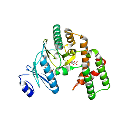
2VJF
 | | Crystal Structure of the MDM2-MDMX RING Domain Heterodimer | | 分子名称: | CITRATE ANION, E3 UBIQUITIN-PROTEIN LIGASE MDM2, MDM4 PROTEIN, ... | | 著者 | Mace, P.D, Linke, K, Smith, C.A, Day, C.L. | | 登録日 | 2007-12-10 | | 公開日 | 2008-05-13 | | 最終更新日 | 2024-05-08 | | 実験手法 | X-RAY DIFFRACTION (2.3 Å) | | 主引用文献 | Structure of the Mdm2/Mdmx Ring Domain Heterodimer Reveals Dimerization is Required for Their Ubiquitylation in Trans.
Cell Death Differ., 15, 2008
|
|
2VJE
 | | Crystal Structure of the MDM2-MDMX RING Domain Heterodimer | | 分子名称: | CITRATE ANION, E3 UBIQUITIN-PROTEIN LIGASE MDM2, MDM4 PROTEIN, ... | | 著者 | Mace, P.D, Linke, K, Smith, C.A, Day, C.L. | | 登録日 | 2007-12-10 | | 公開日 | 2008-05-13 | | 最終更新日 | 2024-05-08 | | 実験手法 | X-RAY DIFFRACTION (2.2 Å) | | 主引用文献 | Structure of the MDM2/MDMX RING domain heterodimer reveals dimerization is required for their ubiquitylation in trans.
Cell Death Differ., 15, 2008
|
|
2PN8
 | | Crystal structure of human peroxiredoxin 4 (thioredoxin peroxidase) | | 分子名称: | Peroxiredoxin-4 | | 著者 | Pilka, E.S, Kavanagh, K.L, Cooper, C, von Delft, F, Sundstrom, M, Arrowsmith, C.A, Weigelt, J, Edwards, A, Oppermann, U, Structural Genomics Consortium (SGC) | | 登録日 | 2007-04-24 | | 公開日 | 2007-05-22 | | 最終更新日 | 2023-08-30 | | 実験手法 | X-RAY DIFFRACTION (1.8 Å) | | 主引用文献 | Crystal structure of human peroxiredoxin 4 (thioredoxin peroxidase).
To be Published
|
|
2Q9V
 | | Crystal structure of the C890S mutant of the 4th PDZ domain of human membrane associated guanylate kinase | | 分子名称: | Membrane-associated guanylate kinase, WW and PDZ domain-containing protein 1 | | 著者 | Pilka, E.S, Hozjan, V, Kavanagh, K.L, Papagrigoriou, E, Cooper, C, Elkins, J.M, Doyle, D.A, von Delft, F, Sundstrom, M, Arrowsmith, C.A, Weigelt, J, Edwards, A, Oppermann, U, Structural Genomics Consortium (SGC) | | 登録日 | 2007-06-14 | | 公開日 | 2007-06-26 | | 最終更新日 | 2023-08-30 | | 実験手法 | X-RAY DIFFRACTION (2 Å) | | 主引用文献 | Crystal structure of the C890S mutant of the 4th PDZ domain of human membrane associated guanylate kinase.
To be Published
|
|
3LEZ
 | |
4PNJ
 | | Recombinant Sperm Whale P6 Myoglobin Solved with Single Pulse Free Electron Laser Data | | 分子名称: | Myoglobin, PROTOPORPHYRIN IX CONTAINING FE, SULFATE ION | | 著者 | Cohen, A, Gonzalez, A, Lam, W, Lyubimov, A, Sauter, N, Tsai, Y, Uervirojnangkoorn, M, Brunger, A, Soltis, M. | | 登録日 | 2014-05-23 | | 公開日 | 2014-11-05 | | 最終更新日 | 2023-09-27 | | 実験手法 | X-RAY DIFFRACTION (1.36 Å) | | 主引用文献 | Goniometer-based femtosecond crystallography with X-ray free electron lasers.
Proc.Natl.Acad.Sci.USA, 111, 2014
|
|
6XPB
 | |
6XPC
 | |
7KQP
 | | Crystal structure of SARS-CoV-2 NSP3 macrodomain in complex with ADP-ribose (P43 crystal form) | | 分子名称: | Non-structural protein 3, [(2R,3S,4R,5R)-5-(6-AMINOPURIN-9-YL)-3,4-DIHYDROXY-OXOLAN-2-YL]METHYL [HYDROXY-[[(2R,3S,4R,5S)-3,4,5-TRIHYDROXYOXOLAN-2-YL]METHOXY]PHOSPHORYL] HYDROGEN PHOSPHATE | | 著者 | Correy, G.J, Young, I.D, Thompson, M.C, Fraser, J.S. | | 登録日 | 2020-11-17 | | 公開日 | 2020-12-09 | | 最終更新日 | 2024-03-06 | | 実験手法 | X-RAY DIFFRACTION (0.88 Å) | | 主引用文献 | Fragment binding to the Nsp3 macrodomain of SARS-CoV-2 identified through crystallographic screening and computational docking.
Sci Adv, 7, 2021
|
|
7KQW
 | | Crystal structure of SARS-CoV-2 NSP3 macrodomain (C2 crystal form, methylated) | | 分子名称: | Non-structural protein 3 | | 著者 | Correy, G.J, Young, I.D, Thompson, M.C, Fraser, J.S. | | 登録日 | 2020-11-17 | | 公開日 | 2020-12-09 | | 最終更新日 | 2023-11-15 | | 実験手法 | X-RAY DIFFRACTION (0.93 Å) | | 主引用文献 | Fragment binding to the Nsp3 macrodomain of SARS-CoV-2 identified through crystallographic screening and computational docking.
Sci Adv, 7, 2021
|
|
7T1M
 | |
7T1S
 | |


