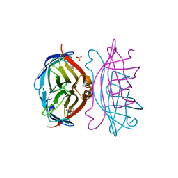
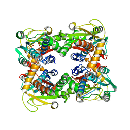
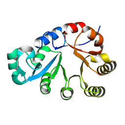
2IZF
 
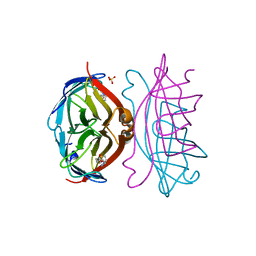 | | STREPTAVIDIN-BIOTIN PH 4.0 I222 COMPLEX | | 分子名称: | BIOTIN, STREPTAVIDIN, SULFATE ION | | 著者 | Katz, B.A. | | 登録日 | 1997-08-13 | | 公開日 | 1998-09-16 | | 最終更新日 | 2024-02-21 | | 実験手法 | X-RAY DIFFRACTION (1.58 Å) | | 主引用文献 | Binding of biotin to streptavidin stabilizes intersubunit salt bridges between Asp61 and His87 at low pH.
J.Mol.Biol., 274, 1997
|
|
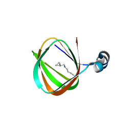
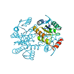
2IZC
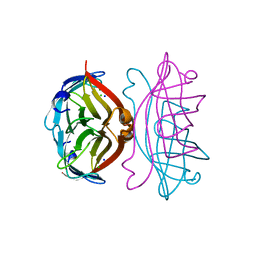 | | APOSTREPTAVIDIN PH 2.0 I222 COMPLEX | | 分子名称: | CHLORIDE ION, SODIUM ION, STREPTAVIDIN | | 著者 | Katz, B.A. | | 登録日 | 1997-08-13 | | 公開日 | 1998-09-16 | | 最終更新日 | 2024-02-21 | | 実験手法 | X-RAY DIFFRACTION (1.4 Å) | | 主引用文献 | Binding of biotin to streptavidin stabilizes intersubunit salt bridges between Asp61 and His87 at low pH.
J.Mol.Biol., 274, 1997
|
|
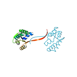
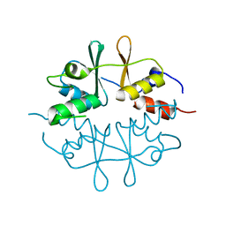
2IZI
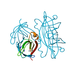 | | STREPTAVIDIN-BIOTIN PH 2.53 I4122 STRUCTURE | | 分子名称: | BIOTIN, STREPTAVIDIN | | 著者 | Katz, B.A. | | 登録日 | 1997-08-13 | | 公開日 | 1998-09-16 | | 最終更新日 | 2024-02-21 | | 実験手法 | X-RAY DIFFRACTION (1.5 Å) | | 主引用文献 | Binding of biotin to streptavidin stabilizes intersubunit salt bridges between Asp61 and His87 at low pH.
J.Mol.Biol., 274, 1997
|
|
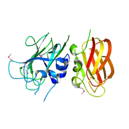
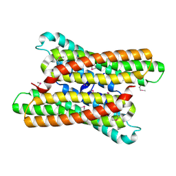
2IZH
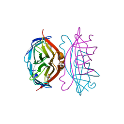 | | STREPTAVIDIN-BIOTIN PH 10.44 I222 COMPLEX | | 分子名称: | BIOTIN, STREPTAVIDIN | | 著者 | Katz, B.A. | | 登録日 | 1997-08-13 | | 公開日 | 1998-09-16 | | 最終更新日 | 2024-02-21 | | 実験手法 | X-RAY DIFFRACTION (1.36 Å) | | 主引用文献 | Binding of biotin to streptavidin stabilizes intersubunit salt bridges between Asp61 and His87 at low pH.
J.Mol.Biol., 274, 1997
|
|
2IZG
 | | STREPTAVIDIN-BIOTIN PH 2.0 I222 COMPLEX | | 分子名称: | BIOTIN, STREPTAVIDIN, SULFATE ION | | 著者 | Katz, B.A. | | 登録日 | 1997-08-13 | | 公開日 | 1998-09-16 | | 最終更新日 | 2024-02-21 | | 実験手法 | X-RAY DIFFRACTION (1.36 Å) | | 主引用文献 | Binding of biotin to streptavidin stabilizes intersubunit salt bridges between Asp61 and His87 at low pH.
J.Mol.Biol., 274, 1997
|
|
1J6U
 | |
1ZKG
 | |
1O5U
 | |
1O4W
 | |
1O59
 | |
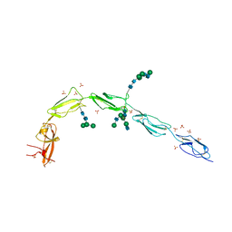
6V06
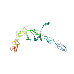 | | Crystal structure of Beta-2 glycoprotein I purified from plasma (pB2GPI) | | 分子名称: | 2-acetamido-2-deoxy-beta-D-glucopyranose-(1-2)-beta-D-mannopyranose-(1-6)-beta-D-mannopyranose-(1-4)-2-acetamido-2-deoxy-beta-D-glucopyranose-(1-4)-2-acetamido-2-deoxy-beta-D-glucopyranose, 4-(2-HYDROXYETHYL)-1-PIPERAZINE ETHANESULFONIC ACID, Beta-2-glycoprotein 1, ... | | 著者 | Chen, Z, Ruben, E.A, Planer, W, Chinnaraj, M, Zuo, X, Pengo, V, Macor, P, Tedesco, F, Pozzi, N. | | 登録日 | 2019-11-18 | | 公開日 | 2020-06-17 | | 最終更新日 | 2023-10-11 | | 実験手法 | X-RAY DIFFRACTION (2.4 Å) | | 主引用文献 | The J-elongated conformation of beta2-glycoprotein I predominates in solution: implications for our understanding of antiphospholipid syndrome.
J.Biol.Chem., 295, 2020
|
|
6V09
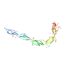 | | Crystal structure of human recombinant Beta-2 glycoprotein I short tag (ST-B2GPI) | | 分子名称: | 2-acetamido-2-deoxy-beta-D-glucopyranose, Beta-2-glycoprotein 1, SULFATE ION, ... | | 著者 | Chen, Z, Ruben, E.A, Planer, W, Chinnaraj, M, Zuo, X, Pengo, V, Macor, P, Tedesco, F, Pozzi, N. | | 登録日 | 2019-11-18 | | 公開日 | 2020-06-17 | | 最終更新日 | 2023-10-11 | | 実験手法 | X-RAY DIFFRACTION (2.99 Å) | | 主引用文献 | The J-elongated conformation of beta2-glycoprotein I predominates in solution: implications for our understanding of antiphospholipid syndrome.
J.Biol.Chem., 295, 2020
|
|
6V08
 | | Crystal structure of human recombinant Beta-2 glycoprotein I (hrB2GPI) | | 分子名称: | 2-acetamido-2-deoxy-beta-D-glucopyranose-(1-4)-2-acetamido-2-deoxy-beta-D-glucopyranose, Beta-2-glycoprotein 1, SULFATE ION, ... | | 著者 | Chen, Z, Ruben, E.A, Planer, W, Chinnaraj, M, Zuo, X, Pengo, V, Macor, P, Tedesco, F, Pozzi, N. | | 登録日 | 2019-11-18 | | 公開日 | 2020-06-17 | | 最終更新日 | 2023-10-11 | | 実験手法 | X-RAY DIFFRACTION (2.58 Å) | | 主引用文献 | The J-elongated conformation of beta2-glycoprotein I predominates in solution: implications for our understanding of antiphospholipid syndrome.
J.Biol.Chem., 295, 2020
|
|
1O58
 | |
1O1Z
 | |
1VJ2
 | |
1O1X
 | |
1O50
 | |
1O5H
 | |
1O20
 | |
1O0X
 | |
1O3U
 | |
1O22
 | |
2AFB
 | |
1ZX8
 | |


