5ZTJ
 
 | | Crystal Structure of GyraseA C-Terminal Domain from Salmonella typhi at 2.4A Resolution | | 分子名称: | DNA gyrase subunit A | | 著者 | Sachdeva, E, Gupta, D, Tiwari, P, Kaur, G, Sharma, S, Singh, T.P, Ethayathulla, A.S, Kaur, P. | | 登録日 | 2018-05-03 | | 公開日 | 2019-05-15 | | 最終更新日 | 2023-11-22 | | 実験手法 | X-RAY DIFFRACTION (2.4 Å) | | 主引用文献 | The pivot point arginines identified in the beta-pinwheel structure of C-terminal domain from Salmonella Typhi DNA Gyrase A subunit.
Sci Rep, 10, 2020
|
|
5ZUG
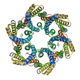 | | Structure of the bacterial acetate channel SatP | | 分子名称: | Succinate-acetate/proton symporter SatP, nonyl beta-D-glucopyranoside | | 著者 | Sun, P.C, Li, J.L, Xiao, Q.J, Guan, Z.Y, Deng, D. | | 登録日 | 2018-05-07 | | 公開日 | 2018-11-21 | | 最終更新日 | 2024-05-29 | | 実験手法 | X-RAY DIFFRACTION (2.802 Å) | | 主引用文献 | Crystal structure of the bacterial acetate transporter SatP reveals that it forms a hexameric channel.
J. Biol. Chem., 293, 2018
|
|
5BRB
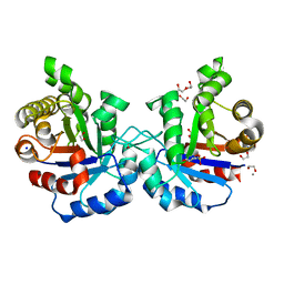
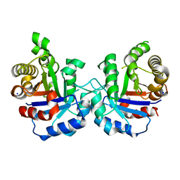 | | Crystal structure of Q64E mutant of Triosephosphate isomerase from Plasmodium falciparum | | 分子名称: | SODIUM ION, Triosephosphate isomerase | | 著者 | Bandyopadhyay, D, Murthy, M.R.N, Balaram, H, Balaram, P. | | 登録日 | 2015-05-30 | | 公開日 | 2015-07-29 | | 最終更新日 | 2023-11-08 | | 実験手法 | X-RAY DIFFRACTION (2.53 Å) | | 主引用文献 | Probing the role of highly conserved residues in triosephosphate isomerase - analysis of site specific mutants at positions 64 and 75 in the Plasmodial enzyme
Febs J., 282, 2015
|
|
6JKG
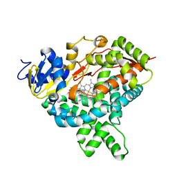
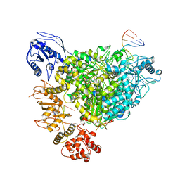
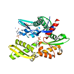
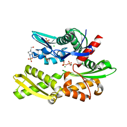
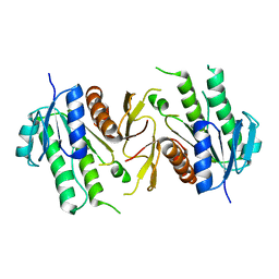 | | The NAD+-free form of human NSDHL | | 分子名称: | Sterol-4-alpha-carboxylate 3-dehydrogenase, decarboxylating | | 著者 | Kim, D, Lee, S.J, Lee, B. | | 登録日 | 2019-02-28 | | 公開日 | 2020-03-04 | | 最終更新日 | 2023-11-22 | | 実験手法 | X-RAY DIFFRACTION (2.9 Å) | | 主引用文献 | Crystal structures of human NSDHL and development of its novel inhibitor with the potential to suppress EGFR activity.
Cell.Mol.Life Sci., 78, 2021
|
|
5BUG
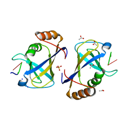
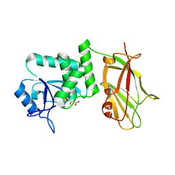 | | Crystal structure of human phosphatase PTEN oxidized by H2O2 | | 分子名称: | L(+)-TARTARIC ACID, Phosphatidylinositol 3,4,5-trisphosphate 3-phosphatase and dual-specificity protein phosphatase PTEN | | 著者 | Lee, C.-U, Bier, D, Hennig, S, Grossmann, T.N. | | 登録日 | 2015-06-03 | | 公開日 | 2015-10-07 | | 最終更新日 | 2024-01-10 | | 実験手法 | X-RAY DIFFRACTION (2.4 Å) | | 主引用文献 | Redox Modulation of PTEN Phosphatase Activity by Hydrogen Peroxide and Bisperoxidovanadium Complexes.
Angew.Chem.Int.Ed.Engl., 54, 2015
|
|
5JL6
 | |
5BT3
 | | Crystal structure of EP300 bromodomain in complex with SGC-CBP30 chemical probe | | 分子名称: | 2-[2-(3-chloro-4-methoxyphenyl)ethyl]-5-(3,5-dimethyl-1,2-oxazol-4-yl)-1-[(2S)-2-(morpholin-4-yl)propyl]-1H-benzimidazole, Histone acetyltransferase p300, ISOPROPYL ALCOHOL | | 著者 | Tallant, C, Hay, D, Krojer, T, Nunez-Alonso, G, Picaud, S, Newman, J.A, Fedorov, O, von Delft, F, Arrowsmith, C.H, Edwards, A.M, Bountra, C, Brennan, P.E, Knapp, S, Structural Genomics Consortium (SGC) | | 登録日 | 2015-06-02 | | 公開日 | 2015-07-01 | | 最終更新日 | 2024-01-10 | | 実験手法 | X-RAY DIFFRACTION (1.05 Å) | | 主引用文献 | Crystal structure of EP300 bromodomain in complex with a 3,5-dimethylisoxazol ligand
To Be Published
|
|
6JLI
 | | Crystal structure of CTLD7 domain of human PLA2R | | 分子名称: | 2-acetamido-2-deoxy-beta-D-glucopyranose, Secretory phospholipase A2 receptor | | 著者 | Yu, B, Hu, Z, Kong, D, Cheng, C, He, Y. | | 登録日 | 2019-03-06 | | 公開日 | 2019-07-17 | | 最終更新日 | 2023-11-22 | | 実験手法 | X-RAY DIFFRACTION (1.778 Å) | | 主引用文献 | Crystal structure of the CTLD7 domain of human M-type phospholipase A2 receptor.
J.Struct.Biol., 207, 2019
|
|
5JLI
 | | Nitric oxide complex of the L16A mutant of cytochrome c prime from Alcaligenes xylosoxidans | | 分子名称: | ASCORBIC ACID, Cytochrome c', HEME C, ... | | 著者 | Kekilli, D, Strange, R.W, Hough, M.A. | | 登録日 | 2016-04-27 | | 公開日 | 2017-03-08 | | 最終更新日 | 2024-01-10 | | 実験手法 | X-RAY DIFFRACTION (1.55 Å) | | 主引用文献 | Engineering proximal vs. distal heme-NO coordination via dinitrosyl dynamics: implications for NO sensor design.
Chem Sci, 8, 2017
|
|
8R6W
 | | Structure of the SFTSV L protein in a transcription-priming state with bound capped RNA [TRANSCRIPTION-PRIMING] | | 分子名称: | RNA (5'-R(*(M7G)*AP*AP*A)-3'), RNA (5'-R(*AP*CP*AP*C)-3'), RNA (5'-R(*AP*CP*AP*CP*AP*GP*AP*GP*AP*CP*GP*CP*CP*CP*AP*G)-3'), ... | | 著者 | Williams, H.M, Thorkelsson, S.R, Vogel, D, Busch, C, Milewski, M, Cusack, S, Grunewald, K, Quemin, E.R.J, Rosenthal, M. | | 登録日 | 2023-11-23 | | 公開日 | 2024-04-24 | | 最終更新日 | 2024-06-19 | | 実験手法 | ELECTRON MICROSCOPY (3.35 Å) | | 主引用文献 | Structural snapshots of phenuivirus cap-snatching and transcription.
Nucleic Acids Res., 52, 2024
|
|
8R6U
 | | Structure of the SFTSV L protein in a transcription-priming state without capped RNA [TRANSCRIPTION-PRIMING (in vitro)] | | 分子名称: | MAGNESIUM ION, RNA (5'-R(P*CP*UP*GP*GP*GP*CP*GP*GP*UP*CP*UP*UP*UP*GP*UP*GP*U)-3'), RNA primer, ... | | 著者 | Williams, H.M, Thorkelsson, S.R, Vogel, D, Busch, C, Milewski, M, Cusack, S, Grunewald, K, Quemin, E.R.J, Rosenthal, M. | | 登録日 | 2023-11-23 | | 公開日 | 2024-04-24 | | 最終更新日 | 2024-06-19 | | 実験手法 | ELECTRON MICROSCOPY (2.98 Å) | | 主引用文献 | Structural snapshots of phenuivirus cap-snatching and transcription.
Nucleic Acids Res., 52, 2024
|
|
5ZYF
 | | Crystal structure of Streptococcus pyogenes type II-A Cas2 | | 分子名称: | 1,2-ETHANEDIOL, CRISPR-associated endoribonuclease Cas2, SULFATE ION | | 著者 | Ka, D, Bae, E. | | 登録日 | 2018-05-24 | | 公開日 | 2018-08-15 | | 最終更新日 | 2024-03-27 | | 実験手法 | X-RAY DIFFRACTION (1.76 Å) | | 主引用文献 | Molecular organization of the type II-A CRISPR adaptation module and its interaction with Cas9 via Csn2
Nucleic Acids Res., 46, 2018
|
|
5JL9
 | |
5B5Y
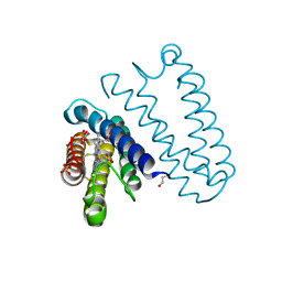
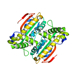 | | Crystal structure of PtLCIB4, a homolog of the limiting CO2-inducible protein LCIB | | 分子名称: | ACETATE ION, PtLCIB4, ZINC ION | | 著者 | Jin, S, Sun, J, Wunder, T, Tang, D, Mueller-Cajar, O.M, Gao, Y. | | 登録日 | 2016-05-24 | | 公開日 | 2016-12-07 | | 最終更新日 | 2023-11-08 | | 実験手法 | X-RAY DIFFRACTION (1.75 Å) | | 主引用文献 | Structural insights into the LCIB protein family reveals a new group of beta-carbonic anhydrases
Proc. Natl. Acad. Sci. U.S.A., 113, 2016
|
|
5BUX
 | |
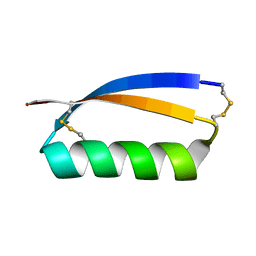
5JI4
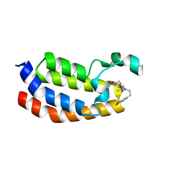
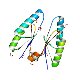
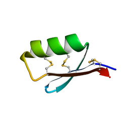 | | Solution structure of the de novo mini protein gEEHE_02 | | 分子名称: | W37 | | 著者 | Buchko, G.W, Bahl, C.D, Pulavarti, S.V, Baker, D, Szyperski, T. | | 登録日 | 2016-04-21 | | 公開日 | 2016-09-28 | | 最終更新日 | 2023-06-14 | | 実験手法 | SOLUTION NMR | | 主引用文献 | Accurate de novo design of hyperstable constrained peptides.
Nature, 538, 2016
|
|
5B5Z
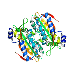 | | Crystal structure of PtLCIB4 H88A mutant, a homolog of the limiting CO2-inducible protein LCIB | | 分子名称: | PtLCIB4 H88A mutant, ZINC ION | | 著者 | Jin, S, Sun, J, Wunder, T, Tang, D, Mueller-Caja, O.M, Gao, Y. | | 登録日 | 2016-05-24 | | 公開日 | 2016-12-07 | | 最終更新日 | 2023-11-08 | | 実験手法 | X-RAY DIFFRACTION (1.6 Å) | | 主引用文献 | Structural insights into the LCIB protein family reveals a new group of beta-carbonic anhydrases
Proc. Natl. Acad. Sci. U.S.A., 113, 2016
|
|
6JHS
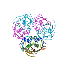
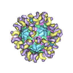 | | The cryo-EM structure of HAV bound to a neutralizing antibody-F7 | | 分子名称: | FAB Heavy Chain, FAB Light Chain, VP1, ... | | 著者 | Cao, L, Liu, P, Yang, P, Gao, Q, Li, H, Sun, Y, Zhu, L, Lin, J, Su, D, Rao, Z, Wang, X. | | 登録日 | 2019-02-19 | | 公開日 | 2020-03-18 | | 実験手法 | ELECTRON MICROSCOPY (3.05 Å) | | 主引用文献 | Structural basis for neutralization of hepatitis A virus informs a rational design of highly potent inhibitors.
Plos Biol., 17, 2019
|
|
1QD1
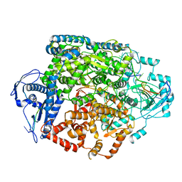
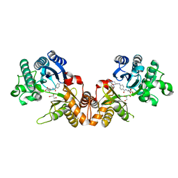 | | THE CRYSTAL STRUCTURE OF THE FORMIMINOTRANSFERASE DOMAIN OF FORMIMINOTRANSFERASE-CYCLODEAMINASE. | | 分子名称: | FORMIMINOTRANSFERASE-CYCLODEAMINASE, GLYCEROL, N-{[4-({[(6R)-2-amino-5-formyl-4-oxo-1,4,5,6,7,8-hexahydropteridin-6-yl]methyl}amino)phenyl]carbonyl}-L-glutamic acid | | 著者 | Kohls, D, Sulea, T, Purisima, E, MacKenzie, R.E, Vrielink, A. | | 登録日 | 1999-07-08 | | 公開日 | 2000-01-12 | | 最終更新日 | 2024-02-14 | | 実験手法 | X-RAY DIFFRACTION (1.7 Å) | | 主引用文献 | The crystal structure of the formiminotransferase domain of formiminotransferase-cyclodeaminase: implications for substrate channeling in a bifunctional enzyme.
Structure Fold.Des., 8, 2000
|
|
5JHI
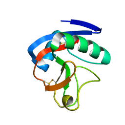
 | | Solution structure of the de novo mini protein gEHE_06 | | 分子名称: | W35 | | 著者 | Buchko, G.W, Bahl, C.D, Gilmore, J.M, Pulavarti, S.V, Baker, D, Szyperski, T. | | 登録日 | 2016-04-21 | | 公開日 | 2016-09-28 | | 最終更新日 | 2023-06-14 | | 実験手法 | SOLUTION NMR | | 主引用文献 | Accurate de novo design of hyperstable constrained peptides.
Nature, 538, 2016
|
|
6JJ0
 | |
5BMW
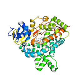
 | | Crystal structure of T75V mutant of Triosephosphate isomerase from Plasmodium falciparum | | 分子名称: | 1,2-ETHANEDIOL, 2-(N-MORPHOLINO)-ETHANESULFONIC ACID, CALCIUM ION, ... | | 著者 | Bandyopadhyay, D, Murthy, M.R.N, Balaram, H, Balaram, P. | | 登録日 | 2015-05-23 | | 公開日 | 2015-07-29 | | 最終更新日 | 2023-11-08 | | 実験手法 | X-RAY DIFFRACTION (1.86 Å) | | 主引用文献 | Probing the role of highly conserved residues in triosephosphate isomerase - analysis of site specific mutants at positions 64 and 75 in the Plasmodial enzyme
Febs J., 282, 2015
|
|
5BN9
 | |
5BN8
 | |
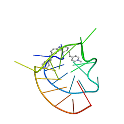
5BNH
 | | Crystal structure of the HLTF HIRAN domain with a ssDNA fragment | | 分子名称: | ACETATE ION, DNA (5'-D(*(GD)P*GP*TP*G)-3'), DNA (5'-D(*(TD)P*TP*G)-3'), ... | | 著者 | Neculai, D, Walker, J.R, Weigelt, J, Bountra, C, Edwards, A.M, Arrowsmith, C.H, Dhe-Paganon, S. | | 登録日 | 2015-05-26 | | 公開日 | 2016-05-25 | | 最終更新日 | 2024-03-20 | | 実験手法 | X-RAY DIFFRACTION (1.7 Å) | | 主引用文献 | Co-crystal structure of the HLTF HIRAN domain with a ssDNA fragment
To Be Published
|
|


