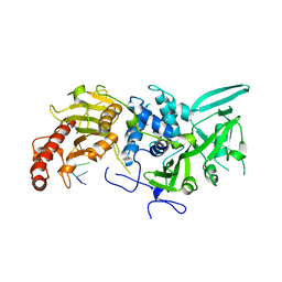
2C71
 
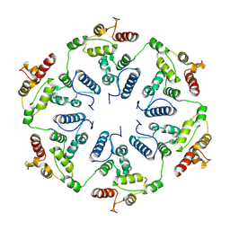
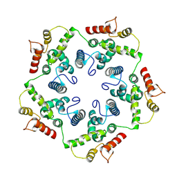
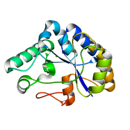 | | The structure of a family 4 acetyl xylan esterase from Clostridium thermocellum in complex with a magnesium ion. | | 分子名称: | GLYCOSIDE HYDROLASE, FAMILY 11:CLOSTRIDIUM CELLULOSOME ENZYME, DOCKERIN TYPE I:POLYSACCHARIDE, ... | | 著者 | Taylor, E.J, Turkenburg, P.J, Vincent, F, Brzozowski, A.M, Gloster, T.M, Dupont, C, Shareck, F, Centeno, M.S.J, Prates, J.A.M, Ferreira, L.M.A, Fontes, C.M.G.A, Biely, P, Davies, G.J. | | 登録日 | 2005-11-17 | | 公開日 | 2006-01-23 | | 最終更新日 | 2024-05-08 | | 実験手法 | X-RAY DIFFRACTION (1.05 Å) | | 主引用文献 | Structure and Activity of Two Metal-Ion Dependent Acetyl Xylan Esterases Involved in Plant Cell-Wall Degradation Reveals a Close Similarity to Peptidoglycan Deacetylases.
J.Biol.Chem., 281, 2006
|
|
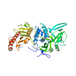
2G81
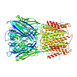
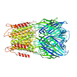
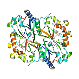
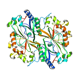
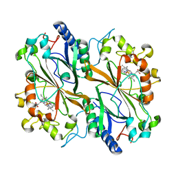
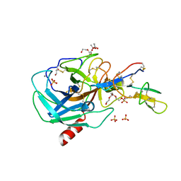 | | Crystal Structure of the Bowman-Birk Inhibitor from Vigna unguiculata Seeds in Complex with Beta-trypsin at 1.55 Angstrons Resolution | | 分子名称: | 1,2-ETHANEDIOL, ACETIC ACID, Bowman-Birk type seed trypsin and chymotrypsin inhibitor, ... | | 著者 | Freitas, S.M, Barbosa, J.A.R.G, Paulino, L.S, Teles, R.C.L, Esteves, G.F, Ventura, M.M. | | 登録日 | 2006-03-01 | | 公開日 | 2007-01-02 | | 最終更新日 | 2023-10-25 | | 実験手法 | X-RAY DIFFRACTION (1.55 Å) | | 主引用文献 | Crystal Structure of the Bowman-Birk Inhibitor from Vigna unguiculata Seeds in Complex with {beta}-Trypsin at 1.55 A Resolution and Its Structural Properties in Association with Proteinases
Biophys.J., 92, 2007
|
|
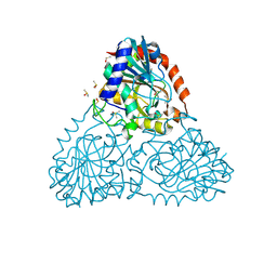
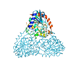
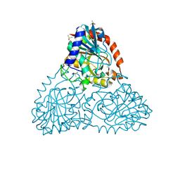
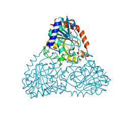
2H5Y
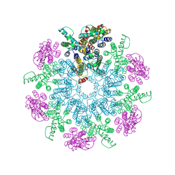
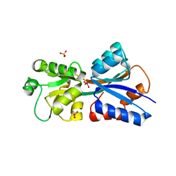 | | Crystallographic structure of the Molybdate-Binding Protein of Xanthomonas citri at 1.7 Ang resolution bound to molybdate | | 分子名称: | MOLYBDATE ION, Molybdate-binding periplasmic protein, SULFATE ION | | 著者 | Balan, A, Santacruz, C.P, Ferreira, L.C.S, Barbosa, J.A.R.G. | | 登録日 | 2006-05-29 | | 公開日 | 2007-06-05 | | 最終更新日 | 2023-10-25 | | 実験手法 | X-RAY DIFFRACTION (1.7 Å) | | 主引用文献 | Crystallization, data collection and phasing of the molybdate-binding protein of the phytopathogen Xanthomonas axonopodis pv. citri
Acta Crystallogr.,Sect.F, 62, 2006
|
|
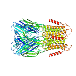
8D64
 | | ELIC with cysteamine in POPC nanodisc | | 分子名称: | 2-AMINO-ETHANETHIOL, Erwinia ligand-gated ion channel | | 著者 | Petroff II, J.T, Deng, Z, Rau, M.J, Fitzpatrick, J.A.J, Yuan, P, Cheng, W.W.L. | | 登録日 | 2022-06-06 | | 公開日 | 2022-11-23 | | 最終更新日 | 2024-06-12 | | 実験手法 | ELECTRON MICROSCOPY (3.14 Å) | | 主引用文献 | Open-channel structure of a pentameric ligand-gated ion channel reveals a mechanism of leaflet-specific phospholipid modulation.
Nat Commun, 13, 2022
|
|
8D67
 | | ELIC3 with cysteamine in 2:1:1 POPC:POPE:POPG nanodisc | | 分子名称: | 2-AMINO-ETHANETHIOL, Erwinia ligand-gated ion channel | | 著者 | Petroff II, J.T, Deng, Z, Rau, M.J, Fitzpatrick, J.A.J, Yuan, P, Cheng, W.W.L. | | 登録日 | 2022-06-06 | | 公開日 | 2022-11-23 | | 最終更新日 | 2024-06-12 | | 実験手法 | ELECTRON MICROSCOPY (3.3 Å) | | 主引用文献 | Open-channel structure of a pentameric ligand-gated ion channel reveals a mechanism of leaflet-specific phospholipid modulation.
Nat Commun, 13, 2022
|
|
8D65
 | | ELIC apo in 2:1:1 POPC:POPE:POPG nanodisc | | 分子名称: | (1R)-2-{[(S)-{[(2S)-2,3-dihydroxypropyl]oxy}(hydroxy)phosphoryl]oxy}-1-[(hexadecanoyloxy)methyl]ethyl (9Z)-octadec-9-enoate, Erwinia ligand-gated ion channel | | 著者 | Petroff II, J.T, Deng, Z, Rau, M.J, Fitzpatrick, J.A.J, Yuan, P, Cheng, W.W.L. | | 登録日 | 2022-06-06 | | 公開日 | 2022-11-23 | | 最終更新日 | 2024-06-12 | | 実験手法 | ELECTRON MICROSCOPY (3.47 Å) | | 主引用文献 | Open-channel structure of a pentameric ligand-gated ion channel reveals a mechanism of leaflet-specific phospholipid modulation.
Nat Commun, 13, 2022
|
|
8D63
 | | ELIC apo in POPC nanodisc | | 分子名称: | 1-palmitoyl-2-oleoyl-sn-glycero-3-phosphocholine, Erwinia ligand-gated ion channel | | 著者 | Petroff II, J.T, Deng, Z, Rau, M.J, Fitzpatrick, J.A.J, Yuan, P, Cheng, W.W.L. | | 登録日 | 2022-06-06 | | 公開日 | 2022-11-23 | | 最終更新日 | 2024-06-12 | | 実験手法 | ELECTRON MICROSCOPY (3.14 Å) | | 主引用文献 | Open-channel structure of a pentameric ligand-gated ion channel reveals a mechanism of leaflet-specific phospholipid modulation.
Nat Commun, 13, 2022
|
|
8D66
 | | ELIC with cysteamine in 2:1:1 POPC:POPE:POPG nanodisc | | 分子名称: | (1R)-2-{[(S)-{[(2S)-2,3-dihydroxypropyl]oxy}(hydroxy)phosphoryl]oxy}-1-[(hexadecanoyloxy)methyl]ethyl (9Z)-octadec-9-enoate, 2-AMINO-ETHANETHIOL, Erwinia ligand-gated ion channel | | 著者 | Petroff II, J.T, Deng, Z, Rau, M.J, Fitzpatrick, J.A.J, Yuan, P, Cheng, W.W.L. | | 登録日 | 2022-06-06 | | 公開日 | 2022-11-23 | | 最終更新日 | 2024-06-12 | | 実験手法 | ELECTRON MICROSCOPY (3.14 Å) | | 主引用文献 | Open-channel structure of a pentameric ligand-gated ion channel reveals a mechanism of leaflet-specific phospholipid modulation.
Nat Commun, 13, 2022
|
|
6TAQ
 | |
6TAU
 | |
6TAR
 | |
6T64
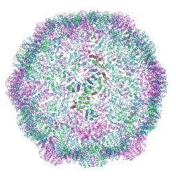
 | | A model of the EIAV CA-SP hexamer (C6) from Gag-deltaMA spheres assembled at pH6 | | 分子名称: | Gag polyprotein | | 著者 | Dick, R.A, Xu, C, Morado, D.R, Kravchuk, V, Ricana, C.L, Lyddon, T.D, Broad, A.M, Feathers, J.R, Johnson, M.C, Vogt, V.M, Perilla, J.R, Briggs, J.A.G, Schur, F.K.M. | | 登録日 | 2019-10-17 | | 公開日 | 2020-01-15 | | 最終更新日 | 2022-03-30 | | 実験手法 | ELECTRON MICROSCOPY (3.7 Å) | | 主引用文献 | Structures of immature EIAV Gag lattices reveal a conserved role for IP6 in lentivirus assembly.
Plos Pathog., 16, 2020
|
|
6Q34
 | | Dye type peroxidase Aa from Streptomyces lividans: 196 kGy structure | | 分子名称: | Deferrochelatase/peroxidase, PROTOPORPHYRIN IX CONTAINING FE | | 著者 | Ebrahim, A, Moreno-Chicano, T, Worrall, J.A.R, Strange, R.W, Axford, D, Sherrell, D.A, Appleby, M, Owen, R.L. | | 登録日 | 2018-12-03 | | 公開日 | 2019-07-31 | | 最終更新日 | 2024-05-15 | | 実験手法 | X-RAY DIFFRACTION (1.82 Å) | | 主引用文献 | Dose-resolved serial synchrotron and XFEL structures of radiation-sensitive metalloproteins.
Iucrj, 6, 2019
|
|
6Q31
 | | Dye type peroxidase Aa from Streptomyces lividans: 156.8 kGy structure | | 分子名称: | Deferrochelatase/peroxidase, PROTOPORPHYRIN IX CONTAINING FE | | 著者 | Ebrahim, A, Moreno-Chicano, T, Worrall, J.A.R, Strange, R.W, Axford, D, Sherrell, D.A, Appleby, M, Owen, R.L. | | 登録日 | 2018-12-03 | | 公開日 | 2019-07-31 | | 最終更新日 | 2024-01-24 | | 実験手法 | X-RAY DIFFRACTION (1.78 Å) | | 主引用文献 | Dose-resolved serial synchrotron and XFEL structures of radiation-sensitive metalloproteins.
Iucrj, 6, 2019
|
|
6Q3D
 | | Dye type peroxidase Aa from Streptomyces lividans: 235.2 kGy structure | | 分子名称: | Deferrochelatase/peroxidase, PROTOPORPHYRIN IX CONTAINING FE | | 著者 | Ebrahim, A, Moreno-Chicano, T, Worrall, J.A.R, Strange, R.W, Axford, D, Sherrell, D.A, Appleby, M, Owen, R.L. | | 登録日 | 2018-12-04 | | 公開日 | 2019-07-31 | | 最終更新日 | 2024-05-15 | | 実験手法 | X-RAY DIFFRACTION (1.93 Å) | | 主引用文献 | Dose-resolved serial synchrotron and XFEL structures of radiation-sensitive metalloproteins.
Iucrj, 6, 2019
|
|
6AWE
 | | Crystal Structure of Purine Nucleoside Phosphorylase Isoform 2 from Schistosoma mansoni in complex with 2-fluoro-6-(2-pyridin-2-ylethylamino)benzonitrile | | 分子名称: | 2-fluoro-6-{[2-(pyridin-2-yl)ethyl]amino}benzonitrile, DIMETHYL SULFOXIDE, Purine nucleoside phosphorylase | | 著者 | Faheem, M, Neto, J.B, Collins, P, Pearce, N.M, Valadares, N.F, Bird, L, Pereira, H.M, Delft, F.V, Barbosa, J.A.R.G. | | 登録日 | 2017-09-05 | | 公開日 | 2018-09-12 | | 最終更新日 | 2023-10-04 | | 実験手法 | X-RAY DIFFRACTION (1.65 Å) | | 主引用文献 | Crystal Structure of Purine Nucleoside Phosphorylase Isoform 2 from Schistosoma mansoni in complex with 2-fluoro-6-(2-pyridin-2-ylethylamino)benzonitrile
To Be Published
|
|
6B37
 | | Crystal Structure of Purine Nucleoside Phosphorylase Isoform 2 from Schistosoma mansoni in complex with 1,3-benzothiazol-2-ol | | 分子名称: | 1,3-benzothiazol-2-ol, DIMETHYL SULFOXIDE, Purine nucleoside phosphorylase | | 著者 | Faheem, M, Neto, J.B, Collins, P, Pearce, N.M, Valadares, N.F, Bird, L, Pereira, H.M, Delft, F.V, Barbosa, J.A.R.G. | | 登録日 | 2017-09-21 | | 公開日 | 2018-09-26 | | 最終更新日 | 2023-10-04 | | 実験手法 | X-RAY DIFFRACTION (1.5 Å) | | 主引用文献 | Crystal Structure of Purine Nucleoside Phosphorylase Isoform 2 from Schistosoma mansoni in complex with 1,3-benzothiazol-2-ol
To Be Published
|
|
6B3L
 | | Crystal Structure of Purine Nucleoside Phosphorylase Isoform 2 from Schistosoma mansoni in complex with 2,3-dihydro-1H-inden-2-amine | | 分子名称: | DIMETHYL SULFOXIDE, INDAN-2-AMINE, Purine nucleoside phosphorylase | | 著者 | Faheem, M, Neto, J.B, Collins, P, Pearce, N.M, Valadares, N.F, Bird, L, Pereira, H.M, Delft, F.V, Barbosa, J.A.R.G. | | 登録日 | 2017-09-22 | | 公開日 | 2018-09-26 | | 実験手法 | X-RAY DIFFRACTION (1.52 Å) | | 主引用文献 | Crystal Structure of Purine Nucleoside Phosphorylase Isoform 2 from Schistosoma mansoni in complex with 2,3-dihydro-1H-inden-2-amine
To Be Published
|
|
6R23
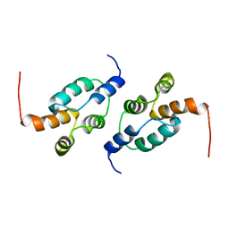 | | The structure of a Ty3 retrotransposon capsid C-terminal domain dimer | | 分子名称: | Transposon Ty3-I Gag-Pol polyprotein | | 著者 | Dodonova, S.O, Prinz, S, Bilanchone, V, Sandmeyer, S, Briggs, J.A.G. | | 登録日 | 2019-03-15 | | 公開日 | 2019-05-08 | | 最終更新日 | 2024-05-15 | | 実験手法 | ELECTRON MICROSCOPY (4.9 Å) | | 主引用文献 | Structure of the Ty3/Gypsy retrotransposon capsid and the evolution of retroviruses.
Proc.Natl.Acad.Sci.USA, 116, 2019
|
|
6R22
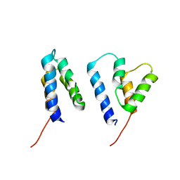 | | The structure of a Ty3 retrotransposon capsid N-terminal domain dimer | | 分子名称: | Transposon Ty3-I Gag-Pol polyprotein | | 著者 | Dodonova, S.O, Prinz, S, Bilanchone, V, Sandmeyer, S, Briggs, J.A.G. | | 登録日 | 2019-03-15 | | 公開日 | 2019-05-08 | | 最終更新日 | 2024-05-15 | | 実験手法 | ELECTRON MICROSCOPY (5.5 Å) | | 主引用文献 | Structure of the Ty3/Gypsy retrotransposon capsid and the evolution of retroviruses.
Proc.Natl.Acad.Sci.USA, 116, 2019
|
|
6R24
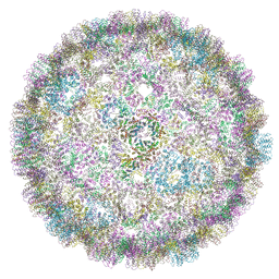 | | The structure of a Ty3 retrotransposon icosahedral capsid | | 分子名称: | Transposon Ty3-I Gag-Pol polyprotein | | 著者 | Dodonova, S.O, Prinz, S, Bilanchone, V, Sandmeyer, S, Briggs, J.A.G. | | 登録日 | 2019-03-15 | | 公開日 | 2019-05-08 | | 最終更新日 | 2024-05-15 | | 実験手法 | ELECTRON MICROSCOPY (7.5 Å) | | 主引用文献 | Structure of the Ty3/Gypsy retrotransposon capsid and the evolution of retroviruses.
Proc.Natl.Acad.Sci.USA, 116, 2019
|
|
6AXA
 | | Crystal Structure of Purine Nucleoside Phosphorylase Isoform 2 from Schistosoma mansoni in complex with (3S)-5-fluoro-3-hydroxy-1,3-dihydroindol-2-one | | 分子名称: | (3S)-5-fluoro-3-hydroxy-1,3-dihydro-2H-indol-2-one, DIMETHYL SULFOXIDE, Purine nucleoside phosphorylase | | 著者 | Faheem, M, Neto, J.B, Collins, P, Pearce, N.M, Valadares, N.F, Bird, L, Pereira, H.M, Delft, F.V, Barbosa, J.A.R.G. | | 登録日 | 2017-09-06 | | 公開日 | 2018-09-12 | | 最終更新日 | 2023-10-04 | | 実験手法 | X-RAY DIFFRACTION (1.59 Å) | | 主引用文献 | Crystal Structure of Purine Nucleoside Phosphorylase Isoform 2 from Schistosoma mansoni in complex with 5-fluoro-3-hydroxy-1,3-dihydroindol-2-one
To Be Published
|
|
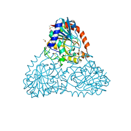
6R9J
 | | Structure of Saccharomyces cerevisiae apo Pan2 pseudoubiquitin hydrolase-RNA exonuclease (UCH-Exo) module in complex with A7 RNA | | 分子名称: | A7 RNA, PAN2-PAN3 deadenylation complex catalytic subunit PAN2 | | 著者 | Tang, T.T.L, Stowell, J.A.W, Hill, C.H, Passmore, L.A. | | 登録日 | 2019-04-03 | | 公開日 | 2019-05-22 | | 最終更新日 | 2024-01-24 | | 実験手法 | X-RAY DIFFRACTION (3.326 Å) | | 主引用文献 | The intrinsic structure of poly(A) RNA determines the specificity of Pan2 and Caf1 deadenylases.
Nat.Struct.Mol.Biol., 26, 2019
|
|
6R9P
 | | Structure of Saccharomyces cerevisiae apo Pan2 pseudoubiquitin hydrolase-RNA exonuclease (UCH-Exo) module in complex with AAUUAA RNA | | 分子名称: | AAUUAA RNA, PAN2-PAN3 deadenylation complex catalytic subunit PAN2 | | 著者 | Tang, T.T.L, Stowell, J.A.W, Hill, C.H, Passmore, L.A. | | 登録日 | 2019-04-03 | | 公開日 | 2019-05-22 | | 最終更新日 | 2024-01-24 | | 実験手法 | X-RAY DIFFRACTION (2.98 Å) | | 主引用文献 | The intrinsic structure of poly(A) RNA determines the specificity of Pan2 and Caf1 deadenylases.
Nat.Struct.Mol.Biol., 26, 2019
|
|
6B56
 | | Crystal Structure of Purine Nucleoside Phosphorylase Isoform 2 from Schistosoma mansoni in complex with 5-butylpyridine-2-carboxylic acid | | 分子名称: | 5-butylpyridine-2-carboxylic acid, DIMETHYL SULFOXIDE, Purine nucleoside phosphorylase | | 著者 | Faheem, M, Neto, J.B, Collins, P, Pearce, N.M, Valadares, N.F, Bird, L, Pereira, H.M, Delft, F.V, Barbosa, J.A.R.G. | | 登録日 | 2017-09-28 | | 公開日 | 2018-10-03 | | 最終更新日 | 2023-10-04 | | 実験手法 | X-RAY DIFFRACTION (1.42 Å) | | 主引用文献 | Crystal Structure of Purine Nucleoside Phosphorylase Isoform 2 from Schistosoma mansoni in complex with 5-butylpyridine-2-carboxylic acid
To Be Published
|
|


