2JDL
 
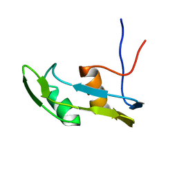
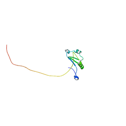
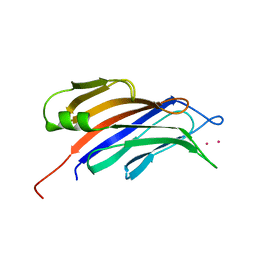
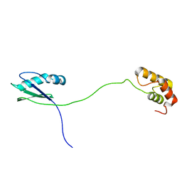
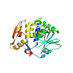 | | Structure of C-terminal region of acidic P2 ribosomal protein complexed with trichosanthin | | 分子名称: | ACIDIC RIBOSOMAL PROTEIN P2, RIBOSOME-INACTIVATING PROTEIN ALPHA-TRICHOSANTHIN | | 著者 | Too, P.H, Mak, A.N, Zhu, G, Au, S.W, Wong, K.B, Shaw, P.C. | | 登録日 | 2007-01-11 | | 公開日 | 2008-02-05 | | 最終更新日 | 2023-12-13 | | 実験手法 | X-RAY DIFFRACTION (2.2 Å) | | 主引用文献 | The C-Terminal Fragment of the Ribosomal P Protein Complexed to Trichosanthin Reveals the Interaction between the Ribosome-Inactivating Protein and the Ribosome.
Nucleic Acids Res., 37, 2009
|
|
2JJR
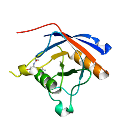
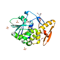 | | V232K, N236D-trichosanthin | | 分子名称: | DI(HYDROXYETHYL)ETHER, RIBOSOME-INACTIVATING PROTEIN ALPHA-TRICHOSANTHIN, SULFATE ION, ... | | 著者 | Too, P.H, Ma, M.K, Mak, A.N, Tung, C.K, Zhu, G, Au, S.W, Wong, K.B, Shaw, P.C. | | 登録日 | 2008-04-21 | | 公開日 | 2008-12-30 | | 最終更新日 | 2023-12-13 | | 実験手法 | X-RAY DIFFRACTION (2.3 Å) | | 主引用文献 | The C-Terminal Fragment of the Ribosomal P Protein Complexed to Trichosanthin Reveals the Interaction between the Ribosome-Inactivating Protein and the Ribosome.
Nucleic Acids Res., 37, 2009
|
|
4HI0
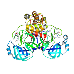
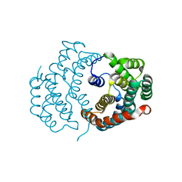
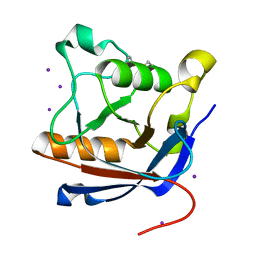
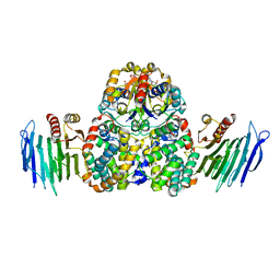 | | Crystal Structure of Helicobacter pylori Urease Accessory Protein UreF/H/G complex | | 分子名称: | GUANOSINE-5'-DIPHOSPHATE, Urease accessory protein UreF, Urease accessory protein UreG, ... | | 著者 | Fong, Y.H, Chen, Y.W, Wong, K.B. | | 登録日 | 2012-10-11 | | 公開日 | 2013-10-16 | | 最終更新日 | 2024-02-28 | | 実験手法 | X-RAY DIFFRACTION (2.35 Å) | | 主引用文献 | Structure of UreG/UreF/UreH complex reveals how urease accessory proteins facilitate maturation of Helicobacter pylori urease.
Plos Biol., 11, 2013
|
|
3SF5
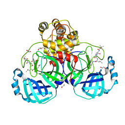
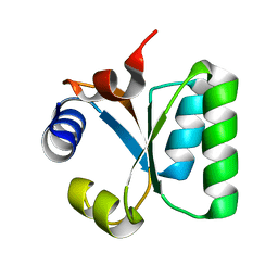
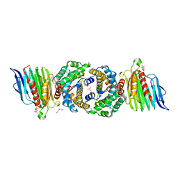 | | Crystal Structure of Helicobacter pylori Urease Accessory Protein UreF/H complex | | 分子名称: | DI(HYDROXYETHYL)ETHER, GLYCEROL, SULFATE ION, ... | | 著者 | Fong, Y.H, Chen, Y.W, Wong, K.B. | | 登録日 | 2011-06-12 | | 公開日 | 2011-11-02 | | 最終更新日 | 2023-11-01 | | 実験手法 | X-RAY DIFFRACTION (2.495 Å) | | 主引用文献 | Assembly of preactivation complex for urease maturation in Helicobacter pylori: crystal structure of UreF-UreH protein complex
J.Biol.Chem., 286, 2011
|
|
6J3L
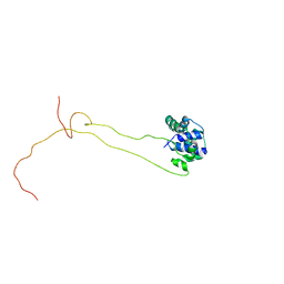
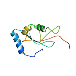
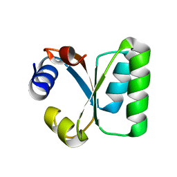
 | | Solution structure of the N-terminal extended protuberant domain of eukaryotic ribosomal stalk protein P0 | | 分子名称: | 60S acidic ribosomal protein P0 | | 著者 | Choi, K.H.A, Lee, K.M, Yang, L, Wing-Heng Yu, C, Banfield, D.K, Ito, K, Uchiumi, T, Wong, K.B. | | 登録日 | 2019-01-04 | | 公開日 | 2019-09-04 | | 最終更新日 | 2024-05-15 | | 実験手法 | SOLUTION NMR | | 主引用文献 | Structural and Mutagenesis Studies Evince the Role of the Extended Protuberant Domain of Ribosomal Protein uL10 in Protein Translation.
Biochemistry, 58, 2019
|
|
2W1O
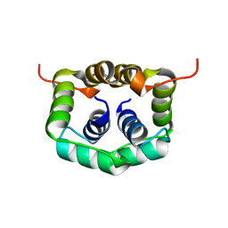 | | NMR structure of dimerization domain of human ribosomal protein P2 | | 分子名称: | 60S ACIDIC RIBOSOMAL PROTEIN P2 | | 著者 | Lee, K.M, Chan, D.S, Sze, K.H, Zhu, G, Shaw, P.C, Wong, K.B. | | 登録日 | 2008-10-20 | | 公開日 | 2009-11-17 | | 最終更新日 | 2024-05-15 | | 実験手法 | SOLUTION NMR | | 主引用文献 | Solution Structure of the Dimerization Domain of Ribosomal Protein P2 Provides Insights for the Structural Organization of Eukaryotic Stalk.
Nucleic Acids Res., 38, 2010
|
|
3VB5
 | | Crystal structure of SARS-CoV 3C-like protease with C4Z | | 分子名称: | 1,2-ETHANEDIOL, 3C-like proteinase, C4Z inhibitor | | 著者 | Chuck, C.P, Wong, K.B. | | 登録日 | 2011-12-31 | | 公開日 | 2012-12-12 | | 最終更新日 | 2023-12-06 | | 実験手法 | X-RAY DIFFRACTION (1.95 Å) | | 主引用文献 | Design, synthesis and crystallographic analysis of nitrile-based broad-spectrum peptidomimetic inhibitors for coronavirus 3C-like proteases
Eur.J.Med.Chem., 59C, 2012
|
|
3VB4
 | | Crystal structure of SARS-CoV 3C-like protease with B4Z | | 分子名称: | 1,2-ETHANEDIOL, 3C-like proteinase, B4Z inhibitor, ... | | 著者 | Chuck, C.P, Wong, K.B. | | 登録日 | 2011-12-31 | | 公開日 | 2012-12-12 | | 最終更新日 | 2023-12-06 | | 実験手法 | X-RAY DIFFRACTION (2.2 Å) | | 主引用文献 | Design, synthesis and crystallographic analysis of nitrile-based broad-spectrum peptidomimetic inhibitors for coronavirus 3C-like proteases
Eur.J.Med.Chem., 59C, 2012
|
|
7XRW
 | | Solution structure of T. brucei RAP1 | | 分子名称: | Repressor activator protein 1 | | 著者 | Yang, X, Pan, X.H, Ji, Z.Y, Wong, K.B, Zhang, M.J, Zhao, Y.X. | | 登録日 | 2022-05-12 | | 公開日 | 2023-03-01 | | 最終更新日 | 2024-05-15 | | 実験手法 | SOLUTION NMR | | 主引用文献 | The RRM-mediated RNA binding activity in T. brucei RAP1 is essential for VSG monoallelic expression.
Nat Commun, 14, 2023
|
|
3O1Q
 | |
4BEH
 | | Solution structure of human ribosomal protein P1.P2 heterodimer | | 分子名称: | 60S ACIDIC RIBOSOMAL PROTEIN P1, 60S ACIDIC RIBOSOMAL PROTEIN P2 | | 著者 | Lee, K.M, Yusa, K, Chu, L.O, Wing-Heng Yu, C, Shaw, P.C, Oono, M, Miyoshi, T, Ito, K, Wong, K.B, Uchiumi, T. | | 登録日 | 2013-03-10 | | 公開日 | 2013-08-14 | | 最終更新日 | 2024-06-19 | | 実験手法 | SOLUTION NMR | | 主引用文献 | Solution Structure of Human P1P2 Heterodimer Provides Insights Into the Role of Eukaryotic Stalk in Recruiting the Ribosome-Inactivating Protein Trichosanthin to the Ribosome.
Nucleic Acids Res., 41, 2013
|
|
2WGL
 | |
7DHT
 | |
1W42
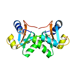
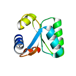 | | T. celer L30e R92A variant | | 分子名称: | 50S RIBOSOMAL PROTEIN L30E | | 著者 | Lee, C.F, Lee, K.M, Chan, S.H, Allen, M.D, Bycroft, M, Wong, K.B. | | 登録日 | 2004-07-22 | | 公開日 | 2005-04-08 | | 最終更新日 | 2023-12-13 | | 実験手法 | X-RAY DIFFRACTION (1.8 Å) | | 主引用文献 | Electrostatic Interactions Contribute to Reduced Heat Capacity Change of Unfolding in a Thermophilic Ribosomal Protein L30E
J.Mol.Biol., 348, 2005
|
|
1W40
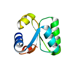 | | T. celer L30e K9A variant | | 分子名称: | 50S RIBOSOMAL PROTEIN L30E | | 著者 | Lee, C.F, Lee, K.M, Chan, S.H, Allen, M.D, Bycroft, M, Wong, K.B. | | 登録日 | 2004-07-22 | | 公開日 | 2005-04-08 | | 最終更新日 | 2023-12-13 | | 実験手法 | X-RAY DIFFRACTION (2.03 Å) | | 主引用文献 | Electrostatic Interactions Contribute to Reduced Heat Capacity Change of Unfolding in a Thermophilic Ribosomal Protein L30E
J.Mol.Biol., 348, 2005
|
|
1W41
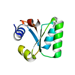 | | T. celer L30e E90A variant | | 分子名称: | 50S RIBOSOMAL PROTEIN L30E | | 著者 | Lee, C.F, Lee, K.M, Chan, S.H, Allen, M.D, Bycroft, M, Wong, K.B. | | 登録日 | 2004-07-22 | | 公開日 | 2005-04-08 | | 最終更新日 | 2023-12-13 | | 実験手法 | X-RAY DIFFRACTION (1.7 Å) | | 主引用文献 | Electrostatic Interactions Contribute to Reduced Heat Capacity Change of Unfolding in a Thermophilic Ribosomal Protein L30E
J.Mol.Biol., 348, 2005
|
|
2JG7
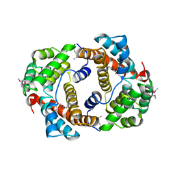
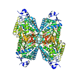 | | Crystal structure of Seabream Antiquitin and Elucidation of its substrate specificity | | 分子名称: | ANTIQUITIN, NICOTINAMIDE-ADENINE-DINUCLEOTIDE | | 著者 | Tang, W.K, Wong, K.B, Cha, S.S, Lee, H.S, Cheng, C.H.K, Fong, W.P. | | 登録日 | 2007-02-09 | | 公開日 | 2008-05-13 | | 最終更新日 | 2023-12-13 | | 実験手法 | X-RAY DIFFRACTION (2.83 Å) | | 主引用文献 | The Crystal Structure of Seabream Antiquitin Reveals the Structural Basis of its Substrate Specificity.
FEBS Lett., 582, 2008
|
|
3RA5
 | |
3TLO
 | |
3RA6
 | |
4RJ9
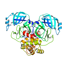
 | | Structure of a plant specific C2 domain protein, OsGAP1 from rice | | 分子名称: | C2 domain-containing protein-like, POTASSIUM ION | | 著者 | Miao, R, Fong, Y.H, Wong, K.B, Lam, H.M. | | 登録日 | 2014-10-08 | | 公開日 | 2015-08-26 | | 最終更新日 | 2023-09-20 | | 実験手法 | X-RAY DIFFRACTION (1.63 Å) | | 主引用文献 | Site-directed Mutagenesis Shows the Significance of Interactions with Phospholipids and the G-protein OsYchF1 for the Physiological Functions of the Rice GTPase-activating Protein 1 (OsGAP1).
J.Biol.Chem., 290, 2015
|
|
4TJX
 | | Crystal structure of protease-associated domain of Arabidopsis VSR1 in complex with aleurain peptide | | 分子名称: | Aleurain peptide, Vacuolar-sorting receptor 1 | | 著者 | Luo, F, Fong, Y.H, Jiang, L.W, Wong, K.B. | | 登録日 | 2014-05-25 | | 公開日 | 2014-12-10 | | 最終更新日 | 2023-09-27 | | 実験手法 | X-RAY DIFFRACTION (1.902 Å) | | 主引用文献 | How vacuolar sorting receptor proteins interact with their cargo proteins: crystal structures of apo and cargo-bound forms of the protease-associated domain from an Arabidopsis vacuolar sorting receptor.
Plant Cell, 26, 2014
|
|
4TJV
 | | Crystal structure of protease-associated domain of Arabidopsis vacuolar sorting receptor 1 | | 分子名称: | IODIDE ION, Vacuolar-sorting receptor 1 | | 著者 | Luo, F, Fong, Y.H, Jiang, L.W, Wong, K.B. | | 登録日 | 2014-05-25 | | 公開日 | 2014-12-10 | | 最終更新日 | 2023-12-27 | | 実験手法 | X-RAY DIFFRACTION (1.651 Å) | | 主引用文献 | How vacuolar sorting receptor proteins interact with their cargo proteins: crystal structures of apo and cargo-bound forms of the protease-associated domain from an Arabidopsis vacuolar sorting receptor.
Plant Cell, 26, 2014
|
|
3LFO
 | |
7VB2
 | |


