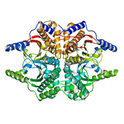
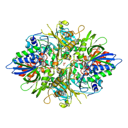
1XAD
 
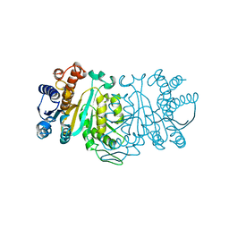 | |
1XAB
 | |
1XAA
 | |
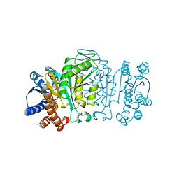
1IYH
 | |
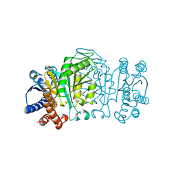
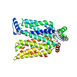
8H4M
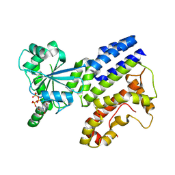 | | Crystal Structure of GTP-bound Irgb6_T95D mutant | | 分子名称: | GUANOSINE-5'-TRIPHOSPHATE, T-cell-specific guanine nucleotide triphosphate-binding protein 2 | | 著者 | Saijo-Hamano, Y, Okuma, H, Sakai, N, Kato, T, Imasaki, T, Nitta, R. | | 登録日 | 2022-10-10 | | 公開日 | 2023-10-18 | | 実験手法 | X-RAY DIFFRACTION (1.68 Å) | | 主引用文献 | Structural basis of Irgb6 inactivation by Toxoplasma gondii through the phosphorylation of switch I
To Be Published
|
|
8H4O
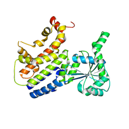 | | Crystal Structure of nucleotide-free Irgb6_T95D mutant | | 分子名称: | T-cell-specific guanine nucleotide triphosphate-binding protein 2 | | 著者 | Saijo-Hamano, Y, Okuma, H, Sakai, N, Kato, T, Imasaki, T, Nitta, R. | | 登録日 | 2022-10-11 | | 公開日 | 2023-10-18 | | 最終更新日 | 2024-07-03 | | 実験手法 | X-RAY DIFFRACTION (2.05 Å) | | 主引用文献 | Structural basis of Irgb6 inactivation by Toxoplasma gondii through the phosphorylation of switch I.
Genes Cells, 29, 2024
|
|
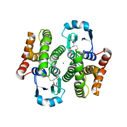
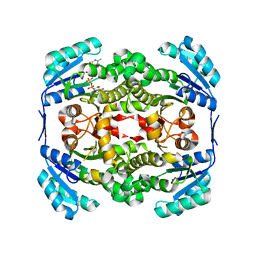
1ZAB
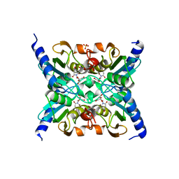 | | Crystal Structure of Mouse Cytidine Deaminase Complexed with 3-Deazauridine | | 分子名称: | 1-((2R,3R,4S,5R)-TETRAHYDRO-3,4-DIHYDROXY-5-(HYDROXYMETHYL)FURAN-2-YL)PYRIDINE-2,4(1H,3H)-DIONE, Cytidine deaminase, SULFATE ION, ... | | 著者 | Teh, A.H. | | 登録日 | 2005-04-06 | | 公開日 | 2006-04-11 | | 最終更新日 | 2023-10-25 | | 実験手法 | X-RAY DIFFRACTION (2.36 Å) | | 主引用文献 | The 1.48 A Resolution Crystal Structure of the Homotetrameric Cytidine Deaminase from Mouse
Biochemistry, 45, 2006
|
|
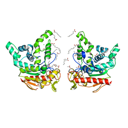
2P39
 | | Crystal structure of human FGF23 | | 分子名称: | 1,3,4,6-tetra-O-sulfo-beta-D-fructofuranose-(2-1)-2,3,4,6-tetra-O-sulfonato-alpha-D-glucopyranose, Fibroblast growth factor 23 | | 著者 | Mohammadi, M. | | 登録日 | 2007-03-08 | | 公開日 | 2007-04-10 | | 最終更新日 | 2023-08-30 | | 実験手法 | X-RAY DIFFRACTION (1.5 Å) | | 主引用文献 | Molecular insights into the klotho-dependent, endocrine mode of action of fibroblast growth factor 19 subfamily members.
Mol.Cell.Biol., 27, 2007
|
|
1DDZ
 | | X-RAY STRUCTURE OF A BETA-CARBONIC ANHYDRASE FROM THE RED ALGA, PORPHYRIDIUM PURPUREUM R-1 | | 分子名称: | CARBONIC ANHYDRASE, ZINC ION | | 著者 | Mitsuhashi, S, Mizushima, T, Yamashita, E, Miyachi, S, Tsukihara, T. | | 登録日 | 1999-11-12 | | 公開日 | 2000-03-08 | | 最終更新日 | 2024-02-07 | | 実験手法 | X-RAY DIFFRACTION (2.2 Å) | | 主引用文献 | X-ray structure of beta-carbonic anhydrase from the red alga, Porphyridium purpureum, reveals a novel catalytic site for CO(2) hydration.
J.Biol.Chem., 275, 2000
|
|
4ZWB
 | | Crystal structure of maltose-bound human GLUT3 in the outward-occluded conformation at 2.4 angstrom | | 分子名称: | Solute carrier family 2, facilitated glucose transporter member 3, alpha-D-glucopyranose-(1-4)-alpha-D-glucopyranose | | 著者 | Deng, D, Sun, P.C, Yan, C.Y, Yan, N. | | 登録日 | 2015-05-19 | | 公開日 | 2015-07-22 | | 最終更新日 | 2023-11-08 | | 実験手法 | X-RAY DIFFRACTION (2.4 Å) | | 主引用文献 | Molecular basis of ligand recognition and transport by glucose transporters
Nature, 526, 2015
|
|
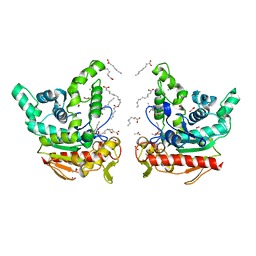
2P23
 | | Crystal structure of human FGF19 | | 分子名称: | Fibroblast growth factor 19 | | 著者 | Mohammadi, M. | | 登録日 | 2007-03-06 | | 公開日 | 2007-04-10 | | 最終更新日 | 2023-08-30 | | 実験手法 | X-RAY DIFFRACTION (1.8 Å) | | 主引用文献 | Molecular insights into the klotho-dependent, endocrine mode of action of fibroblast growth factor 19 subfamily members.
Mol.Cell.Biol., 27, 2007
|
|
6GTL
 | | Achromobacter cycloclastes copper nitrite reductase at pH 6.0 | | 分子名称: | COPPER (II) ION, Copper-containing nitrite reductase, MALONATE ION, ... | | 著者 | Halsted, T.P, Eady, R.R, Antonyuk, S.V, Hasnain, S.S. | | 登録日 | 2018-06-18 | | 公開日 | 2019-07-03 | | 最終更新日 | 2024-05-15 | | 実験手法 | X-RAY DIFFRACTION (1.5 Å) | | 主引用文献 | Catalytically important damage-free structures of a copper nitrite reductase obtained by femtosecond X-ray laser and room-temperature neutron crystallography.
Iucrj, 6, 2019
|
|
6GTN
 | | Achromobacter cycloclastes copper nitrite reductase at pH 6.5 | | 分子名称: | COPPER (II) ION, Copper-containing nitrite reductase, MALONATE ION, ... | | 著者 | Halsted, T.P, Eady, R.R, Antonyuk, S.V, Hasnain, S.S. | | 登録日 | 2018-06-18 | | 公開日 | 2019-07-03 | | 最終更新日 | 2024-05-15 | | 実験手法 | X-RAY DIFFRACTION (1.5 Å) | | 主引用文献 | Catalytically important damage-free structures of a copper nitrite reductase obtained by femtosecond X-ray laser and room-temperature neutron crystallography.
Iucrj, 6, 2019
|
|
6GTI
 | | Achromobacter cycloclastes copper nitrite reductase at pH 5.0 | | 分子名称: | COPPER (II) ION, Copper-containing nitrite reductase, MALONATE ION, ... | | 著者 | Halsted, T.P, Eady, R.R, Antonyuk, S.V, Hasnain, S.S. | | 登録日 | 2018-06-18 | | 公開日 | 2019-07-03 | | 最終更新日 | 2024-01-17 | | 実験手法 | X-RAY DIFFRACTION (1.5 Å) | | 主引用文献 | Catalytically important damage-free structures of a copper nitrite reductase obtained by femtosecond X-ray laser and room-temperature neutron crystallography.
Iucrj, 6, 2019
|
|
6GTK
 | | Achromobacter cycloclastes copper nitrite reductase at pH 5.5 | | 分子名称: | COPPER (II) ION, Copper-containing nitrite reductase, MALONATE ION, ... | | 著者 | Halsted, T.P, Eady, R.R, Antonyuk, S.V, Hasnain, S.S. | | 登録日 | 2018-06-18 | | 公開日 | 2019-07-03 | | 最終更新日 | 2024-05-15 | | 実験手法 | X-RAY DIFFRACTION (1.5 Å) | | 主引用文献 | Catalytically important damage-free structures of a copper nitrite reductase obtained by femtosecond X-ray laser and room-temperature neutron crystallography.
Iucrj, 6, 2019
|
|
6GTJ
 | | Neutron crystal structure for copper nitrite reductase from Achromobacter Cycloclastes at 1.8 A resolution | | 分子名称: | COPPER (II) ION, Copper-containing nitrite reductase | | 著者 | Antonyuk, S.V, Blakeley, M.P, Halsted, T.P, Eady, R.R, Hasnain, S.S. | | 登録日 | 2018-06-18 | | 公開日 | 2019-07-03 | | 最終更新日 | 2024-01-17 | | 実験手法 | NEUTRON DIFFRACTION (1.801 Å) | | 主引用文献 | Catalytically important damage-free structures of a copper nitrite reductase obtained by femtosecond X-ray laser and room-temperature neutron crystallography.
Iucrj, 6, 2019
|
|
2YR5
 | | Crystal structure of L-phenylalanine oxidase from Psuedomonas sp.P501 | | 分子名称: | FLAVIN-ADENINE DINUCLEOTIDE, GLYCEROL, Pro-enzyme of L-phenylalanine oxidase, ... | | 著者 | Ida, K, Kurabayashi, M, Suguro, M, Hikima, T, Yamamoto, M, Suzuki, H. | | 登録日 | 2007-04-02 | | 公開日 | 2008-04-15 | | 最終更新日 | 2023-10-25 | | 実験手法 | X-RAY DIFFRACTION (1.25 Å) | | 主引用文献 | Structural basis of proteolytic activation of L-phenylalanine oxidase from Pseudomonas sp. P-501.
J.Biol.Chem., 283, 2008
|
|
2YW9
 | |
8K7P
 | | Staphylococcus aureus lipase -PSA complex | | 分子名称: | ACETIC ACID, CALCIUM ION, CHLORIDE ION, ... | | 著者 | Kitadokoro, J, Kamitani, S, Kitadokoro, K. | | 登録日 | 2023-07-27 | | 公開日 | 2024-06-05 | | 最終更新日 | 2024-06-19 | | 実験手法 | X-RAY DIFFRACTION (2.19 Å) | | 主引用文献 | Crystal structure of Staphylococcus aureus lipase complex with unsaturated petroselinic acid.
Febs Open Bio, 14, 2024
|
|
8K7Q
 | | Staphylococcus aureus lipase S116A inactive mutant-PSA complex | | 分子名称: | ACETIC ACID, CALCIUM ION, CHLORIDE ION, ... | | 著者 | Kitadokoro, J, Kamitani, S, Kitadokoro, K. | | 登録日 | 2023-07-27 | | 公開日 | 2024-06-05 | | 最終更新日 | 2024-06-19 | | 実験手法 | X-RAY DIFFRACTION (2.02 Å) | | 主引用文献 | Crystal structure of Staphylococcus aureus lipase complex with unsaturated petroselinic acid.
Febs Open Bio, 14, 2024
|
|
1F88
 | | CRYSTAL STRUCTURE OF BOVINE RHODOPSIN | | 分子名称: | 2-acetamido-2-deoxy-beta-D-glucopyranose-(1-4)-2-acetamido-2-deoxy-beta-D-glucopyranose, MERCURY (II) ION, RETINAL, ... | | 著者 | Okada, T, Palczewski, K, Stenkamp, R.E, Miyano, M. | | 登録日 | 2000-06-29 | | 公開日 | 2000-08-04 | | 最終更新日 | 2020-07-29 | | 実験手法 | X-RAY DIFFRACTION (2.8 Å) | | 主引用文献 | Crystal structure of rhodopsin: A G protein-coupled receptor.
Science, 289, 2000
|
|
5YWY
 | | Crystal structure of the human prostaglandin E receptor EP4 in complex with Fab and ONO-AE3-208 | | 分子名称: | 4-[4-cyano-2-[[(2R)-2-(4-fluoranylnaphthalen-1-yl)propanoyl]amino]phenyl]butanoic acid, Heavy chain of Fab fragment, Light chain of Fab fragment, ... | | 著者 | Toyoda, Y, Morimoto, K, Suno, R, Horita, S, Iwata, S, Kobayashi, T. | | 登録日 | 2017-11-30 | | 公開日 | 2018-12-05 | | 最終更新日 | 2018-12-19 | | 実験手法 | X-RAY DIFFRACTION (3.2 Å) | | 主引用文献 | Ligand binding to human prostaglandin E receptor EP4at the lipid-bilayer interface.
Nat. Chem. Biol., 15, 2019
|
|
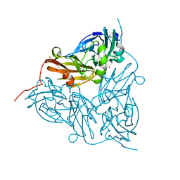
3WTW
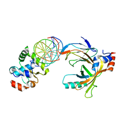 | | Crystal structure of the complex comprised of ETS1(K167A), RUNX1, CBFBETA, and the tcralpha gene enhancer DNA | | 分子名称: | Core-binding factor subunit beta, DNA (5'-D(*AP*GP*AP*GP*GP*AP*TP*GP*TP*GP*GP*CP*TP*TP*C)-3'), DNA (5'-D(*GP*AP*AP*GP*CP*CP*AP*CP*AP*TP*CP*CP*TP*CP*T)-3'), ... | | 著者 | Shiina, M, Hamada, K, Ogata, K. | | 登録日 | 2014-04-21 | | 公開日 | 2014-08-20 | | 最終更新日 | 2023-11-08 | | 実験手法 | X-RAY DIFFRACTION (2.9 Å) | | 主引用文献 | A novel allosteric mechanism on protein-DNA interactions underlying the phosphorylation-dependent regulation of Ets1 target gene expressions.
J.Mol.Biol., 427, 2015
|
|
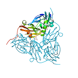
3WTX
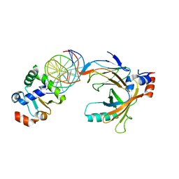 | | Crystal structure of the complex comprised of ETS1(Y329A), RUNX1, CBFBETA, and the tcralpha gene enhancer DNA | | 分子名称: | Core-binding factor subunit beta, DNA (5'-D(*AP*GP*AP*GP*GP*AP*TP*GP*TP*GP*GP*CP*TP*TP*C)-3'), DNA (5'-D(*GP*AP*AP*GP*CP*CP*AP*CP*AP*TP*CP*CP*TP*CP*T)-3'), ... | | 著者 | Shiina, M, Hamada, K, Ogata, K. | | 登録日 | 2014-04-21 | | 公開日 | 2014-08-13 | | 最終更新日 | 2023-11-08 | | 実験手法 | X-RAY DIFFRACTION (2.8 Å) | | 主引用文献 | A novel allosteric mechanism on protein-DNA interactions underlying the phosphorylation-dependent regulation of Ets1 target gene expressions.
J.Mol.Biol., 427, 2015
|
|
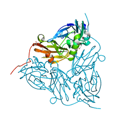
3WTV
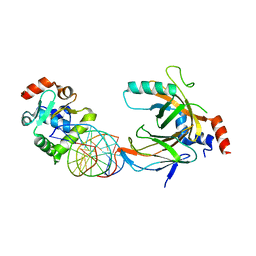 | | Crystal structure of the complex comprised of ETS1(V170G), RUNX1, CBFBETA, and the tcralpha gene enhancer DNA | | 分子名称: | Core-binding factor subunit beta, DNA (5'-D(*AP*GP*AP*GP*GP*AP*TP*GP*TP*GP*GP*CP*TP*TP*C)-3'), DNA (5'-D(*GP*AP*AP*GP*CP*CP*AP*CP*AP*TP*CP*CP*TP*CP*T)-3'), ... | | 著者 | Shiina, M, Hamada, K, Ogata, K. | | 登録日 | 2014-04-21 | | 公開日 | 2014-08-20 | | 最終更新日 | 2023-11-08 | | 実験手法 | X-RAY DIFFRACTION (2.7 Å) | | 主引用文献 | A novel allosteric mechanism on protein-DNA interactions underlying the phosphorylation-dependent regulation of Ets1 target gene expressions.
J.Mol.Biol., 427, 2015
|
|


