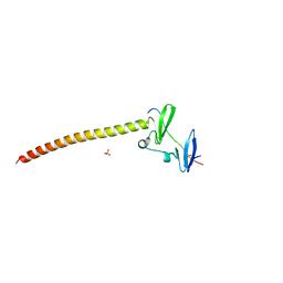
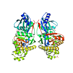
7V56
 
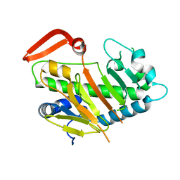 | | Structure of AdaV | | 分子名称: | AdaV, FE (III) ION | | 著者 | Zhang, Z.Y, Chen, W.Q, Zhai, G.Q, Zhang, M. | | 登録日 | 2021-08-16 | | 公開日 | 2022-08-24 | | 最終更新日 | 2024-05-29 | | 実験手法 | X-RAY DIFFRACTION (2.27 Å) | | 主引用文献 | Structural Insight into the Catalytic Mechanism of Non-Heme Iron Halogenase AdaV in 2'-Chloropentostatin Biosynthesis
Acs Catalysis, 12, 2022
|
|
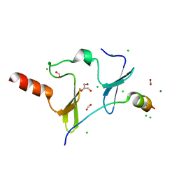
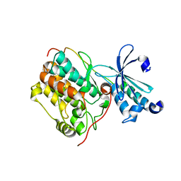
7V7X
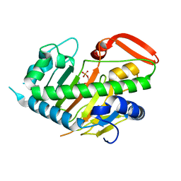 | | Structure of H194A AdaV | | 分子名称: | 2'-DEOXYADENOSINE-5'-MONOPHOSPHATE, AdaV | | 著者 | Zhang, Z.Y, Chen, W.Q, Zhai, G.Q, Zhang, M. | | 登録日 | 2021-08-22 | | 公開日 | 2022-08-31 | | 最終更新日 | 2024-05-29 | | 実験手法 | X-RAY DIFFRACTION (2.7 Å) | | 主引用文献 | Structural Insight into the Catalytic Mechanism of Non-Heme Iron Halogenase AdaV in 2'-Chloropentostatin Biosynthesis
Acs Catalysis, 12, 2022
|
|
4FYP
 | | Crystal Structure of Plant Vegetative Storage Protein | | 分子名称: | MAGNESIUM ION, Vegetative storage protein 1 | | 著者 | Chen, Y, Wei, J, Wang, M, Gong, W, Zhang, M. | | 登録日 | 2012-07-05 | | 公開日 | 2013-06-26 | | 実験手法 | X-RAY DIFFRACTION (1.8 Å) | | 主引用文献 | The crystal structure of Arabidopsis VSP1 reveals the plant class C-like phosphatase structure of the DDDD superfamily of phosphohydrolases
Plos One, 7, 2012
|
|
1D1P
 | | CRYSTAL STRUCTURE OF A YEAST LOW MOLECULAR WEIGHT PROTEIN TYROSINE PHOSPHATASE (LTP1) | | 分子名称: | 4-(2-HYDROXYETHYL)-1-PIPERAZINE ETHANESULFONIC ACID, TYROSINE PHOSPHATASE | | 著者 | Wang, S, Tabernero, L, Zhang, M, Harms, E, Van Etten, R.L, Stauffacher, C.V. | | 登録日 | 1999-09-20 | | 公開日 | 2000-03-08 | | 最終更新日 | 2023-08-09 | | 実験手法 | X-RAY DIFFRACTION (2.2 Å) | | 主引用文献 | Crystal structures of a low-molecular weight protein tyrosine phosphatase from Saccharomyces cerevisiae and its complex with the substrate p-nitrophenyl phosphate.
Biochemistry, 39, 2000
|
|
1D1Q
 | | CRYSTAL STRUCTURE OF A YEAST LOW MOLECULAR WEIGHT PROTEIN TYROSINE PHOSPHATASE (LTP1) COMPLEXED WITH THE SUBSTRATE PNPP | | 分子名称: | 4-NITROPHENYL PHOSPHATE, GLYCEROL, PHOSPHATE ION, ... | | 著者 | Wang, S, Tabernero, L, Zhang, M, Harms, E, Van Etten, R.L, Staufacher, C.V. | | 登録日 | 1999-09-20 | | 公開日 | 2000-03-08 | | 最終更新日 | 2024-02-07 | | 実験手法 | X-RAY DIFFRACTION (1.7 Å) | | 主引用文献 | Crystal structures of a low-molecular weight protein tyrosine phosphatase from Saccharomyces cerevisiae and its complex with the substrate p-nitrophenyl phosphate.
Biochemistry, 39, 2000
|
|
2HQ4
 | | Crystal Structure of ORF 1580 a hypothetical protein from Pyrococcus horikoshii | | 分子名称: | Hypothetical protein PH1570 | | 著者 | Li, Y, Marshall, M, Chang, J, Zhao, M, Zhang, M, Xu, H, Liu, Z.J, Rose, J.P, Wang, B.C, Southeast Collaboratory for Structural Genomics, Southeast Collaboratory for Structural Genomics (SECSG) | | 登録日 | 2006-07-18 | | 公開日 | 2006-09-19 | | 最終更新日 | 2024-03-06 | | 実験手法 | X-RAY DIFFRACTION (1.99 Å) | | 主引用文献 | Crystal Structure of ORF 1580 a hypothetical protein from Pyrococcus horikoshii
To be Published
|
|
2FQD
 | | Crystal Structures of E. coli Laccase CueO under different copper binding situations | | 分子名称: | Blue copper oxidase cueO, CITRIC ACID, COPPER (II) ION, ... | | 著者 | Li, X, Wei, Z, Zhang, M, Teng, M, Gong, W. | | 登録日 | 2006-01-18 | | 公開日 | 2007-01-30 | | 最終更新日 | 2024-03-13 | | 実験手法 | X-RAY DIFFRACTION (2.4 Å) | | 主引用文献 | Crystal structures of E. coli laccase CueO at different copper concentrations.
Biochem.Biophys.Res.Commun., 354, 2007
|
|
2FQE
 | | Crystal Structures of E. coli Laccase CueO under different copper binding situations | | 分子名称: | Blue copper oxidase cueO, CITRIC ACID, COPPER (II) ION, ... | | 著者 | Li, X, Wei, Z, Zhang, M, Teng, M, Gong, W. | | 登録日 | 2006-01-18 | | 公開日 | 2007-01-30 | | 最終更新日 | 2024-03-13 | | 実験手法 | X-RAY DIFFRACTION (1.92 Å) | | 主引用文献 | Crystal structures of E. coli laccase CueO at different copper concentrations.
Biochem.Biophys.Res.Commun., 354, 2007
|
|
2FQG
 | | Crystal Structures of E. coli Laccase CueO under different copper binding situations | | 分子名称: | Blue copper oxidase cueO, CITRIC ACID, COPPER (II) ION, ... | | 著者 | Li, X, Wei, Z, Zhang, M, Teng, M, Gong, W. | | 登録日 | 2006-01-18 | | 公開日 | 2007-01-30 | | 最終更新日 | 2024-03-13 | | 実験手法 | X-RAY DIFFRACTION (2.3 Å) | | 主引用文献 | Crystal structures of E. coli laccase CueO at different copper concentrations.
Biochem.Biophys.Res.Commun., 354, 2007
|
|
2FQF
 | | Crystal Structures of E. coli Laccase CueO under different copper binding situations | | 分子名称: | Blue copper oxidase cueO, CITRIC ACID, COPPER (II) ION, ... | | 著者 | Li, X, Wei, Z, Zhang, M, Teng, M, Gong, W. | | 登録日 | 2006-01-18 | | 公開日 | 2007-01-30 | | 最終更新日 | 2024-03-13 | | 実験手法 | X-RAY DIFFRACTION (2 Å) | | 主引用文献 | Crystal structures of E. coli laccase CueO at different copper concentrations.
Biochem.Biophys.Res.Commun., 354, 2007
|
|
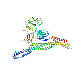
6IRR
 | | Solution structure of DISC1/ATF4 complex | | 分子名称: | Disrupted in schizophrenia 1 homolog,Cyclic AMP-dependent transcription factor ATF-4 | | 著者 | Ye, F, Yu, C, Zhang, M. | | 登録日 | 2018-11-14 | | 公開日 | 2019-09-25 | | 最終更新日 | 2024-05-15 | | 実験手法 | SOLUTION NMR | | 主引用文献 | Structural interaction between DISC1 and ATF4 underlying transcriptional and synaptic dysregulation in an iPSC model of mental disorders.
Mol. Psychiatry, 2019
|
|
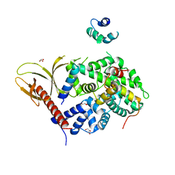
6J68
 | | Structure of KIBRA and LATS1 Complex | | 分子名称: | Peptide from Serine/threonine-protein kinase LATS1, Protein KIBRA | | 著者 | Lin, Z, Yang, Z, Ji, Z, Zhang, M. | | 登録日 | 2019-01-14 | | 公開日 | 2019-09-25 | | 最終更新日 | 2024-03-27 | | 実験手法 | X-RAY DIFFRACTION (2.495 Å) | | 主引用文献 | Decoding WW domain tandem-mediated target recognitions in tissue growth and cell polarity.
Elife, 8, 2019
|
|
6JJY
 | | Crystal Structure of KIBRA and beta-Dystroglycan | | 分子名称: | Peptide from Dystroglycan, Protein KIBRA, SULFATE ION | | 著者 | Lin, Z, Yang, Z, Ji, Z, Zhang, M. | | 登録日 | 2019-02-27 | | 公開日 | 2019-09-25 | | 最終更新日 | 2023-11-22 | | 実験手法 | X-RAY DIFFRACTION (2.298 Å) | | 主引用文献 | Decoding WW domain tandem-mediated target recognitions in tissue growth and cell polarity.
Elife, 8, 2019
|
|
6JJW
 | | Crystal Structure of KIBRA and PTPN14 complex | | 分子名称: | CHLORIDE ION, FORMIC ACID, GLYCEROL, ... | | 著者 | Lin, Z, Yang, Z, Ji, Z, Zhang, M. | | 登録日 | 2019-02-27 | | 公開日 | 2019-09-25 | | 最終更新日 | 2023-11-22 | | 実験手法 | X-RAY DIFFRACTION (2.4 Å) | | 主引用文献 | Decoding WW domain tandem-mediated target recognitions in tissue growth and cell polarity.
Elife, 8, 2019
|
|
6JJX
 | | Crystal Structure of KIBRA and Angiomotin complex | | 分子名称: | Peptide from Angiomotin, Protein KIBRA | | 著者 | Lin, Z, Yang, Z, Ji, Z, Zhang, M. | | 登録日 | 2019-02-27 | | 公開日 | 2019-09-25 | | 最終更新日 | 2023-11-22 | | 実験手法 | X-RAY DIFFRACTION (2 Å) | | 主引用文献 | Decoding WW domain tandem-mediated target recognitions in tissue growth and cell polarity.
Elife, 8, 2019
|
|
6JLE
 | | Crystal structure of MORN4/Myo3a complex | | 分子名称: | CITRIC ACID, GLYCEROL, MORN repeat-containing protein 4, ... | | 著者 | Li, J, Liu, H, Raval, M.H, Wan, J, Yengo, C.M, Liu, W, Zhang, M. | | 登録日 | 2019-03-05 | | 公開日 | 2019-07-24 | | 最終更新日 | 2024-03-27 | | 実験手法 | X-RAY DIFFRACTION (1.55 Å) | | 主引用文献 | Structure of the MORN4/Myo3a Tail Complex Reveals MORN Repeats as Protein Binding Modules.
Structure, 27, 2019
|
|
2IDG
 | | Crystal Structure of hypothetical protein AF0160 from Archaeoglobus fulgidus | | 分子名称: | Hypothetical protein AF0160 | | 著者 | Zhao, M, Zhang, M, Chang, J, Chen, L, Xu, H, Li, Y, Liu, Z.J, Rose, J.P, Wang, B.C, Southeast Collaboratory for Structural Genomics (SECSG) | | 登録日 | 2006-09-15 | | 公開日 | 2006-11-14 | | 最終更新日 | 2017-09-13 | | 実験手法 | X-RAY DIFFRACTION (2.69 Å) | | 主引用文献 | Crystal structure of Hypothetical Protein AF0160 from Archaeoglobus fulgidus at 2.69 Angstrom resolution
To be Published
|
|
6GR2
 | | Structure of human galactokinase in complex with galactose and ADP | | 分子名称: | ADENOSINE-5'-DIPHOSPHATE, Galactokinase, SULFATE ION, ... | | 著者 | Bezerra, G.A, Mackinnon, S, Williams, E, Zhang, M, Arrowsmith, C, Edwards, A, Bountra, C, Lai, K, Yue, W.W, Structural Genomics Consortium (SGC) | | 登録日 | 2018-06-08 | | 公開日 | 2019-06-19 | | 最終更新日 | 2024-01-17 | | 実験手法 | X-RAY DIFFRACTION (2.49 Å) | | 主引用文献 | Structure of human galactokinase in complex with galactose and ADP
To Be Published
|
|
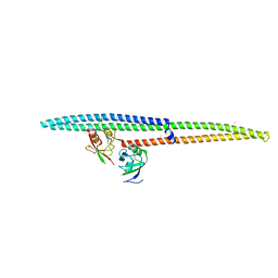
6LNM
 | |
6M3Q
 | | Crystal structure of AnkB/beta4-spectrin complex | | 分子名称: | Ankyrin-2, Spectrin beta chain | | 著者 | Li, J, Chen, K, Zhu, R, Zhang, M. | | 登録日 | 2020-03-04 | | 公開日 | 2020-05-13 | | 最終更新日 | 2023-11-29 | | 実験手法 | X-RAY DIFFRACTION (3.436 Å) | | 主引用文献 | Structural Basis Underlying Strong Interactions between Ankyrins and Spectrins.
J.Mol.Biol., 432, 2020
|
|
6M3R
 | | Crystal structure of AnkG/beta4-spectrin complex | | 分子名称: | Ankyrin-3, Spectrin beta chain | | 著者 | Li, J, Chen, K, Zhu, R, Zhang, M. | | 登録日 | 2020-03-04 | | 公開日 | 2020-05-13 | | 最終更新日 | 2023-11-29 | | 実験手法 | X-RAY DIFFRACTION (4.313 Å) | | 主引用文献 | Structural Basis Underlying Strong Interactions between Ankyrins and Spectrins.
J.Mol.Biol., 432, 2020
|
|
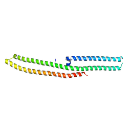
7VMB
 | |
6IRD
 | | Complex structure of INADL PDZ89 and PLCb4 C-terminal CC-PBM | | 分子名称: | 1-phosphatidylinositol 4,5-bisphosphate phosphodiesterase, GOLD ION, InaD-like protein | | 著者 | Ye, F, Li, J, Huang, Y, Liu, W, Zhang, M. | | 登録日 | 2018-11-12 | | 公開日 | 2019-01-23 | | 最終更新日 | 2023-11-22 | | 実験手法 | X-RAY DIFFRACTION (2.813 Å) | | 主引用文献 | An unexpected INAD PDZ tandem-mediated plc beta binding in Drosophila photo receptors.
Elife, 7, 2018
|
|
6IRC
 | | C-terminal domain of Drosophila phospholipase b NORPA, methylated | | 分子名称: | 1-phosphatidylinositol 4,5-bisphosphate phosphodiesterase | | 著者 | Ye, F, Li, J, Huang, Y, Liu, W, Zhang, M. | | 登録日 | 2018-11-12 | | 公開日 | 2019-01-02 | | 最終更新日 | 2020-10-28 | | 実験手法 | X-RAY DIFFRACTION (3.538 Å) | | 主引用文献 | An unexpected INAD PDZ tandem-mediated plc beta binding in Drosophila photo receptors.
Elife, 7, 2018
|
|
7W3W
 | | X-ray structure of apo-VmFbpA, a ferric ion-binding protein from Vibrio metschnikovii | | 分子名称: | Iron-utilization periplasmic protein | | 著者 | Lu, P, Sui, M, Zhang, M, Nagata, K. | | 登録日 | 2021-11-26 | | 公開日 | 2021-12-15 | | 最終更新日 | 2023-11-29 | | 実験手法 | X-RAY DIFFRACTION (1.858 Å) | | 主引用文献 | Rosmarinic Acid and Sodium Citrate Have a Synergistic Bacteriostatic Effect against Vibrio Species by Inhibiting Iron Uptake.
Int J Mol Sci, 22, 2021
|
|


