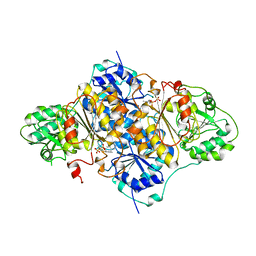
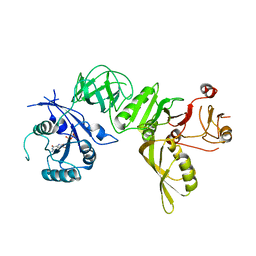
2RDE
 
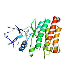
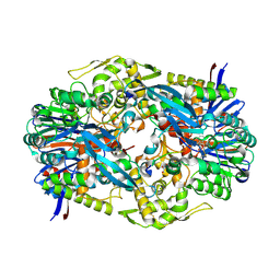
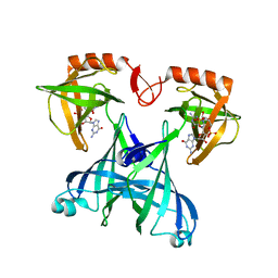 | | Crystal structure of VCA0042 complexed with c-di-GMP | | 分子名称: | 9,9'-[(2R,3R,3aS,5S,7aR,9R,10R,10aS,12S,14aR)-3,5,10,12-tetrahydroxy-5,12-dioxidooctahydro-2H,7H-difuro[3,2-d:3',2'-j][1,3,7,9,2,8]tetraoxadiphosphacyclododecine-2,9-diyl]bis(2-amino-1,9-dihydro-6H-purin-6-one), Uncharacterized protein VCA0042 | | 著者 | Benach, J, Swaminathan, S.S, Tamayo, R, Seetharaman, J, Handelman, S, Forouhar, F, Neely, H, Camilli, A, Hunt, J.F, Northeast Structural Genomics Consortium (NESG) | | 登録日 | 2007-09-22 | | 公開日 | 2007-10-30 | | 最終更新日 | 2023-08-30 | | 実験手法 | X-RAY DIFFRACTION (1.92 Å) | | 主引用文献 | The structural basis of cyclic diguanylate signal transduction by PilZ domains.
Embo J., 26, 2007
|
|
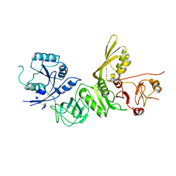
4ZJD
 | |
4ZJA
 | |
5HI4
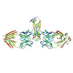
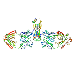 | | Binding site elucidation and structure guided design of macrocyclic IL-17A antagonists | | 分子名称: | (9'S,17'R)-6'-chloro-N-methyl-9'-{[(1-methyl-1H-pyrazol-5-yl)carbonyl]amino}-10',19'-dioxo-2'-oxa-11',18'-diazaspiro[cyclopentane-1,21'-tetracyclo[20.2.2.2~12,15~.1~3,7~]nonacosane]-1'(24'),3'(29'),4',6',12',14',22',25',27'-nonaene-17'-carboxamide, CAT-2000 FAB heavy chain, CAT-2000 FAB light chain, ... | | 著者 | Liu, S. | | 登録日 | 2016-01-11 | | 公開日 | 2016-08-31 | | 最終更新日 | 2023-09-27 | | 実験手法 | X-RAY DIFFRACTION (1.8 Å) | | 主引用文献 | Binding site elucidation and structure guided design of macrocyclic IL-17A antagonists.
Sci Rep, 6, 2016
|
|
3D0E
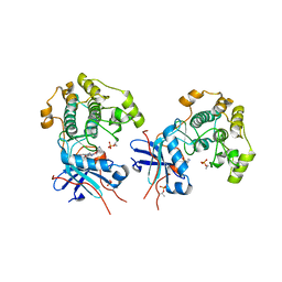 | | Crystal structure of human Akt2 in complex with GSK690693 | | 分子名称: | 4-{2-(4-amino-1,2,5-oxadiazol-3-yl)-1-ethyl-7-[(3S)-piperidin-3-ylmethoxy]-1H-imidazo[4,5-c]pyridin-4-yl}-2-methylbut-3-yn-2-ol, RAC-beta serine/threonine-protein kinase | | 著者 | Concha, N.O, Smallwood, A. | | 登録日 | 2008-05-01 | | 公開日 | 2008-10-21 | | 最終更新日 | 2017-10-25 | | 実験手法 | X-RAY DIFFRACTION (2 Å) | | 主引用文献 | Identification of 4-(2-(4-amino-1,2,5-oxadiazol-3-yl)-1-ethyl-7-{[(3S)-3-piperidinylmethyl]oxy}-1H-imidazo[4,5-c]pyridin-4-yl)-2-methyl-3-butyn-2-ol (GSK690693), a novel inhibitor of AKT kinase.
J.Med.Chem., 51, 2008
|
|
4JOA
 | | Crystal Structure of Human Anaplastic Lymphoma Kinase in complex with 7-azaindole based inhibitor | | 分子名称: | 3-[1-(2,5-difluorobenzyl)-1H-pyrazol-4-yl]-5-(1-methyl-1H-pyrazol-4-yl)-1H-pyrrolo[2,3-b]pyridine, ALK tyrosine kinase receptor | | 著者 | Hosahalli, S, Krishnamurthy, N.R, Lakshminarasimhan, A. | | 登録日 | 2013-03-18 | | 公開日 | 2013-07-17 | | 最終更新日 | 2023-11-08 | | 実験手法 | X-RAY DIFFRACTION (2.7 Å) | | 主引用文献 | Discovery of 7-azaindole based anaplastic lymphoma kinase (ALK) inhibitors: wild type and mutant (L1196M) active compounds with unique binding mode
Bioorg.Med.Chem.Lett., 23, 2013
|
|
4ZJ9
 | |
1XG6
 | |
5HI3
 | |
1PYD
 | |
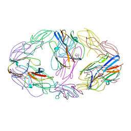
8SW4
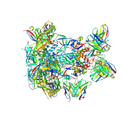 | | BG505 GT1.1 SOSIP in complex with NHP Fabs 21N13, 21M20 and RM20A3 | | 分子名称: | 2-acetamido-2-deoxy-beta-D-glucopyranose, 2-acetamido-2-deoxy-beta-D-glucopyranose-(1-4)-2-acetamido-2-deoxy-beta-D-glucopyranose, 21M20 heavy chain variable region, ... | | 著者 | Ozorowski, G, Torres, J.L, Zhang, S, Ward, A.B. | | 登録日 | 2023-05-17 | | 公開日 | 2024-05-29 | | 実験手法 | ELECTRON MICROSCOPY (2.85 Å) | | 主引用文献 | Neutralizing antibodies induced in non-human primates by germline targeting Env trimer
To Be Published
|
|
2TDX
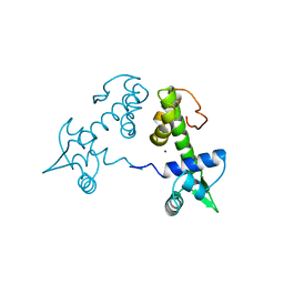 | | DIPHTHERIA TOX REPRESSOR (C102D MUTANT) COMPLEXED WITH NICKEL | | 分子名称: | DIPHTHERIA TOX REPRESSOR, NICKEL (II) ION | | 著者 | White, A, Ding, X, Zheng, H, Schiering, N, Ringe, D, Murphy, J.R. | | 登録日 | 1998-06-22 | | 公開日 | 1998-10-14 | | 最終更新日 | 2024-05-22 | | 実験手法 | X-RAY DIFFRACTION (2.4 Å) | | 主引用文献 | Structure of the metal-ion-activated diphtheria toxin repressor/tox operator complex.
Nature, 394, 1998
|
|
5F1R
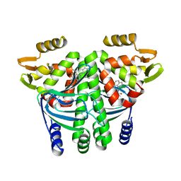 | | The Transcriptional Regulator PrfA from Listeria Monocytogenes in complex with a ring-fused 2-pyridone (C10) | | 分子名称: | (3~{R})-8-cyclopropyl-7-(naphthalen-1-ylmethyl)-5-oxidanylidene-2,3-dihydro-[1,3]thiazolo[3,2-a]pyridine-3-carboxylic acid, Listeriolysin regulatory protein | | 著者 | Begum, A, Grundstrom, C, Good, J.A.D, Andersson, C, ALmqvist, F, Johansson, J, Sauer, U.H, Sauer-Eriksson, A.E. | | 登録日 | 2015-11-30 | | 公開日 | 2016-03-16 | | 最終更新日 | 2024-01-10 | | 実験手法 | X-RAY DIFFRACTION (2.25 Å) | | 主引用文献 | Attenuating Listeria monocytogenes Virulence by Targeting the Regulatory Protein PrfA.
Cell Chem Biol, 23, 2016
|
|
5A9X
 | | Structure of GDP bound BipA | | 分子名称: | GTP-BINDING PROTEIN, GUANOSINE-5'-DIPHOSPHATE | | 著者 | Kumar, V, Chen, Y, Ero, R, Li, Z, Gao, Y.-G. | | 登録日 | 2015-07-23 | | 公開日 | 2015-08-26 | | 最終更新日 | 2024-05-08 | | 実験手法 | X-RAY DIFFRACTION (3.8 Å) | | 主引用文献 | Structure of Bipa in GTP Form Bound to the Ratcheted Ribosome.
Proc.Natl.Acad.Sci.USA, 112, 2015
|
|
5A9V
 | | Structure of apo BipA | | 分子名称: | GTP-BINDING PROTEIN | | 著者 | Kumar, V, Chen, Y, Ero, R, Li, Z, Gao, Y. | | 登録日 | 2015-07-23 | | 公開日 | 2015-09-02 | | 最終更新日 | 2024-01-10 | | 実験手法 | X-RAY DIFFRACTION (3.31 Å) | | 主引用文献 | Structure of Bipa in GTP Form Bound to the Ratcheted Ribosome.
Proc.Natl.Acad.Sci.USA, 112, 2015
|
|
1CYI
 | | CYTOCHROME C6 | | 分子名称: | CADMIUM ION, CYTOCHROME C6, HEME C | | 著者 | Kerfeld, C.A, Yeates, T.O. | | 登録日 | 1995-05-09 | | 公開日 | 1996-01-29 | | 最終更新日 | 2021-03-10 | | 実験手法 | X-RAY DIFFRACTION (1.9 Å) | | 主引用文献 | The structure of chloroplast cytochrome c6 at 1.9 A resolution: evidence for functional oligomerization.
J.Mol.Biol., 250, 1995
|
|
2K0U
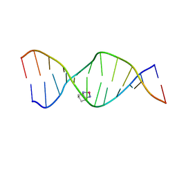 | | High Resolution Solution NMR Structures of Oxaliplatin-DNA Adduct | | 分子名称: | CYCLOHEXANE-1(R),2(R)-DIAMINE-PLATINUM(II), DNA (5'-D(*DCP*DCP*DTP*DCP*DTP*DGP*DGP*DTP*DCP*DTP*DCP*DC)-3'), DNA (5'-D(*DGP*DGP*DAP*DGP*DAP*DCP*DCP*DAP*DGP*DAP*DGP*DG)-3') | | 著者 | Bhattacharyya, D, King, C.L, Chaney, S.G, Campbell, S.L. | | 登録日 | 2008-02-15 | | 公開日 | 2009-02-03 | | 最終更新日 | 2024-05-01 | | 実験手法 | SOLUTION NMR | | 主引用文献 | Flanking Bases Influence the Nature of DNA Distortion by Platinum 1,2-Intrastrand (GG) Cross-Links.
Plos One, 6, 2011
|
|
2K0V
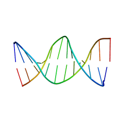 | | High Resolution Solution NMR Structures of Undamaged DNA Dodecamer Duplex | | 分子名称: | DNA (5'-D(*DCP*DCP*DTP*DCP*DTP*DGP*DGP*DTP*DCP*DTP*DCP*DC)-3'), DNA (5'-D(*DGP*DGP*DAP*DGP*DAP*DCP*DCP*DAP*DGP*DAP*DGP*DG)-3') | | 著者 | Bhattacharyya, D, King, C.L, Chaney, S.G, Campbell, S.L. | | 登録日 | 2008-02-15 | | 公開日 | 2009-02-03 | | 最終更新日 | 2024-05-01 | | 実験手法 | SOLUTION NMR | | 主引用文献 | Flanking Bases Influence the Nature of DNA Distortion by Platinum 1,2-Intrastrand (GG) Cross-Links.
Plos One, 6, 2011
|
|
4YFB
 | |
8WXR
 | |
8WXU
 | |
8WXV
 | |
6K08
 | |
8WXQ
 | |
8WXT
 | |


