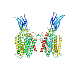
8XLD
 
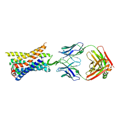
 | | Structure of the GFP:GFP-nanobody complex from Biortus. | | 分子名称: | 1,2-ETHANEDIOL, Nanobody(Staygold-S2G10)-Nanobody(Staygold-S4F1), ZINC ION, ... | | 著者 | Wang, F, Cheng, W, Yuan, Z, Lin, D, Bao, C. | | 登録日 | 2023-12-25 | | 公開日 | 2024-03-06 | | 実験手法 | X-RAY DIFFRACTION (2.1 Å) | | 主引用文献 | Structure of the GFP:GFP-nanobody complex from Biortus.
To Be Published
|
|
8W2L
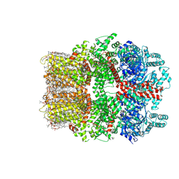 | | TRPM7 structure in complex with anticancer agent CCT128930 in closed state | | 分子名称: | (2S)-3-(hexadecanoyloxy)-2-[(9Z)-octadec-9-enoyloxy]propyl 2-(trimethylammonio)ethyl phosphate, 2-[2-[(1~{S},2~{S},4~{S},5'~{R},6~{R},7~{S},8~{R},9~{S},12~{S},13~{R},16~{S})-5',7,9,13-tetramethylspiro[5-oxapentacyclo[10.8.0.0^{2,9}.0^{4,8}.0^{13,18}]icos-18-ene-6,2'-oxane]-16-yl]oxyethyl]propane-1,3-diol, 4-(4-chlorobenzyl)-1-(7H-pyrrolo[2,3-d]pyrimidin-4-yl)piperidin-4-aminium, ... | | 著者 | Nadezhdin, K.D, Sobolevsky, A.I. | | 登録日 | 2024-02-20 | | 公開日 | 2024-04-17 | | 最終更新日 | 2024-04-24 | | 実験手法 | ELECTRON MICROSCOPY (2.45 Å) | | 主引用文献 | Structural basis of selective TRPM7 inhibition by the anticancer agent CCT128930.
Cell Rep, 43, 2024
|
|
8UBG
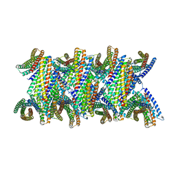 | | DpHF19 filament | | 分子名称: | DpHF19,Green fluorescent protein (Fragment) | | 著者 | Lynch, E.M, Shen, H, Kollman, J.M, Baker, D. | | 登録日 | 2023-09-22 | | 公開日 | 2024-04-10 | | 実験手法 | ELECTRON MICROSCOPY (3.5 Å) | | 主引用文献 | De novo design of pH-responsive self-assembling helical protein filaments.
Nat Nanotechnol, 2024
|
|
8UB6
 | |
8U24
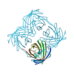
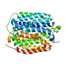
 | | A Highly Stable Variant of Corynactis Californica Green Fluorescent Protein, ccGFP 9 | | 分子名称: | Green Fluorescent Protein Variant #9, ccGFP 9 | | 著者 | Hung, L.-W, Terwilliger, T.C, Waldo, G, Nguyen, H.B. | | 登録日 | 2023-09-05 | | 公開日 | 2024-01-31 | | 最終更新日 | 2024-02-07 | | 実験手法 | X-RAY DIFFRACTION (1.85 Å) | | 主引用文献 | Engineering highly stable variants of Corynactis californica green fluorescent proteins.
Protein Sci., 33, 2024
|
|
8U23
 | | A Highly Stable Variant of Corynactis Californica Green Fluorescent Protein, ccGFP 8 | | 分子名称: | Green Fluorescent Protein Variant #8, ccGFP 8 | | 著者 | Hung, L.-W, Terwilliger, T.C, Waldo, G, Nguyen, H.B. | | 登録日 | 2023-09-05 | | 公開日 | 2024-01-31 | | 最終更新日 | 2024-02-07 | | 実験手法 | X-RAY DIFFRACTION (1.78 Å) | | 主引用文献 | Engineering highly stable variants of Corynactis californica green fluorescent proteins.
Protein Sci., 33, 2024
|
|
8U22
 | | A Highly Stable Variant of Corynactis Californica Green Fluorescent Protein, ccGFP 7 | | 分子名称: | Green Fluorescent Protein Variant #7, ccGFP 7 | | 著者 | Hung, L.-W, Terwilliger, T.C, Waldo, G, Nguyen, H.B. | | 登録日 | 2023-09-05 | | 公開日 | 2024-01-31 | | 最終更新日 | 2024-02-07 | | 実験手法 | X-RAY DIFFRACTION (1.8 Å) | | 主引用文献 | Engineering highly stable variants of Corynactis californica green fluorescent proteins.
Protein Sci., 33, 2024
|
|
8U21
 | | A Highly Stable Variant of Corynactis Californica Green Fluorescent Protein, ccGFP E6 | | 分子名称: | Green Fluorescent Protein Variant E6, ccGFP E6 | | 著者 | Hung, L.-W, Terwilliger, T.C, Waldo, G, Nguyen, H.B. | | 登録日 | 2023-09-05 | | 公開日 | 2024-01-31 | | 最終更新日 | 2024-02-07 | | 実験手法 | X-RAY DIFFRACTION (1.96 Å) | | 主引用文献 | Engineering highly stable variants of Corynactis californica green fluorescent proteins.
Protein Sci., 33, 2024
|
|
8U20
 | | A Highly Stable Variant of Corynactis Californica Green Fluorescent Protein, ccGFP 5 | | 分子名称: | Green Fluorescent Protein Variant #5, ccGFP 5 | | 著者 | Hung, L.-W, Terwilliger, T.C, Waldo, G, Nguyen, H.B. | | 登録日 | 2023-09-05 | | 公開日 | 2024-01-31 | | 最終更新日 | 2024-02-07 | | 実験手法 | X-RAY DIFFRACTION (1.9 Å) | | 主引用文献 | Engineering highly stable variants of Corynactis californica green fluorescent proteins.
Protein Sci., 33, 2024
|
|
8TU9
 | | Cryo-EM structure of HGSNAT-acetyl-CoA complex at pH 7.5 | | 分子名称: | 2-acetamido-2-deoxy-beta-D-glucopyranose, ACETYL COENZYME *A, Enhanced green fluorescent protein,Heparan-alpha-glucosaminide N-acetyltransferase,Isoform 2 of Heparan-alpha-glucosaminide N-acetyltransferase | | 著者 | Navratna, V, Kumar, A, Mosalaganti, S. | | 登録日 | 2023-08-15 | | 公開日 | 2024-02-07 | | 実験手法 | ELECTRON MICROSCOPY (3.26 Å) | | 主引用文献 | Structure of the human heparan-alpha-glucosaminide N-acetyltransferase (HGSNAT)
eLife, 13, 2024
|
|
8TLM
 | | Structure of a class A GPCR/Fab complex | | 分子名称: | C-C chemokine receptor type 8, Green fluorescent protein fusion, Fab heavy chain, ... | | 著者 | Sun, D, Johnson, M, Masureel, M. | | 登録日 | 2023-07-27 | | 公開日 | 2023-12-20 | | 実験手法 | ELECTRON MICROSCOPY (2.9 Å) | | 主引用文献 | Structural basis of antibody inhibition and chemokine activation of the human CC chemokine receptor 8.
Nat Commun, 14, 2023
|
|
8THR
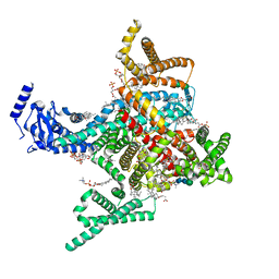
 | | Structure of the human vesicular monoamine transporter 2 (VMAT2) bound to tetrabenazine in an occluded conformation | | 分子名称: | (3S,5R,11bS)-9,10-dimethoxy-3-(2-methylpropyl)-1,3,4,6,7,11b-hexahydro-2H-pyrido[2,1-a]isoquinolin-2-one, fluorescent protein mVenus,Synaptic vesicular amine transporter,GFP nano body,Synaptic vesicular amine transporter,Synaptic vesicular amine transporter | | 著者 | Dalton, M.P, Coleman, J.A. | | 登録日 | 2023-07-17 | | 公開日 | 2023-10-25 | | 実験手法 | ELECTRON MICROSCOPY (3.12 Å) | | 主引用文献 | Structure of the human vesicular monoamine transporter 2 (VMAT2) bound to tetrabenazine in an occluded conformation
To Be Published
|
|
8TGP
 | |
8T6L
 | | Cryo-EM structure of rat cardiac sodium channel NaV1.5 with batrachotoxin analog BTX-B | | 分子名称: | (1R)-1-[(5aR,7aR,9R,11aS,11bS,12R,13aR)-9,12-dihydroxy-2,11a-dimethyl-1,2,3,4,7a,8,9,10,11,11a,12,13-dodecahydro-7H-9,11b-epoxy-13a,5a-prop[1]enophenanthro[2,1-f][1,4]oxazepin-14-yl]ethyl benzoate, (3beta,14beta,17beta,25R)-3-[4-methoxy-3-(methoxymethyl)butoxy]spirost-5-en, 1-palmitoyl-2-oleoyl-sn-glycero-3-phosphocholine, ... | | 著者 | Tonggu, L, Wisedchaisri, G, Gamal El-Din, T.M, Zheng, N, Catterall, W.A. | | 登録日 | 2023-06-16 | | 公開日 | 2024-03-06 | | 最終更新日 | 2024-03-27 | | 実験手法 | ELECTRON MICROSCOPY (3.3 Å) | | 主引用文献 | Dual receptor-sites reveal the structural basis for hyperactivation of sodium channels by poison-dart toxin batrachotoxin.
Nat Commun, 15, 2024
|
|
8SMU
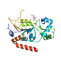
 | | Integral fusion of the HtaA CR2 domain from Corynebacterium diphtheriae within EGFP | | 分子名称: | CHLORIDE ION, GLYCEROL, HtaACR2 integral fusion within enhanced green fluorescent protein, ... | | 著者 | Mahoney, B.J, Cascio, D, Clubb, R.T. | | 登録日 | 2023-04-26 | | 公開日 | 2023-09-27 | | 最終更新日 | 2023-11-15 | | 実験手法 | X-RAY DIFFRACTION (2.45 Å) | | 主引用文献 | Development and atomic structure of a new fluorescence-based sensor to probe heme transfer in bacterial pathogens.
J.Inorg.Biochem., 249, 2023
|
|
8PN2
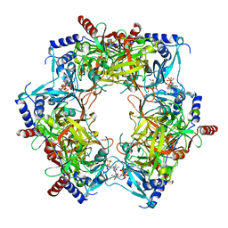 | | CryoEM structure of Nal1 protein, allele IR64, from Oryza sativa indica cultivar | | 分子名称: | ADENOSINE-5'-TRIPHOSPHATE, MAGNESIUM ION, Protein NARROW LEAF 1 | | 著者 | Huang, L.Y, Rety, S, Xi, X.G. | | 登録日 | 2023-06-29 | | 公開日 | 2024-04-17 | | 最終更新日 | 2024-04-24 | | 実験手法 | ELECTRON MICROSCOPY (2.63 Å) | | 主引用文献 | The catalytic triad of rice NARROW LEAF1 involves H234.
Nat.Plants, 2024
|
|
8PN1
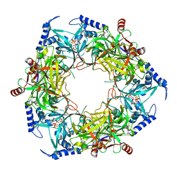 | | CryoEM structure of Nal1 protein, allele SPIKE, from Oryza sativa japonica group | | 分子名称: | ADENOSINE-5'-TRIPHOSPHATE, MAGNESIUM ION, Protein NARROW LEAF 1 | | 著者 | Huang, L.Y, Rety, S, Xi, X.G. | | 登録日 | 2023-06-29 | | 公開日 | 2024-04-17 | | 最終更新日 | 2024-04-24 | | 実験手法 | ELECTRON MICROSCOPY (2.4 Å) | | 主引用文献 | The catalytic triad of rice NARROW LEAF1 involves H234.
Nat.Plants, 2024
|
|
8PMM
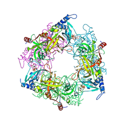 | | Structure of Nal1 protein, allele SPIKE from japonica rice, construct 31-458 | | 分子名称: | ADENOSINE-5'-TRIPHOSPHATE, MAGNESIUM ION, PHOSPHATE ION, ... | | 著者 | Huang, L.Y, Rety, S, Xi, X.G. | | 登録日 | 2023-06-29 | | 公開日 | 2024-04-17 | | 最終更新日 | 2024-04-24 | | 実験手法 | X-RAY DIFFRACTION (1.75 Å) | | 主引用文献 | The catalytic triad of rice NARROW LEAF1 involves H234.
Nat.Plants, 2024
|
|
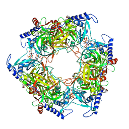
8PML
 | | Structure of Nal1 protein , SPIKE allele from japonica rice, construct 46-458 | | 分子名称: | ADENOSINE-5'-TRIPHOSPHATE, MAGNESIUM ION, Protein NARROW LEAF 1 | | 著者 | Huang, L.Y, Rety, S, Xi, X.G. | | 登録日 | 2023-06-29 | | 公開日 | 2024-04-17 | | 最終更新日 | 2024-04-24 | | 実験手法 | X-RAY DIFFRACTION (2.95 Å) | | 主引用文献 | The catalytic triad of rice NARROW LEAF1 involves H234.
Nat.Plants, 2024
|
|
8PMI
 | | Structure of Nal1 indica cultivar IR64, construct 36-458 in presence of peptide from FZP protein | | 分子名称: | ADENOSINE-5'-TRIPHOSPHATE, AMMONIUM ION, MAGNESIUM ION, ... | | 著者 | Huang, L.Y, Rety, S, Xi, X.G. | | 登録日 | 2023-06-28 | | 公開日 | 2024-04-17 | | 最終更新日 | 2024-04-24 | | 実験手法 | X-RAY DIFFRACTION (2.83 Å) | | 主引用文献 | The catalytic triad of rice NARROW LEAF1 involves H234.
Nat.Plants, 2024
|
|
8PMG
 | | Structure of Nal1 indica cultivar IR64, construct 36-458 | | 分子名称: | ADENOSINE-5'-TRIPHOSPHATE, MAGNESIUM ION, PHOSPHATE ION, ... | | 著者 | Huang, L.Y, Rety, S, Xi, X.G. | | 登録日 | 2023-06-28 | | 公開日 | 2024-04-17 | | 最終更新日 | 2024-04-24 | | 実験手法 | X-RAY DIFFRACTION (3.29 Å) | | 主引用文献 | The catalytic triad of rice NARROW LEAF1 involves H234.
Nat.Plants, 2024
|
|
8OVY
 | | Structure of analogue of superfolded GFP | | 分子名称: | Green fluorescent protein | | 著者 | Dunkelmann, D, Fiedler, M, Bellini, D, Alvira, C.P, Chin, J.W. | | 登録日 | 2023-04-26 | | 公開日 | 2024-01-10 | | 最終更新日 | 2024-01-31 | | 実験手法 | X-RAY DIFFRACTION (1.537 Å) | | 主引用文献 | Adding alpha , alpha-disubstituted and beta-linked monomers to the genetic code of an organism.
Nature, 625, 2024
|
|
8OVP
 | | X-ray structure of the iAspSnFR in complex with L-aspartate | | 分子名称: | ACETATE ION, ASPARTIC ACID, MAGNESIUM ION, ... | | 著者 | Tarnawski, M, Hellweg, L, Bergner, A, Hiblot, J, Leippe, P, Johnsson, K. | | 登録日 | 2023-04-26 | | 公開日 | 2023-05-17 | | 実験手法 | X-RAY DIFFRACTION (1.7 Å) | | 主引用文献 | X-ray structure of the SF-iAspSnFR in complex with L-aspartate
To Be Published
|
|
8OVO
 | | X-ray structure of the SF-iGluSnFR-S72A in complex with L-aspartate | | 分子名称: | ASPARTIC ACID, Putative periplasmic binding transport protein,Green fluorescent protein | | 著者 | Tarnawski, M, Hellweg, L, Bergner, A, Hiblot, J, Leippe, P, Johnsson, K. | | 登録日 | 2023-04-26 | | 公開日 | 2023-05-17 | | 実験手法 | X-RAY DIFFRACTION (1.7 Å) | | 主引用文献 | X-ray structure of the SF-iGluSnFR-S72A in complex with L-aspartate
To Be Published
|
|
8OVN
 | | X-ray structure of the SF-iGluSnFR-S72A | | 分子名称: | CITRIC ACID, Putative periplasmic binding transport protein,Green fluorescent protein | | 著者 | Tarnawski, M, Hellweg, L, Bergner, A, Hiblot, J, Leippe, P, Johnsson, K. | | 登録日 | 2023-04-26 | | 公開日 | 2023-05-17 | | 実験手法 | X-RAY DIFFRACTION (2.6 Å) | | 主引用文献 | X-ray structure of the SF-iGluSnFR-S72A
To Be Published
|
|


