Loading
PDBj
MenuPDBj@FacebookPDBj@TwitterPDBj@YouTubewwPDB FoundationwwPDB
RCSB PDBPDBeBMRBAdv. SearchSearch help

5DPH
DownloadVisualize
BU of 5dph by Molmil
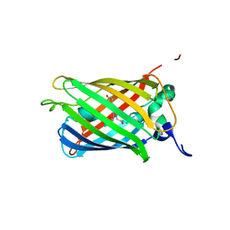
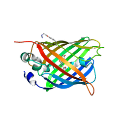
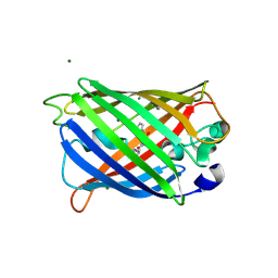
sfGFP mutant - 149 p-cyano-L-phenylalanine
Descriptor: CARBON DIOXIDE, Green fluorescent protein, MAGNESIUM ION
Authors:Dippel, A.B, Olenginski, G.M, Maurici, N, Liskov, M.T, Brewer, S.H, Phillips-Piro, C.M.
Deposit date:2015-09-12
Release date:2016-01-13
Last modified:2023-11-15
Method:X-RAY DIFFRACTION (1.42 Å)
Cite:Probing the effectiveness of spectroscopic reporter unnatural amino acids: a structural study.
Acta Crystallogr D Struct Biol, 72, 2016
3DQI
DownloadVisualize
BU of 3dqi by Molmil
Structure of the Yellow Fluorescent Protein Citrine Frozen at 1000 Atmospheres Number 4: Structure 8 in a Series of 26 High Pressure Structures
Descriptor: Green fluorescent protein
Authors:Barstow, B, Kim, C.U.
Deposit date:2008-07-09
Release date:2008-09-23
Last modified:2023-11-15
Method:X-RAY DIFFRACTION (1.42 Å)
Cite:Alteration of citrine structure by hydrostatic pressure explains the accompanying spectral shift.
Proc.Natl.Acad.Sci.Usa, 105, 2008
3DQU
DownloadVisualize
BU of 3dqu by Molmil
Structure of the Yellow Fluorescent Protein Citrine Frozen at 2000 Atmospheres Number 1: Structure 20 in a Series of 26 High Pressure Structures
Descriptor: Green fluorescent protein
Authors:Barstow, B, Kim, C.U.
Deposit date:2008-07-09
Release date:2008-09-23
Last modified:2023-11-15
Method:X-RAY DIFFRACTION (1.42 Å)
Cite:Alteration of citrine structure by hydrostatic pressure explains the accompanying spectral shift.
Proc.Natl.Acad.Sci.Usa, 105, 2008
7PNN
DownloadVisualize
BU of 7pnn by Molmil
mVenus released from fusion protein.
Descriptor: CHLORIDE ION, mVenus
Authors:Bloch, Y, Savvides, S.N.
Deposit date:2021-09-07
Release date:2021-11-03
Last modified:2024-01-31
Method:X-RAY DIFFRACTION (1.43 Å)
Cite:mVenus released from fusion protein.
To Be Published
3DQE
DownloadVisualize
BU of 3dqe by Molmil
Structure of the Yellow Fluorescent Protein Citrine Frozen at 1250 Atmospheres Number 1: Structure 11 in a Series of 26 High Pressure Structures
Descriptor: Green fluorescent protein
Authors:Barstow, B, Kim, C.U.
Deposit date:2008-07-09
Release date:2008-09-23
Last modified:2023-11-15
Method:X-RAY DIFFRACTION (1.43 Å)
Cite:Alteration of citrine structure by hydrostatic pressure explains the accompanying spectral shift.
Proc.Natl.Acad.Sci.Usa, 105, 2008
3DQA
DownloadVisualize
BU of 3dqa by Molmil
Structure of the Yellow Fluorescent Protein Citrine Frozen at 1250 Atmospheres Number 4: Structure 14 in a Series of 26 High Pressure Structures
Descriptor: Green fluorescent protein
Authors:Barstow, B, Kim, C.U.
Deposit date:2008-07-09
Release date:2008-09-23
Last modified:2023-11-15
Method:X-RAY DIFFRACTION (1.44 Å)
Cite:Alteration of citrine structure by hydrostatic pressure explains the accompanying spectral shift.
Proc.Natl.Acad.Sci.Usa, 105, 2008
3DQM
DownloadVisualize
BU of 3dqm by Molmil
Structure of the Yellow Fluorescent Protein Citrine Frozen at 750 Atmospheres: Structure 4 in a Series of 26 High Pressure Structures
Descriptor: Green fluorescent protein
Authors:Barstow, B, Kim, C.U.
Deposit date:2008-07-09
Release date:2008-09-23
Last modified:2023-11-15
Method:X-RAY DIFFRACTION (1.44 Å)
Cite:Alteration of citrine structure by hydrostatic pressure explains the accompanying spectral shift.
Proc.Natl.Acad.Sci.Usa, 105, 2008
3DQN
DownloadVisualize
BU of 3dqn by Molmil
Structure of the Yellow Fluorescent Protein Citrine Frozen at 500 Atmospheres: Structure 3 in a Series of 26 High Pressure Structures
Descriptor: Green fluorescent protein
Authors:Barstow, B, Kim, C.U.
Deposit date:2008-07-09
Release date:2008-09-23
Last modified:2023-11-15
Method:X-RAY DIFFRACTION (1.44 Å)
Cite:Alteration of citrine structure by hydrostatic pressure explains the accompanying spectral shift.
Proc.Natl.Acad.Sci.Usa, 105, 2008
1KYS
DownloadVisualize
BU of 1kys by Molmil
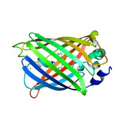
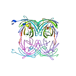
Crystal Structure of a Zn-bound Green Fluorescent Protein Biosensor
Descriptor: Green Fluorescent Protein, ZINC ION
Authors:Barondeau, D.P, Kassmann, C.J, Tainer, J.A, Getzoff, E.D.
Deposit date:2002-02-05
Release date:2002-04-10
Last modified:2023-11-15
Method:X-RAY DIFFRACTION (1.44 Å)
Cite:Structural chemistry of a green fluorescent protein Zn biosensor.
J.Am.Chem.Soc., 124, 2002
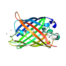
6L26
DownloadVisualize
BU of 6l26 by Molmil
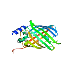
Neutron crystal structure of the mutant green fluorescent protein (EGFP)
Descriptor: Green fluorescent protein, trideuteriooxidanium
Authors:Adachi, M, Shimizu, R, Shibazaki, C, Kagotani, Y, Ostermann, A, Schrader, T.E.
Deposit date:2019-10-02
Release date:2020-04-08
Last modified:2023-11-22
Method:NEUTRON DIFFRACTION (1.444 Å)
Cite:Direct Observation of the Protonation States in the Mutant Green Fluorescent Protein.
J Phys Chem Lett, 11, 2020
3SSL
DownloadVisualize
BU of 3ssl by Molmil
Engineered high-affinity halide-binding protein derived from YFP: iodide complex
Descriptor: Green fluorescent protein, IODIDE ION
Authors:Wang, W, Grimley, J.S, Beese, L.S, Hellinga, H.W.
Deposit date:2011-07-08
Release date:2012-07-11
Last modified:2023-11-15
Method:X-RAY DIFFRACTION (1.449 Å)
Cite:Determination of engineered chloride-binding site structures in fluorescent proteins reveals principles of halide recognition
To be published
2A54
DownloadVisualize
BU of 2a54 by Molmil
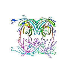
fluorescent protein asFP595, A143S, on-state, 1min irradiation
Descriptor: CHLORIDE ION, GFP-like non-fluorescent chromoprotein FP595 chain 1, GFP-like non-fluorescent chromoprotein FP595 chain 2
Authors:Andresen, M, Wahl, M.C, Stiel, A.C, Graeter, F, Schaefer, L, Trowitzsch, S, Weber, G, Eggeling, C, Grubmueller, H, Hell, S.W, Jakobs, S.
Deposit date:2005-06-30
Release date:2005-08-16
Last modified:2021-11-10
Method:X-RAY DIFFRACTION (1.45 Å)
Cite:Structure and mechanism of the reversible photoswitch of a fluorescent protein
Proc.Natl.Acad.Sci.Usa, 102, 2005
4B5Y
DownloadVisualize
BU of 4b5y by Molmil
X-ray structure of the cyan fluorescent protein mTurquoise-GL (K206A mutant) in space group C222(1)
Descriptor: GREEN FLUORESCENT PROTEIN
Authors:von Stetten, D, Lelimousin, M, Oost, K, Noirclerc-Savoye, M, Gadella, T.W.J, Goedhart, J, Royant, A.
Deposit date:2012-08-08
Release date:2013-08-28
Last modified:2023-12-20
Method:X-RAY DIFFRACTION (1.45 Å)
Cite:Influence of the H148G Mutation on Fluorescence Properties of Cyan Fluorescent Proteins
To be Published
2A53
DownloadVisualize
BU of 2a53 by Molmil
fluorescent protein asFP595, A143S, off-state
Descriptor: CHLORIDE ION, GFP-like non-fluorescent chromoprotein FP595 chain 1, GFP-like non-fluorescent chromoprotein FP595 chain 2
Authors:Andresen, M, Wahl, M.C, Stiel, A.C, Graeter, F, Schaefer, L, Trowitzsch, S, Weber, G, Eggeling, C, Grubmueller, H, Hell, S.W, Jakobs, S.
Deposit date:2005-06-30
Release date:2005-08-16
Last modified:2021-11-10
Method:X-RAY DIFFRACTION (1.45 Å)
Cite:Structure and mechanism of the reversible photoswitch of a fluorescent protein
Proc.Natl.Acad.Sci.Usa, 102, 2005
4H48
DownloadVisualize
BU of 4h48 by Molmil
1.45 angstrom CyPet Structure at pH7.0
Descriptor: 2-AMINO-2-HYDROXYMETHYL-PROPANE-1,3-DIOL, Green fluorescent protein
Authors:Hu, X.-J, Liu, R.
Deposit date:2012-09-17
Release date:2013-09-18
Last modified:2023-12-06
Method:X-RAY DIFFRACTION (1.45 Å)
Cite:Structure insight of the fluorescent state of CyPet
To be Published
2O24
DownloadVisualize
BU of 2o24 by Molmil
Spectroscopic and Structural Study of the Heterotropic Linkage between Halide and Proton Ion Binding to Gfp Proteins: E2(GFP)-Cl Complex
Descriptor: CHLORIDE ION, Green fluorescent protein
Authors:Garau, G.
Deposit date:2006-11-29
Release date:2007-05-15
Last modified:2023-11-15
Method:X-RAY DIFFRACTION (1.45 Å)
Cite:Spectroscopic and Structural Study of Proton and Halide Ion Cooperative Binding to GFP.
Biophys.J., 93, 2007
6PFT
DownloadVisualize
BU of 6pft by Molmil
rsEGFP2 with a chlorinated chromophore in the non-fluorescent off-state
Descriptor: Green fluorescent protein, SULFATE ION
Authors:Chang, J, Romei, M.G, Boxer, S.G.
Deposit date:2019-06-22
Release date:2019-08-07
Last modified:2023-11-15
Method:X-RAY DIFFRACTION (1.45 Å)
Cite:Structural Evidence of Photoisomerization Pathways in Fluorescent Proteins.
J.Am.Chem.Soc., 141, 2019
5DTX
DownloadVisualize
BU of 5dtx by Molmil
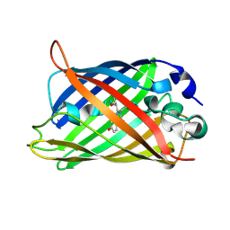
Crystal structure of rsEGFP2 in the fluorescent on-state
Descriptor: Green fluorescent protein
Authors:Adam, V, Martins, A.
Deposit date:2015-09-18
Release date:2016-01-20
Last modified:2024-01-10
Method:X-RAY DIFFRACTION (1.45 Å)
Cite:Rational design of ultrastable and reversibly photoswitchable fluorescent proteins for super-resolution imaging of the bacterial periplasm.
Sci Rep, 6, 2016
1Q4A
DownloadVisualize
BU of 1q4a by Molmil
S65T Q80R Green Fluorescent Protein (GFP) pH 8.5
Descriptor: Green Fluorescent Protein
Authors:Jain, R.K, Ranganathan, R.
Deposit date:2003-08-02
Release date:2004-02-03
Last modified:2023-11-15
Method:X-RAY DIFFRACTION (1.45 Å)
Cite:Local complexity of amino acid interactions in a protein core.
Proc.Natl.Acad.Sci.USA, 101, 2004
5EHU
DownloadVisualize
BU of 5ehu by Molmil
sfGFP mutant with unnatural amino acid 4-azidoethoxy-L-phenylalanine incorporated at the 149 site
Descriptor: Green fluorescent protein
Authors:Tookmanian, E.M, Phillips-Piro, C.M, Fenlon, E.E, Brewer, S.B.
Deposit date:2015-10-28
Release date:2015-12-23
Last modified:2023-11-15
Method:X-RAY DIFFRACTION (1.45 Å)
Cite:Azidoethoxyphenylalanine as a Vibrational Reporter and Click Chemistry Partner in Proteins.
Chemistry, 21, 2015
3DQH
DownloadVisualize
BU of 3dqh by Molmil
Structure of the Yellow Fluorescent Protein Citrine Frozen at 1000 Atmospheres Number 5: Structure 9 in a Series of 26 High Pressure Structures
Descriptor: Green fluorescent protein
Authors:Barstow, B, Kim, C.U.
Deposit date:2008-07-09
Release date:2008-09-23
Last modified:2023-11-15
Method:X-RAY DIFFRACTION (1.45 Å)
Cite:Alteration of citrine structure by hydrostatic pressure explains the accompanying spectral shift.
Proc.Natl.Acad.Sci.Usa, 105, 2008
3EVP
DownloadVisualize
BU of 3evp by Molmil
crystal structure of circular-permutated EGFP
Descriptor: Green fluorescent protein,Green fluorescent protein
Authors:Wang, Q, Shui, B, Kotlikoff, M.I, Sondermann, H.
Deposit date:2008-10-13
Release date:2008-12-09
Last modified:2023-12-27
Method:X-RAY DIFFRACTION (1.453 Å)
Cite:Structural Basis for Calcium Sensing by GCaMP2.
Structure, 16, 2008
7QLK
DownloadVisualize
BU of 7qlk by Molmil
Cis structure intermediate of rsKiiro Illuminated at 200 K
Descriptor: GLYCEROL, SULFATE ION, rsKiiro
Authors:van Thor, J.J, Baxter, J.M.
Deposit date:2021-12-20
Release date:2023-07-05
Last modified:2023-11-15
Method:X-RAY DIFFRACTION (1.458 Å)
Cite:Optical control of ultrafast structural dynamics in a fluorescent protein.
Nat.Chem., 15, 2023
6QQ8
DownloadVisualize
BU of 6qq8 by Molmil
Cryogenic temperature structure of the fluorescent protein Cerulean recorded after an accumulated dose of 290 kGy
Descriptor: Green fluorescent protein
Authors:Gotthard, G, Aumonier, S, Royant, A.
Deposit date:2019-02-18
Release date:2019-06-19
Last modified:2024-01-24
Method:X-RAY DIFFRACTION (1.46 Å)
Cite:Specific radiation damage is a lesser concern at room temperature.
Iucrj, 6, 2019
3DQF
DownloadVisualize
BU of 3dqf by Molmil
Structure of the Yellow Fluorescent Protein Citrine Frozen at 1000 Atmospheres Number 6: Structure 10 in a Series of 26 High Pressure Structures
Descriptor: Green fluorescent protein
Authors:Barstow, B, Kim, C.U.
Deposit date:2008-07-09
Release date:2008-09-23
Last modified:2023-11-15
Method:X-RAY DIFFRACTION (1.46 Å)
Cite:Alteration of citrine structure by hydrostatic pressure explains the accompanying spectral shift.
Proc.Natl.Acad.Sci.Usa, 105, 2008

218853

PDB entries from 2024-04-24

PDB statisticsPDBj update infoContact PDBjnumon