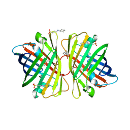
Loading
PDBj
MenuPDBj@FacebookPDBj@TwitterPDBj@YouTubewwPDB FoundationwwPDB
RCSB PDBPDBeBMRBAdv. SearchSearch help

2C9I
DownloadVisualize
BU of 2c9i by Molmil
Structure of the fluorescent protein asFP499 from Anemonia sulcata
Descriptor: GREEN FLUORESCENT PROTEIN ASFP499
Authors:Renzi, F, Nienhaus, K, Wiedenmann, J, Vallone, B, Nienhaus, G.U.
Deposit date:2005-12-12
Release date:2007-01-16
Last modified:2023-11-15
Method:X-RAY DIFFRACTION (1.82 Å)
Cite:Chromophore-Protein Interactions in the Anthozoan Green Fluorescent Protein Asfp499
Biophys.J., 91, 2006
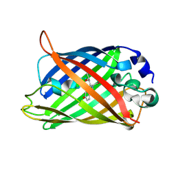
2C9J
DownloadVisualize
BU of 2c9j by Molmil
Structure of the fluorescent protein cmFP512 at 1.35A from Cerianthus membranaceus
Descriptor: GREEN FLUORESCENT PROTEIN FP512
Authors:Renzi, F, Nienhaus, K, Wiedenmann, J, Vallone, B, Nienhaus, G.U.
Deposit date:2005-12-12
Release date:2006-10-30
Last modified:2023-11-15
Method:X-RAY DIFFRACTION (1.35 Å)
Cite:Exploring Chromophore-Protein Interactions in Fluorescent Protein Cmfp512 from Cerianthus Membranaceus: X-Ray Structure Analysis and Optical Spectroscopy.
Biochemistry, 45, 2006
2DD7
DownloadVisualize
BU of 2dd7 by Molmil
A GFP-like protein from marine copepod, Chiridius poppei
Descriptor: 3-CYCLOHEXYL-1-PROPYLSULFONIC ACID, CHLORIDE ION, green fluorescent protein
Authors:Suto, K, Masuda, H, Takenaka, Y, Mizuno, H.
Deposit date:2006-01-23
Release date:2007-01-23
Last modified:2011-07-13
Method:X-RAY DIFFRACTION (1.9 Å)
Cite:Structural basis for red-shifted emission of a GFP-like protein from the marine copepod Chiridius poppei
Genes Cells, 14, 2009
2DD9
DownloadVisualize
BU of 2dd9 by Molmil
A mutant of GFP-like protein from Chiridius poppei
Descriptor: 3-CYCLOHEXYL-1-PROPYLSULFONIC ACID, CHLORIDE ION, green fluorescent protein
Authors:Suto, K, Masuda, H, Takenaka, Y, Mizuno, H.
Deposit date:2006-01-24
Release date:2007-01-23
Last modified:2021-11-10
Method:X-RAY DIFFRACTION (2.3 Å)
Cite:Structural basis for red-shifted emission of a GFP-like protein from the marine copepod Chiridius poppei
Genes Cells, 14, 2009
2DUE
DownloadVisualize
BU of 2due by Molmil
crystal structure of a green fluorescent protein variant S65T/H148D at pH 10
Descriptor: Green fluorescent protein
Authors:Shu, X, Remington, S.J.
Deposit date:2006-07-23
Release date:2007-07-10
Last modified:2021-11-10
Method:X-RAY DIFFRACTION (1.24 Å)
Cite:Ultrafast excited-state dynamics in the green fluorescent protein variant S65T/H148D. 1. Mutagenesis and structural studies.
Biochemistry, 46, 2007
2DUF
DownloadVisualize
BU of 2duf by Molmil
crystal structure of a green fluorescent protein variant S65T/H148D at pH 5.6
Descriptor: Green fluorescent protein
Authors:Shu, X, Remington, S.J.
Deposit date:2006-07-23
Release date:2007-07-10
Last modified:2021-11-10
Method:X-RAY DIFFRACTION (1.5 Å)
Cite:Ultrafast excited-state dynamics in the green fluorescent protein variant S65T/H148D. 1. Mutagenesis and structural studies.
Biochemistry, 46, 2007
2DUG
DownloadVisualize
BU of 2dug by Molmil
crystal structure of a green fluorescent protein S65T/H148N at pH 5
Descriptor: Green fluorescent protein
Authors:Shu, X, Remington, S.J.
Deposit date:2006-07-23
Release date:2007-07-10
Last modified:2021-11-10
Method:X-RAY DIFFRACTION (1.4 Å)
Cite:Ultrafast excited-state dynamics in the green fluorescent protein variant S65T/H148D. 1. Mutagenesis and structural studies.
Biochemistry, 46, 2007
2DUH
DownloadVisualize
BU of 2duh by Molmil
crystal structure of a green fluorescent protein variant S65T/H148N at pH 9.5
Descriptor: Green fluorescent protein
Authors:Shu, X, Remington, S.J.
Deposit date:2006-07-23
Release date:2007-07-10
Last modified:2021-11-10
Method:X-RAY DIFFRACTION (1.2 Å)
Cite:Ultrafast excited-state dynamics in the green fluorescent protein variant S65T/H148D. 1. Mutagenesis and structural studies.
Biochemistry, 46, 2007
2DUI
DownloadVisualize
BU of 2dui by Molmil
crystal structure of a green fluorescent protein variant H148D at pH 9
Descriptor: Green fluorescent protein
Authors:Shu, X, Remington, S.J.
Deposit date:2006-07-23
Release date:2007-07-10
Last modified:2023-11-15
Method:X-RAY DIFFRACTION (1.36 Å)
Cite:Ultrafast excited-state dynamics in the green fluorescent protein variant S65T/H148D. 1. Mutagenesis and structural studies.
Biochemistry, 46, 2007
2EMD
DownloadVisualize
BU of 2emd by Molmil
GREEN FLUORESCENT PROTEIN FROM AEQUOREA VICTORIA, MUTANT
Descriptor: GREEN FLUORESCENT PROTEIN
Authors:Palm, G, Zdanov, A, Wlodawer, A.
Deposit date:1997-03-31
Release date:1997-08-20
Last modified:2023-08-09
Method:X-RAY DIFFRACTION (2 Å)
Cite:The structural basis for spectral variations in green fluorescent protein.
Nat.Struct.Biol., 4, 1997
2EMN
DownloadVisualize
BU of 2emn by Molmil
GREEN FLUORESCENT PROTEIN FROM AEQUOREA VICTORIA, MUTANT
Descriptor: GREEN FLUORESCENT PROTEIN
Authors:Palm, G, Zdanov, A, Wlodawer, A.
Deposit date:1997-03-31
Release date:1997-08-20
Last modified:2023-08-09
Method:X-RAY DIFFRACTION (2.3 Å)
Cite:The structural basis for spectral variations in green fluorescent protein.
Nat.Struct.Biol., 4, 1997
2EMO
DownloadVisualize
BU of 2emo by Molmil
GREEN FLUORESCENT PROTEIN FROM AEQUOREA VICTORIA, MUTANT
Descriptor: GREEN FLUORESCENT PROTEIN
Authors:Palm, G, Zdanov, A, Wlodawer, A.
Deposit date:1997-03-31
Release date:1997-08-20
Last modified:2023-08-09
Method:X-RAY DIFFRACTION (2.6 Å)
Cite:The structural basis for spectral variations in green fluorescent protein.
Nat.Struct.Biol., 4, 1997
2FL1
DownloadVisualize
BU of 2fl1 by Molmil
Crystal structure of red fluorescent protein from Zoanthus, zRFP574, at 2.4A resolution
Descriptor: Red fluorescent protein zoanRFP, SULFATE ION
Authors:Pletnev, V, Pletneva, N, Martynov, V, Tikhonova, T, Popov, B, Pletnev, S.
Deposit date:2006-01-05
Release date:2007-01-09
Last modified:2023-11-15
Method:X-RAY DIFFRACTION (2.4 Å)
Cite:Structure of a red fluorescent protein from Zoanthus, zRFP574, reveals a novel chromophore
Acta Crystallogr.,Sect.D, 62, 2006
2FWQ
DownloadVisualize
BU of 2fwq by Molmil
Reduced enolate chromophore intermediate for Y66H GFP variant
Descriptor: Green fluorescent protein, MAGNESIUM ION
Authors:Barondeau, D.P, Tainer, J.A, Getzoff, E.D.
Deposit date:2006-02-02
Release date:2006-03-14
Last modified:2023-11-15
Method:X-RAY DIFFRACTION (1.4 Å)
Cite:Structural evidence for an enolate intermediate in GFP fluorophore biosynthesis.
J.Am.Chem.Soc., 128, 2006
2FZU
DownloadVisualize
BU of 2fzu by Molmil
Reduced enolate chromophore intermediate for GFP variant
Descriptor: 1,2-ETHANEDIOL, Green fluorescent protein, MAGNESIUM ION
Authors:Barondeau, D.P, Tainer, J.A, Getzoff, E.D.
Deposit date:2006-02-10
Release date:2006-03-14
Last modified:2023-11-15
Method:X-RAY DIFFRACTION (1.25 Å)
Cite:Structural evidence for an enolate intermediate in GFP fluorophore biosynthesis.
J.Am.Chem.Soc., 128, 2006
2G16
DownloadVisualize
BU of 2g16 by Molmil
Structure of S65A Y66S GFP variant after backbone fragmentation
Descriptor: Green fluorescent protein
Authors:Barondeau, D.P.
Deposit date:2006-02-13
Release date:2006-04-18
Last modified:2023-11-15
Method:X-RAY DIFFRACTION (2 Å)
Cite:Understanding GFP Posttranslational Chemistry: Structures of Designed Variants that Achieve Backbone Fragmentation, Hydrolysis, and Decarboxylation.
J.Am.Chem.Soc., 128, 2006
2G2S
DownloadVisualize
BU of 2g2s by Molmil
Structure of S65G Y66S GFP variant after spontaneous peptide hydrolysis
Descriptor: Green fluorescent protein, MAGNESIUM ION
Authors:Barondeau, D.P.
Deposit date:2006-02-16
Release date:2006-04-18
Last modified:2023-08-30
Method:X-RAY DIFFRACTION (1.2 Å)
Cite:Understanding GFP Posttranslational Chemistry: Structures of Designed Variants that Achieve Backbone Fragmentation, Hydrolysis, and Decarboxylation.
J.Am.Chem.Soc., 128, 2006
2G3D
DownloadVisualize
BU of 2g3d by Molmil
Structure of S65G Y66A GFP variant after spontaneous peptide hydrolysis
Descriptor: Green fluorescent protein, MAGNESIUM ION
Authors:Barondeau, D.P.
Deposit date:2006-02-17
Release date:2006-04-18
Last modified:2023-08-30
Method:X-RAY DIFFRACTION (1.35 Å)
Cite:Understanding GFP Posttranslational Chemistry: Structures of Designed Variants that Achieve Backbone Fragmentation, Hydrolysis, and Decarboxylation.
J.Am.Chem.Soc., 128, 2006
2G3O
DownloadVisualize
BU of 2g3o by Molmil
The 2.1A crystal structure of copGFP
Descriptor: green fluorescent protein 2
Authors:Wilmann, P.G.
Deposit date:2006-02-20
Release date:2006-08-15
Last modified:2017-10-18
Method:X-RAY DIFFRACTION (2.1 Å)
Cite:The 2.1A crystal structure of copGFP, a representative member of the copepod clade within the green fluorescent protein superfamily
J.Mol.Biol., 359, 2006
2G5Z
DownloadVisualize
BU of 2g5z by Molmil
Structure of S65G Y66S GFP variant after spontaneous peptide hydrolysis and decarboxylation
Descriptor: Green fluorescent protein, MAGNESIUM ION
Authors:Barondeau, D.P, Kassmann, C.J, Tainer, J.A, Getzoff, E.D.
Deposit date:2006-02-23
Release date:2006-04-18
Last modified:2023-08-30
Method:X-RAY DIFFRACTION (1.8 Å)
Cite:Understanding GFP Posttranslational Chemistry: Structures of Designed Variants that Achieve Backbone Fragmentation, Hydrolysis, and Decarboxylation.
J.Am.Chem.Soc., 128, 2006
2G6E
DownloadVisualize
BU of 2g6e by Molmil
Structure of cyclized F64L S65A Y66S GFP variant
Descriptor: Green fluorescent protein, MAGNESIUM ION
Authors:Barondeau, D.P.
Deposit date:2006-02-24
Release date:2006-04-18
Last modified:2023-11-15
Method:X-RAY DIFFRACTION (1.3 Å)
Cite:Understanding GFP Posttranslational Chemistry: Structures of Designed Variants that Achieve Backbone Fragmentation, Hydrolysis, and Decarboxylation.
J.Am.Chem.Soc., 128, 2006
2G6X
DownloadVisualize
BU of 2g6x by Molmil
Crystal structure of a novel green fluorescent protein from marine copepod Pontellina plumata
Descriptor: green fluorescent protein 2
Authors:Evdokimov, A.G, Pokross, M.E, Chudakov, D.M.
Deposit date:2006-02-26
Release date:2006-03-28
Last modified:2021-10-20
Method:X-RAY DIFFRACTION (2 Å)
Cite:Structural basis for the fast maturation of Arthropoda green fluorescent protein.
Embo Rep., 7, 2006
2G6Y
DownloadVisualize
BU of 2g6y by Molmil
Crystal structure of the novel green fluorescent protein from marine copepod Pontellina plumata
Descriptor: green fluorescent protein 2
Authors:Evdokimov, A.G, Pokross, M.E, Chudakov, D.M.
Deposit date:2006-02-26
Release date:2006-03-28
Last modified:2021-10-20
Method:X-RAY DIFFRACTION (1.6 Å)
Cite:Structural basis for the fast maturation of Arthropoda green fluorescent protein.
Embo Rep., 7, 2006
2GX0
DownloadVisualize
BU of 2gx0 by Molmil
Crystal structural and functional analysis of GFP-like fluorescent protein
Descriptor: fluorescent protein Dronpa
Authors:Hwang, K.Y, Nam, K.-H, Park, S.-Y, Sugiyama, K.
Deposit date:2006-05-08
Release date:2007-05-08
Last modified:2023-11-15
Method:X-RAY DIFFRACTION (1.9 Å)
Cite:Structural characterization of the photoswitchable fluorescent protein Dronpa-C62S
Biochem.Biophys.Res.Commun., 354, 2007
2GX2
DownloadVisualize
BU of 2gx2 by Molmil
Crystal structural and functional analysis of GFP-like fluorescent protein Dronpa
Descriptor: MAGNESIUM ION, fluorescent protein Dronpa
Authors:Hwang, K.Y, Nam, K.-H, Park, S.-Y, Sugiyama, K.
Deposit date:2006-05-08
Release date:2007-05-08
Last modified:2023-11-15
Method:X-RAY DIFFRACTION (1.8 Å)
Cite:Structural characterization of the photoswitchable fluorescent protein Dronpa-C62S
Biochem.Biophys.Res.Commun., 354, 2007

218500

PDB entries from 2024-04-17

PDB statisticsPDBj update infoContact PDBjnumon