Loading
PDBj
MenuPDBj@FacebookPDBj@TwitterPDBj@YouTubewwPDB FoundationwwPDB
RCSB PDBPDBeBMRBAdv. SearchSearch help

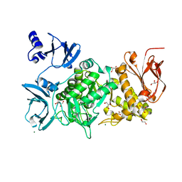
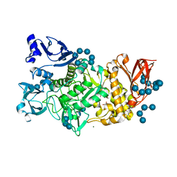
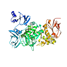
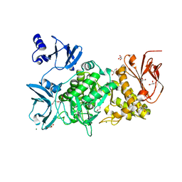
6JFJ
DownloadVisualize
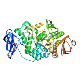
BU of 6jfj by Molmil
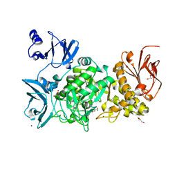
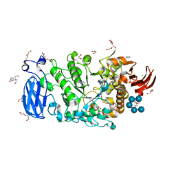
Crystal structure of Pullulanase from Paenibacillus barengoltzii complex with maltohexaose and alpha-cyclodextrin
Descriptor: CALCIUM ION, CHLORIDE ION, DI(HYDROXYETHYL)ETHER, ...
Authors:Wu, S.W, Yang, S.Q, Qin, Z, You, X, Huang, P, Jiang, Z.Q.
Deposit date:2019-02-09
Release date:2019-02-20
Last modified:2023-11-22
Method:X-RAY DIFFRACTION (1.932 Å)
Cite:Structural basis of carbohydrate binding in domain C of a type I pullulanase from Paenibacillus barengoltzii.
Acta Crystallogr D Struct Biol, 76, 2020
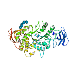
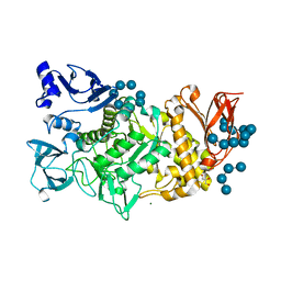
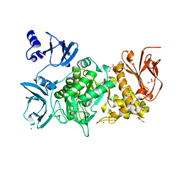
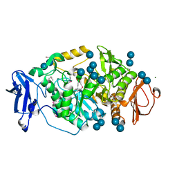
6KLF
DownloadVisualize
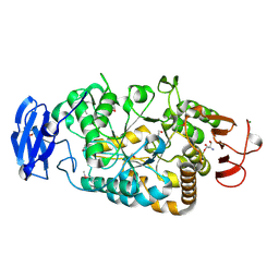
BU of 6klf by Molmil
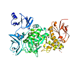
Crystal structure of branching enzyme D434A mutant from Cyanothece sp. ATCC 51142
Descriptor: 1,4-alpha-glucan branching enzyme GlgB, GLYCEROL, MAGNESIUM ION
Authors:Suzuki, R, Suzuki, E.
Deposit date:2019-07-30
Release date:2020-08-05
Last modified:2023-11-22
Method:X-RAY DIFFRACTION (2.5 Å)
Cite:Cyanobacterial branching enzymes bind to alpha-glucan via surface binding sites
Arch.Biochem.Biophys., 702, 2021
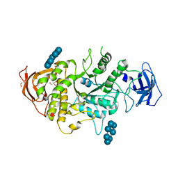
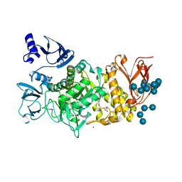
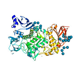
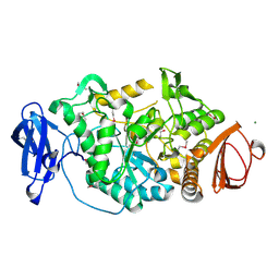
5E70
DownloadVisualize
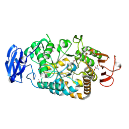
BU of 5e70 by Molmil
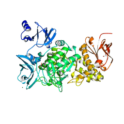
Crystal structure of Ecoli Branching Enzyme with gamma cyclodextrin
Descriptor: 1,4-alpha-glucan branching enzyme GlgB, Cyclooctakis-(1-4)-(alpha-D-glucopyranose), GLYCEROL
Authors:Feng, L, Nosrati, M, Geiger, J.H.
Deposit date:2015-10-11
Release date:2015-12-16
Last modified:2023-09-27
Method:X-RAY DIFFRACTION (2.33 Å)
Cite:Crystal structures of Escherichia coli branching enzyme in complex with cyclodextrins.
Acta Crystallogr D Struct Biol, 72, 2016
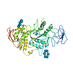
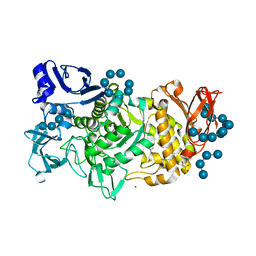
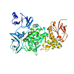
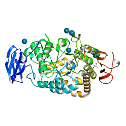
5E6Z
DownloadVisualize
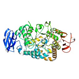
BU of 5e6z by Molmil
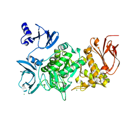
Crystal structure of Ecoli Branching Enzyme with beta cyclodextrin
Descriptor: 1,4-alpha-glucan branching enzyme GlgB, Cycloheptakis-(1-4)-(alpha-D-glucopyranose), GLYCEROL
Authors:Feng, L, Nosrati, M, Geiger, J.H.
Deposit date:2015-10-11
Release date:2015-12-16
Last modified:2023-09-27
Method:X-RAY DIFFRACTION (1.878 Å)
Cite:Crystal structures of Escherichia coli branching enzyme in complex with cyclodextrins.
Acta Crystallogr D Struct Biol, 72, 2016
5E6Y
DownloadVisualize
BU of 5e6y by Molmil
Crystal structure of E.Coli branching enzyme in complex with alpha cyclodextrin
Descriptor: 1,4-alpha-glucan branching enzyme GlgB, Cyclohexakis-(1-4)-(alpha-D-glucopyranose), GLYCEROL
Authors:Feng, L, Nosrati, M, Geiger, J.H.
Deposit date:2015-10-11
Release date:2015-12-16
Last modified:2023-09-27
Method:X-RAY DIFFRACTION (2.6 Å)
Cite:Crystal structures of Escherichia coli branching enzyme in complex with cyclodextrins.
Acta Crystallogr D Struct Biol, 72, 2016
5GR0
DownloadVisualize
BU of 5gr0 by Molmil
Crystal structure of branching enzyme D501A mutant from Cyanothece sp. ATCC 51142
Descriptor: 1,4-alpha-glucan branching enzyme GlgB, GLYCEROL, MAGNESIUM ION
Authors:Suzuki, R, Suzuki, E.
Deposit date:2016-08-08
Release date:2017-08-16
Last modified:2023-11-08
Method:X-RAY DIFFRACTION (1.95 Å)
Cite:Structural basis for substrate binding and catalysis of branching enzyme from Cyanothece sp. ATCC 51142
To be published
5GQU
DownloadVisualize
BU of 5gqu by Molmil
Crystal structure of branching enzyme from Cyanothece sp. ATCC 51142
Descriptor: 1,4-alpha-glucan branching enzyme GlgB, GLYCEROL, MAGNESIUM ION
Authors:Suzuki, R, Suzuki, E.
Deposit date:2016-08-08
Release date:2017-02-22
Last modified:2023-11-08
Method:X-RAY DIFFRACTION (1.85 Å)
Cite:Bound Substrate in the Structure of Cyanobacterial Branching Enzyme Supports a New Mechanistic Model
J. Biol. Chem., 292, 2017
5GR6
DownloadVisualize
BU of 5gr6 by Molmil
Crystal structure of branching enzyme Y500A/D501A double mutant from Cyanothece sp. ATCC 51142
Descriptor: 1,4-alpha-glucan branching enzyme GlgB, GLYCEROL, MAGNESIUM ION
Authors:Suzuki, R, Suzuki, E.
Deposit date:2016-08-08
Release date:2017-08-16
Last modified:2023-11-08
Method:X-RAY DIFFRACTION (2 Å)
Cite:Structural basis for substrate binding and catalysis of branching enzyme from Cyanothece sp. ATCC 51142
To be published
5GR3
DownloadVisualize
BU of 5gr3 by Molmil
Crystal structure of branching enzyme L541A/W655A mutant from Cyanothece sp. ATCC 51142
Descriptor: 1,4-alpha-glucan branching enzyme GlgB, GLYCEROL, MAGNESIUM ION
Authors:Suzuki, R, Suzuki, E.
Deposit date:2016-08-08
Release date:2017-08-16
Last modified:2023-11-08
Method:X-RAY DIFFRACTION (2.6 Å)
Cite:Structural basis for substrate binding and catalysis of branching enzyme from Cyanothece sp. ATCC 51142
To be published
5GQV
DownloadVisualize
BU of 5gqv by Molmil
Crystal structure of branching enzyme from Cyanothece sp. ATCC 51142 in complex with maltohexaose
Descriptor: 1,4-alpha-glucan branching enzyme GlgB, GLYCEROL, MAGNESIUM ION, ...
Authors:Suzuki, R, Suzuki, E.
Deposit date:2016-08-08
Release date:2017-02-22
Last modified:2023-11-08
Method:X-RAY DIFFRACTION (3 Å)
Cite:Bound Substrate in the Structure of Cyanobacterial Branching Enzyme Supports a New Mechanistic Model
J. Biol. Chem., 292, 2017
5GR4
DownloadVisualize
BU of 5gr4 by Molmil
Crystal structure of branching enzyme L541A mutant from Cyanothece sp. ATCC 51142 in complex with maltoheptaose
Descriptor: 1,4-alpha-glucan branching enzyme GlgB, GLYCEROL, MAGNESIUM ION, ...
Authors:Suzuki, R, Suzuki, E.
Deposit date:2016-08-08
Release date:2017-08-16
Last modified:2024-03-20
Method:X-RAY DIFFRACTION (2 Å)
Cite:Structural basis for substrate binding and catalysis of branching enzyme from Cyanothece sp. ATCC 51142
To be published
5GR1
DownloadVisualize
BU of 5gr1 by Molmil
Crystal structure of branching enzyme Y500A/D501A mutant from Cyanothece sp. ATCC 51142 in complex with maltoheptaose
Descriptor: 1,4-alpha-glucan branching enzyme GlgB, GLYCEROL, MAGNESIUM ION, ...
Authors:Suzuki, R, Suzuki, E.
Deposit date:2016-08-08
Release date:2017-08-16
Last modified:2024-03-20
Method:X-RAY DIFFRACTION (2.5 Å)
Cite:Structural basis for substrate binding and catalysis of branching enzyme from Cyanothece sp. ATCC 51142
To be published
5GQY
DownloadVisualize
BU of 5gqy by Molmil
Crystal structure of branching enzyme from Cyanothece sp. ATCC 51142 in complex with maltoheptaose
Descriptor: 1,4-alpha-glucan branching enzyme GlgB, GLYCEROL, MAGNESIUM ION, ...
Authors:Suzuki, R, Suzuki, E.
Deposit date:2016-08-08
Release date:2017-08-16
Last modified:2023-11-08
Method:X-RAY DIFFRACTION (2 Å)
Cite:Structural basis for substrate binding and catalysis of branching enzyme from Cyanothece sp. ATCC 51142
To be published
5GR2
DownloadVisualize
BU of 5gr2 by Molmil
Crystal structure of branching enzyme L541A mutant from Cyanothece sp. ATCC 51142
Descriptor: 1,4-alpha-glucan branching enzyme GlgB, GLYCEROL, MAGNESIUM ION
Authors:Suzuki, R, Suzuki, E.
Deposit date:2016-08-08
Release date:2017-08-16
Last modified:2023-11-08
Method:X-RAY DIFFRACTION (1.95 Å)
Cite:Structural basis for substrate binding and catalysis of branching enzyme from Cyanothece sp. ATCC 51142
To be published
5GR5
DownloadVisualize
BU of 5gr5 by Molmil
Crystal structure of branching enzyme W610A mutant from Cyanothece sp. ATCC 51142
Descriptor: 1,4-alpha-glucan branching enzyme GlgB, GLYCEROL, MAGNESIUM ION
Authors:Suzuki, R, Suzuki, E.
Deposit date:2016-08-08
Release date:2017-08-16
Last modified:2023-11-08
Method:X-RAY DIFFRACTION (2.4 Å)
Cite:Structural basis for substrate binding and catalysis of branching enzyme from Cyanothece sp. ATCC 51142
To be published
5GQX
DownloadVisualize
BU of 5gqx by Molmil
Crystal structure of branching enzyme W610N mutant from Cyanothece sp. ATCC 51142 in complex with maltoheptaose
Descriptor: 1,4-alpha-glucan branching enzyme GlgB, GLYCEROL, MAGNESIUM ION, ...
Authors:Suzuki, R, Suzuki, E.
Deposit date:2016-08-08
Release date:2017-02-22
Last modified:2023-11-08
Method:X-RAY DIFFRACTION (2.3 Å)
Cite:Bound Substrate in the Structure of Cyanobacterial Branching Enzyme Supports a New Mechanistic Model
J. Biol. Chem., 292, 2017
5GQW
DownloadVisualize
BU of 5gqw by Molmil
Crystal structure of branching enzyme W610N mutant from Cyanothece sp. ATCC 51142
Descriptor: 1,4-alpha-glucan branching enzyme GlgB, GLYCEROL, MAGNESIUM ION
Authors:Suzuki, R, Suzuki, E.
Deposit date:2016-08-08
Release date:2017-02-22
Last modified:2023-11-08
Method:X-RAY DIFFRACTION (1.8 Å)
Cite:Bound Substrate in the Structure of Cyanobacterial Branching Enzyme Supports a New Mechanistic Model
J. Biol. Chem., 292, 2017
5GQZ
DownloadVisualize
BU of 5gqz by Molmil
Crystal structure of branching enzyme Y500A mutant from Cyanothece sp. ATCC 51142
Descriptor: 1,4-alpha-glucan branching enzyme GlgB, GLYCEROL, MAGNESIUM ION
Authors:Suzuki, R, Suzuki, E.
Deposit date:2016-08-08
Release date:2017-08-16
Last modified:2023-11-08
Method:X-RAY DIFFRACTION (1.85 Å)
Cite:Structural basis for substrate binding and catalysis of branching enzyme from Cyanothece sp. ATCC 51142
To be published
2BHY
DownloadVisualize
BU of 2bhy by Molmil
Crystal structure of Deinococcus radiodurans maltooligosyltrehalose trehalohydrolase in complex with trehalose
Descriptor: 2-AMINO-2-HYDROXYMETHYL-PROPANE-1,3-DIOL, BETA-MERCAPTOETHANOL, MAGNESIUM ION, ...
Authors:Timmins, J, Leiros, H.-K.S, Leonard, G, Leiros, I, McSweeney, S.
Deposit date:2005-01-20
Release date:2005-03-31
Last modified:2020-07-29
Method:X-RAY DIFFRACTION (1.5 Å)
Cite:Crystal Structure of Maltooligosyltrehalose Trehalohydrolase from Deinococcus Radiodurans in Complex with Disaccharides
J.Mol.Biol., 347, 2005
2BY3
DownloadVisualize
BU of 2by3 by Molmil
Is radiation damage dependent on the dose-rate used during macromolecular crystallography data collection
Descriptor: 2-AMINO-2-HYDROXYMETHYL-PROPANE-1,3-DIOL, BETA-MERCAPTOETHANOL, MAGNESIUM ION, ...
Authors:Leiros, H.-K.S, Timmins, J, Ravelli, R.B.G, McSweeney, S.M.
Deposit date:2005-07-28
Release date:2006-02-06
Last modified:2020-07-29
Method:X-RAY DIFFRACTION (1.5 Å)
Cite:Is Radiation Damage Dependent on the Dose-Rate Used During Macromolecular Crystallography Data Collection?
Acta Crystallogr.,Sect.D, 62, 2006
2BXY
DownloadVisualize
BU of 2bxy by Molmil
Is radiation damage dependent on the dose-rate used during macromolecular crystallography data collection
Descriptor: 2-AMINO-2-HYDROXYMETHYL-PROPANE-1,3-DIOL, BETA-MERCAPTOETHANOL, MAGNESIUM ION, ...
Authors:Leiros, H.-K.S, Timmins, J, Ravelli, R.B.G, McSweeney, S.M.
Deposit date:2005-07-28
Release date:2006-02-06
Last modified:2020-07-29
Method:X-RAY DIFFRACTION (1.75 Å)
Cite:Is Radiation Damage Dependent on the Dose-Rate Used During Macromolecular Crystallography Data Collection?
Acta Crystallogr.,Sect.D, 62, 2006
2BY2
DownloadVisualize
BU of 2by2 by Molmil
Is radiation damage dependent on the dose-rate used during macromolecular crystallography data collection
Descriptor: 2-AMINO-2-HYDROXYMETHYL-PROPANE-1,3-DIOL, BETA-MERCAPTOETHANOL, MAGNESIUM ION, ...
Authors:Leiros, H.-K.S, Timmins, J, Ravelli, R.B.G, McSweeney, S.M.
Deposit date:2005-07-28
Release date:2006-02-06
Last modified:2020-07-29
Method:X-RAY DIFFRACTION (1.5 Å)
Cite:Is Radiation Damage Dependent on the Dose-Rate Used During Macromolecular Crystallography Data Collection?
Acta Crystallogr.,Sect.D, 62, 2006
2BY1
DownloadVisualize
BU of 2by1 by Molmil
Is radiation damage dependent on the dose-rate used during macromolecular crystallography data collection
Descriptor: 2-AMINO-2-HYDROXYMETHYL-PROPANE-1,3-DIOL, BETA-MERCAPTOETHANOL, MAGNESIUM ION, ...
Authors:Leiros, H.-K.S, Timmins, J, Ravelli, R.B.G, McSweeney, S.M.
Deposit date:2005-07-28
Release date:2006-02-06
Last modified:2020-07-29
Method:X-RAY DIFFRACTION (1.55 Å)
Cite:Is Radiation Damage Dependent on the Dose-Rate Used During Macromolecular Crystallography Data Collection?
Acta Crystallogr.,Sect.D, 62, 2006
2BY0
DownloadVisualize
BU of 2by0 by Molmil
Is radiation damage dependent on the dose-rate used during macromolecular crystallography data collection
Descriptor: 2-AMINO-2-HYDROXYMETHYL-PROPANE-1,3-DIOL, BETA-MERCAPTOETHANOL, MAGNESIUM ION, ...
Authors:Leiros, H.-K.S, Timmins, J, Ravelli, R.B.G, McSweeney, S.M.
Deposit date:2005-07-28
Release date:2006-02-06
Last modified:2020-07-29
Method:X-RAY DIFFRACTION (1.55 Å)
Cite:Is Radiation Damage Dependent on the Dose-Rate Used During Macromolecular Crystallography Data Collection?
Acta Crystallogr.,Sect.D, 62, 2006
2BXZ
DownloadVisualize
BU of 2bxz by Molmil
Is radiation damage dependent on the dose-rate used during macromolecular crystallography data collection
Descriptor: 2-AMINO-2-HYDROXYMETHYL-PROPANE-1,3-DIOL, BETA-MERCAPTOETHANOL, MAGNESIUM ION, ...
Authors:Leiros, H.-K.S, Timmins, J, Ravelli, R.B.G, McSweeney, S.M.
Deposit date:2005-07-28
Release date:2006-02-06
Last modified:2020-07-29
Method:X-RAY DIFFRACTION (1.75 Å)
Cite:Is Radiation Damage Dependent on the Dose-Rate Used During Macromolecular Crystallography Data Collection?
Acta Crystallogr.,Sect.D, 62, 2006

218500

PDB entries from 2024-04-17

PDB statisticsPDBj update infoContact PDBjnumon