Loading
PDBj
MenuPDBj@FacebookPDBj@TwitterPDBj@YouTubewwPDB FoundationwwPDB
RCSB PDBPDBeBMRBAdv. SearchSearch help

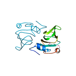
2O8Q
DownloadVisualize
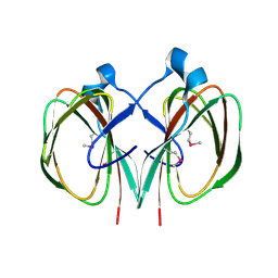
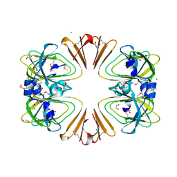
BU of 2o8q by Molmil
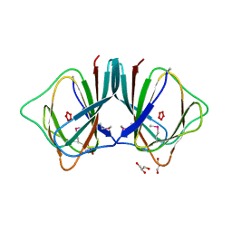
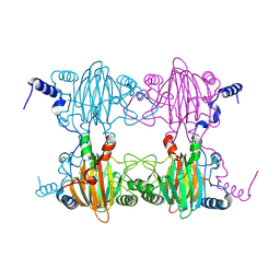
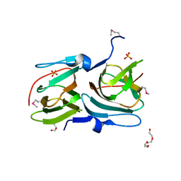
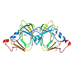
CRYSTAL STRUCTURE OF A PROTEIN WITH A CUPIN-LIKE FOLD AND UNKNOWN FUNCTION (BXE_C0505) FROM BURKHOLDERIA XENOVORANS LB400 AT 1.55 A RESOLUTION
Descriptor: 1,2-ETHANEDIOL, Hypothetical protein, NICKEL (II) ION
Authors:Joint Center for Structural Genomics (JCSG)
Deposit date:2006-12-12
Release date:2006-12-26
Last modified:2023-01-25
Method:X-RAY DIFFRACTION (1.55 Å)
Cite:Crystal structure of hypothetical protein (YP_555756.1) from Burkholderia xenovorans LB400 at 1.55 A resolution
To be published
2OA2
DownloadVisualize
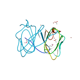
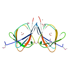
BU of 2oa2 by Molmil
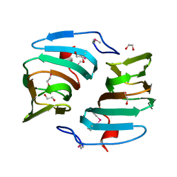
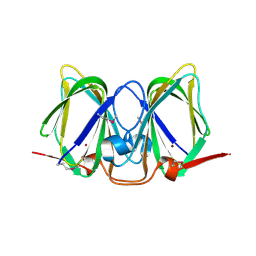
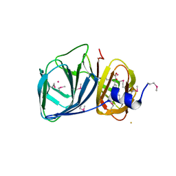
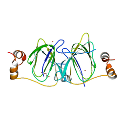
Crystal structure of BH2720 (10175341) from Bacillus halodurans at 1.41 A resolution
Descriptor: BH2720 protein
Authors:Joint Center for Structural Genomics (JCSG)
Deposit date:2006-12-14
Release date:2007-01-16
Last modified:2023-01-25
Method:X-RAY DIFFRACTION (1.41 Å)
Cite:Crystal structure of BH2720 (10175341) from Bacillus halodurans at 1.41 A resolution
To be published
2OPK
DownloadVisualize
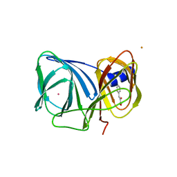
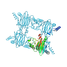
BU of 2opk by Molmil
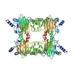
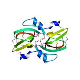
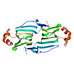
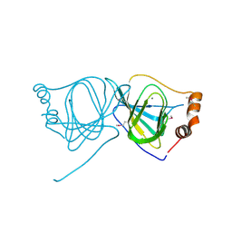
CRYSTAL STRUCTURE OF A PUTATIVE MANNOSE-6-PHOSPHATE ISOMERASE (REUT_A1446) FROM RALSTONIA EUTROPHA JMP134 AT 2.10 A RESOLUTION
Descriptor: CHLORIDE ION, GLYCEROL, Hypothetical protein, ...
Authors:Joint Center for Structural Genomics (JCSG)
Deposit date:2007-01-29
Release date:2007-02-13
Last modified:2023-01-25
Method:X-RAY DIFFRACTION (2.1 Å)
Cite:Crystal structure of hypothetical protein (YP_295660.1) from Ralstonia eutropha JMP134 at 2.10 A resolution
To be published
2PFW
DownloadVisualize
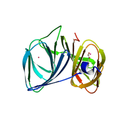
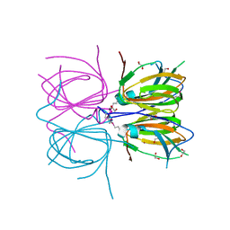
BU of 2pfw by Molmil
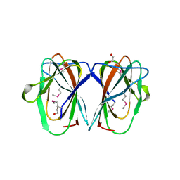
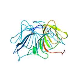
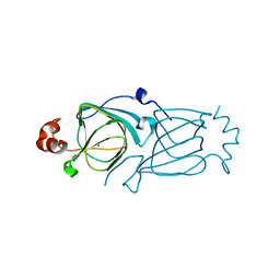
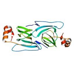
CRYSTAL STRUCTURE OF A RMLC-LIKE CUPIN (SFRI_3105) FROM SHEWANELLA FRIGIDIMARINA NCIMB 400 AT 1.90 A RESOLUTION
Descriptor: 1,2-ETHANEDIOL, Cupin 2, conserved barrel domain protein
Authors:Joint Center for Structural Genomics (JCSG)
Deposit date:2007-04-05
Release date:2007-04-17
Last modified:2023-01-25
Method:X-RAY DIFFRACTION (1.9 Å)
Cite:Crystal structure of Cupin 2 conserved barrel domain protein (YP_751781.1) from Shewanella frigidimarina NCIMB 400 at 1.90 A resolution
To be published
2PHD
DownloadVisualize
BU of 2phd by Molmil
Crystal Structure Determination of a Salicylate 1,2-Dioxygenase from Pseudaminobacter salicylatoxidans
Descriptor: ACETATE ION, CHLORIDE ION, FE (III) ION, ...
Authors:Matera, I, Ferraroni, M, Briganti, F.
Deposit date:2007-04-11
Release date:2008-04-22
Last modified:2023-08-30
Method:X-RAY DIFFRACTION (2.9 Å)
Cite:Crystal Structure Determination of a Salicylate 1,2-Dioxygenase from Pseudaminobacter salicylatoxidans
To be Published
2Q30
DownloadVisualize
BU of 2q30 by Molmil
Crystal structure of a rmlc-like cupin protein (dde_2303) from desulfovibrio desulfuricans subsp. at 1.94 A resolution
Descriptor: 1,2-ETHANEDIOL, SULFATE ION, Uncharacterized protein
Authors:Joint Center for Structural Genomics (JCSG)
Deposit date:2007-05-29
Release date:2007-06-19
Last modified:2023-01-25
Method:X-RAY DIFFRACTION (1.94 Å)
Cite:Crystal structure of uncharacterized protein (YP_388795.1) from Desulfovibrio desulfuricans G20 at 1.94 A resolution
To be published
3BU7
DownloadVisualize
BU of 3bu7 by Molmil
Crystal Structure and Biochemical Characterization of GDOsp, a Gentisate 1,2-Dioxygenase from Silicibacter Pomeroyi
Descriptor: FE (II) ION, Gentisate 1,2-dioxygenase
Authors:Chen, J, Wang, M.Z, Zhu, G.Y, Zhang, X.C, Rao, Z.H.
Deposit date:2008-01-02
Release date:2008-08-12
Last modified:2023-08-30
Method:X-RAY DIFFRACTION (2.8 Å)
Cite:Crystal structure and mutagenic analysis of GDOsp, a gentisate 1,2-dioxygenase from Silicibacter pomeroyi.
Protein Sci., 17, 2008
3CEW
DownloadVisualize
BU of 3cew by Molmil
Crystal structure of a cupin protein (BF4112) from Bacteroides fragilis. Northeast Structural Genomics Consortium target BfR205
Descriptor: Uncharacterized cupin protein, ZINC ION
Authors:Forouhar, F, Chen, Y, Seetharaman, J, Mao, L, Xiao, R, Ciccosanti, C, Foote, E.L, Maglaqui, M, Wang, H, Baran, M.C, Acton, T.B, Montelione, G.T, Hunt, J.F, Tong, L, Northeast Structural Genomics Consortium (NESG)
Deposit date:2008-02-29
Release date:2008-03-25
Last modified:2011-07-13
Method:X-RAY DIFFRACTION (2.31 Å)
Cite:Crystal structure of a cupin protein (BF4112) from Bacteroides fragilis. Northeast Structural Genomics Consortium target BfR205.
To be Published
3D82
DownloadVisualize
BU of 3d82 by Molmil
Crystal structure of a cupin-2 domain containing protein (sfri_3543) from shewanella frigidimarina ncimb 400 at 2.05 A resolution
Descriptor: Cupin 2, conserved barrel domain protein, GLYCEROL, ...
Authors:Joint Center for Structural Genomics (JCSG)
Deposit date:2008-05-22
Release date:2008-06-10
Last modified:2023-02-01
Method:X-RAY DIFFRACTION (2.05 Å)
Cite:Crystal structure of Domain of Unknown Function with a Cupin Fold (YP_752209.1) from SHEWANELLA FRIGIDIMARINA NCIMB 400 at 2.05 A resolution
To be published
3ES1
DownloadVisualize
BU of 3es1 by Molmil
Crystal structure of protein with a cupin-like fold and unknown function (YP_001165807.1) from NOVOSPHINGOBIUM AROMATICIVORANS DSM 12444 at 1.91 A resolution
Descriptor: CHLORIDE ION, Cupin 2, conserved barrel domain protein
Authors:Joint Center for Structural Genomics (JCSG)
Deposit date:2008-10-03
Release date:2008-10-14
Last modified:2023-02-01
Method:X-RAY DIFFRACTION (1.91 Å)
Cite:Crystal structure of protein with a cupin-like fold and unknown function (YP_001165807.1) from NOVOSPHINGOBIUM AROMATICIVORANS DSM 12444 at 1.91 A resolution
To be published
3FJS
DownloadVisualize
BU of 3fjs by Molmil
CRYSTAL STRUCTURE OF A PUTATIVE BIOSYNTHETIC PROTEIN WITH RMLC-LIKE CUPIN FOLD (REUT_B4087) FROM RALSTONIA EUTROPHA JMP134 AT 1.90 A RESOLUTION
Descriptor: uncharacterized protein with RmlC-like cupin fold
Authors:Joint Center for Structural Genomics (JCSG)
Deposit date:2008-12-15
Release date:2009-01-13
Last modified:2023-02-01
Method:X-RAY DIFFRACTION (1.9 Å)
Cite:Crystal structure of protein of unknown function with RmlC-like cupin fold (YP_298287.1) from RALSTONIA EUTROPHA JMP134 at 1.90 A resolution
To be published
3H50
DownloadVisualize
BU of 3h50 by Molmil
CRYSTAL STRUCTURE OF A TETRACENOMYCIN POLYKETIDE SYNTHESIS PROTEIN (TCMJ) FROM XANTHOMONAS CAMPESTRIS PV. CAMPESTRIS AT 1.60 A RESOLUTION
Descriptor: ACETATE ION, Tetracenomycin polyketide synthesis protein, ZINC ION
Authors:Joint Center for Structural Genomics (JCSG)
Deposit date:2009-04-21
Release date:2009-05-05
Last modified:2023-02-01
Method:X-RAY DIFFRACTION (1.6 Å)
Cite:Conformational changes associated with the binding of zinc acetate at the putative active site of XcTcmJ, a cupin from Xanthomonas campestris pv. campestris.
Acta Crystallogr.,Sect.F, 66, 2010
3H7J
DownloadVisualize
BU of 3h7j by Molmil
Crystal structure of BacB, an enzyme involved in Bacilysin synthesis, in monoclinic form
Descriptor: 3-PHENYLPYRUVIC ACID, Bacilysin biosynthesis protein bacB, COBALT (II) ION, ...
Authors:Rajavel, M, Gopal, B.
Deposit date:2009-04-27
Release date:2009-09-22
Last modified:2023-11-01
Method:X-RAY DIFFRACTION (1.87 Å)
Cite:Role of Bacillus subtilis BacB in the synthesis of bacilysin
J.Biol.Chem., 284, 2009
3H7Y
DownloadVisualize
BU of 3h7y by Molmil
Crystal structure of BacB, an enzyme involved in Bacilysin synthesis, in tetragonal form
Descriptor: 3-PHENYLPYRUVIC ACID, Bacilysin biosynthesis protein bacB, COBALT (II) ION, ...
Authors:Rajavel, M, Gopal, B.
Deposit date:2009-04-28
Release date:2009-09-22
Last modified:2024-03-20
Method:X-RAY DIFFRACTION (2.22 Å)
Cite:Role of Bacillus subtilis BacB in the synthesis of bacilysin
J.Biol.Chem., 284, 2009
3H8U
DownloadVisualize
BU of 3h8u by Molmil
Crystal structure of uncharacterized conserved protein with double-stranded beta-helix domain (YP_001338853.1) from Klebsiella pneumoniae subsp. pneumoniae MGH 78578 at 1.80 A resolution
Descriptor: GLYCEROL, NONAETHYLENE GLYCOL, SULFATE ION, ...
Authors:Joint Center for Structural Genomics (JCSG)
Deposit date:2009-04-29
Release date:2009-05-12
Last modified:2023-02-01
Method:X-RAY DIFFRACTION (1.8 Å)
Cite:Crystal structure of uncharacterized conserved protein with double-stranded beta-helix domain (YP_001338853.1) from Klebsiella pneumoniae subsp. pneumoniae MGH 78578 at 1.80 A resolution
To be published
3H9A
DownloadVisualize
BU of 3h9a by Molmil
Crystal structure of BacB, an enzyme involved in Bacilysin synthesis, in triclinic form
Descriptor: 3-PHENYLPYRUVIC ACID, Bacilysin biosynthesis protein bacB, COBALT (II) ION, ...
Authors:Rajavel, M, Gopal, B.
Deposit date:2009-04-30
Release date:2010-03-02
Last modified:2023-11-22
Method:X-RAY DIFFRACTION (2.04 Å)
Cite:Analysis of multiple crystal forms of Bacillus subtilis BacB suggests a role for a metal ion as a nucleant for crystallization
Acta Crystallogr.,Sect.D, 66, 2010
3HT2
DownloadVisualize
BU of 3ht2 by Molmil
Zink containing polyketide cyclase RemF from Streptomyces resistomycificus
Descriptor: RemF protein, ZINC ION
Authors:Silvennoinen, L, Sandalova, T, Schneider, G.
Deposit date:2009-06-11
Release date:2009-10-13
Last modified:2024-04-03
Method:X-RAY DIFFRACTION (2 Å)
Cite:The polyketide cyclase RemF from Streptomyces resistomycificus contains an unusual octahedral zinc binding site
Febs Lett., 583, 2009
3HT1
DownloadVisualize
BU of 3ht1 by Molmil
1.2A structure of the polyketide cyclase RemF from Streptomyces resistomycificus
Descriptor: NICKEL (II) ION, RemF protein
Authors:Silvennoinen, L, Sandalova, T, Schneider, G.
Deposit date:2009-06-11
Release date:2009-10-13
Last modified:2024-03-20
Method:X-RAY DIFFRACTION (1.2 Å)
Cite:The polyketide cyclase RemF from Streptomyces resistomycificus contains an unusual octahedral zinc binding site
Febs Lett., 583, 2009
3I7D
DownloadVisualize
BU of 3i7d by Molmil
Crystal structure of sugar phosphate isomerase from a cupin superfamily SPO2919 from Silicibacter pomeroyi (YP_168127.1) from SILICIBACTER POMEROYI DSS-3 at 2.30 A resolution
Descriptor: ACETATE ION, CACODYLATE ION, CHLORIDE ION, ...
Authors:Joint Center for Structural Genomics (JCSG)
Deposit date:2009-07-08
Release date:2009-07-21
Last modified:2023-02-01
Method:X-RAY DIFFRACTION (2.3 Å)
Cite:Crystal structure of sugar phosphate isomerase from a cupin superfamily SPO2919 from Silicibacter pomeroyi (YP_168127.1) from SILICIBACTER POMEROYI DSS-3 at 2.30 A resolution
To be published
3IBM
DownloadVisualize
BU of 3ibm by Molmil
CRYSTAL STRUCTURE OF cupin 2 domain-containing protein Hhal_0468 FROM Halorhodospira halophila
Descriptor: Cupin 2, conserved barrel domain protein, GLYCEROL, ...
Authors:Patskovsky, Y, Toro, R, Freeman, J, Miller, S, Sauder, J.M, Burley, S.K, Almo, S.C, New York SGX Research Center for Structural Genomics (NYSGXRC)
Deposit date:2009-07-16
Release date:2009-07-28
Last modified:2024-02-21
Method:X-RAY DIFFRACTION (2 Å)
Cite:CRYSTAL STRUCTURE OF cupin 2 domain-containing PROTEIN Hhal_0468 FROM Halorhodospira halophila
To be Published
3JZV
DownloadVisualize
BU of 3jzv by Molmil
Crystal structure of Rru_A2000 from Rhodospirillum rubrum: A cupin-2 domain.
Descriptor: MANGANESE (II) ION, uncharacterized protein Rru_A2000
Authors:Ramagopal, U.A, Toro, R, Burley, S.K, Almo, S.C, New York SGX Research Center for Structural Genomics (NYSGXRC)
Deposit date:2009-09-24
Release date:2009-10-13
Last modified:2021-02-10
Method:X-RAY DIFFRACTION (2.3 Å)
Cite:Crystal structure of Rru_A2000 from Rhodospirillum rubrum: A cupin-2 domain.
To be Published
3KGZ
DownloadVisualize
BU of 3kgz by Molmil
Crystal structure of a cupin 2 conserved barrel domain protein from Rhodopseudomonas palustris
Descriptor: Cupin 2 conserved barrel domain protein, MANGANESE (II) ION
Authors:Bonanno, J.B, Freeman, J, Bain, K.T, Do, J, Romero, R, Wasserman, S, Sauder, J.M, Burley, S.K, Almo, S.C, New York SGX Research Center for Structural Genomics (NYSGXRC)
Deposit date:2009-10-29
Release date:2009-11-24
Last modified:2024-02-21
Method:X-RAY DIFFRACTION (1.85 Å)
Cite:Crystal structure of a cupin 2 conserved barrel domain protein from Rhodopseudomonas palustris
To be Published
3L2H
DownloadVisualize
BU of 3l2h by Molmil
Crystal structure of Putative sugar phosphate isomerase (AFE_0303) from Acidithiobacillus ferrooxidans ATCC 23270 at 1.85 A resolution
Descriptor: 1,2-ETHANEDIOL, 3-CYCLOHEXYL-1-PROPYLSULFONIC ACID, CHLORIDE ION, ...
Authors:Joint Center for Structural Genomics (JCSG)
Deposit date:2009-12-15
Release date:2009-12-29
Last modified:2023-02-01
Method:X-RAY DIFFRACTION (1.85 Å)
Cite:Crystal structure of Putative sugar phosphate isomerase (AFE_0303) from Acidithiobacillus ferrooxidans ATCC 23270 at 1.85 A resolution
To be published
3LAG
DownloadVisualize
BU of 3lag by Molmil
The crystal structure of a functionally unknown protein RPA4178 from Rhodopseudomonas palustris CGA009
Descriptor: CALCIUM ION, FORMIC ACID, NICKEL (II) ION, ...
Authors:Tan, K, Evdokimova, E, Kudritska, M, Savchenko, A, Edwards, A, Joachimiak, A, Midwest Center for Structural Genomics (MCSG)
Deposit date:2010-01-06
Release date:2010-01-19
Last modified:2011-07-13
Method:X-RAY DIFFRACTION (1.15 Å)
Cite:The crystal structure of a functionally unknown protein RPA4178 from Rhodopseudomonas palustris CGA009
To be Published
3NJZ
DownloadVisualize
BU of 3njz by Molmil
Crystal Structure of Salicylate 1,2-dioxygenase from Pseudoaminobacter salicylatoxidans Adducts with salicylate
Descriptor: 2-HYDROXYBENZOIC ACID, FE (II) ION, GLYCEROL, ...
Authors:Ferraroni, M, Briganti, F, Matera, I.
Deposit date:2010-06-18
Release date:2011-07-13
Last modified:2024-02-21
Method:X-RAY DIFFRACTION (2.1 Å)
Cite:Crystal structures of salicylate 1,2-dioxygenase-substrates adducts: A step towards the comprehension of the structural basis for substrate selection in class III ring cleaving dioxygenases.
J.Struct.Biol., 177, 2012

218853

PDB entries from 2024-04-24

PDB statisticsPDBj update infoContact PDBjnumon