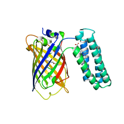
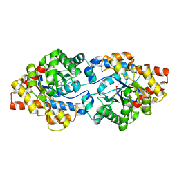
3U0L
 
 | | Crystal structure of the engineered fluorescent protein mRuby, crystal form 1, pH 4.5 | | 分子名称: | ACETATE ION, mRuby | | 著者 | Akerboom, J, Looger, L.L, Schreiter, E.R. | | 登録日 | 2011-09-28 | | 公開日 | 2012-10-03 | | 最終更新日 | 2023-12-06 | | 実験手法 | X-RAY DIFFRACTION (1.25 Å) | | 主引用文献 | Genetically encoded calcium indicators for multi-color neural activity imaging and combination with optogenetics.
Front Mol Neurosci, 6, 2013
|
|
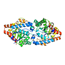
3U0M
 | |
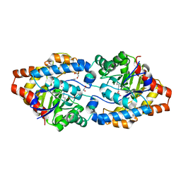
3U0N
 | |
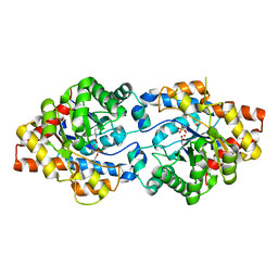
3U8P
 | | Cytochrome b562 integral fusion with EGFP | | 分子名称: | Cytochrome b562 integral fusion with enhanced green fluorescent protein, PROTOPORPHYRIN IX CONTAINING FE | | 著者 | Arpino, J, Czapinska, H, Piasecka, A, Edwards, W.R, Barker, P, Gajda, M, Bochtler, M, Jones, D.D. | | 登録日 | 2011-10-17 | | 公開日 | 2012-08-29 | | 最終更新日 | 2023-12-06 | | 実験手法 | X-RAY DIFFRACTION (2.75 Å) | | 主引用文献 | Structural basis for efficient chromophore communication and energy transfer in a constructed didomain protein scaffold.
J.Am.Chem.Soc., 134, 2012
|
|
3UFZ
 | | Crystal structure of a Trp-less green fluorescent protein translated by the universal genetic code | | 分子名称: | Green fluorescent protein | | 著者 | Kawahara-Kobayashi, A, Araiso, Y, Matsuda, T, Yokoyama, S, Kigawa, T, Nureki, O, Kiga, D. | | 登録日 | 2011-11-02 | | 公開日 | 2012-10-17 | | 最終更新日 | 2023-12-06 | | 実験手法 | X-RAY DIFFRACTION (1.85 Å) | | 主引用文献 | Simplification of the genetic code: restricted diversity of genetically encoded amino acids.
Nucleic Acids Res., 40, 2012
|
|
3UG0
 | | Crystal structure of a Trp-less green fluorescent protein translated by the simplified genetic code | | 分子名称: | Green fluorescent protein | | 著者 | Kawahara-Kobayashi, A, Araiso, Y, Matsuda, T, Yokoyama, S, Kigawa, T, Nureki, O, Kiga, D. | | 登録日 | 2011-11-02 | | 公開日 | 2012-10-17 | | 最終更新日 | 2023-12-06 | | 実験手法 | X-RAY DIFFRACTION (2.093 Å) | | 主引用文献 | Simplification of the genetic code: restricted diversity of genetically encoded amino acids.
Nucleic Acids Res., 40, 2012
|
|
3UPM
 | | Crystal Structure of PTE mutant H254Q/H257F/K185R/I274N | | 分子名称: | COBALT (II) ION, Parathion hydrolase | | 著者 | Tsai, P, Fox, N.G, Li, Y, Barondeau, D.P, Raushel, F.M. | | 登録日 | 2011-11-18 | | 公開日 | 2012-08-01 | | 最終更新日 | 2023-12-06 | | 実験手法 | X-RAY DIFFRACTION (1.95 Å) | | 主引用文献 | Enzymes for the homeland defense: optimizing phosphotriesterase for the hydrolysis of organophosphate nerve agents.
Biochemistry, 51, 2012
|
|
3UR2
 | | Crystal Structure of PTE mutant H254G/H257W/L303T/K185R/I274N/A80V | | 分子名称: | 1,2-ETHANEDIOL, COBALT (II) ION, IMIDAZOLE, ... | | 著者 | Tsai, P, Fox, N.G, Li, Y, Barondeau, D.P, Raushel, F.M. | | 登録日 | 2011-11-21 | | 公開日 | 2012-08-01 | | 最終更新日 | 2023-12-06 | | 実験手法 | X-RAY DIFFRACTION (2 Å) | | 主引用文献 | Enzymes for the homeland defense: optimizing phosphotriesterase for the hydrolysis of organophosphate nerve agents.
Biochemistry, 51, 2012
|
|
3UR5
 | | Crystal Structure of PTE mutant K185R/I274N | | 分子名称: | COBALT (II) ION, DIETHYL HYDROGEN PHOSPHATE, Parathion hydrolase | | 著者 | Tsai, P, Fox, N.G, Li, Y, Barondeau, D.P, Raushel, F.M. | | 登録日 | 2011-11-21 | | 公開日 | 2012-08-01 | | 最終更新日 | 2023-12-06 | | 実験手法 | X-RAY DIFFRACTION (1.6 Å) | | 主引用文献 | Enzymes for the homeland defense: optimizing phosphotriesterase for the hydrolysis of organophosphate nerve agents.
Biochemistry, 51, 2012
|
|
3URA
 | | Crystal Structure of PTE mutant H254G/H257W/L303T/K185R/I274N/A80V/S61T | | 分子名称: | COBALT (II) ION, IMIDAZOLE, Parathion hydrolase | | 著者 | Tsai, P, Fox, N.G, Li, Y, Barondeau, D.P, Raushel, F.M. | | 登録日 | 2011-11-21 | | 公開日 | 2012-08-01 | | 最終更新日 | 2023-12-06 | | 実験手法 | X-RAY DIFFRACTION (1.88 Å) | | 主引用文献 | Enzymes for the homeland defense: optimizing phosphotriesterase for the hydrolysis of organophosphate nerve agents.
Biochemistry, 51, 2012
|
|
3URB
 | | Crystal Structure of PTE mutant H254G/H257W/L303T/M317L/I106C/F132I/L271I/K185R/I274N/A80V/R67H | | 分子名称: | COBALT (II) ION, DIETHYL HYDROGEN PHOSPHATE, IMIDAZOLE, ... | | 著者 | Tsai, P, Fox, N.G, Li, Y, Barondeau, D.P, Raushel, F.M. | | 登録日 | 2011-11-21 | | 公開日 | 2012-08-01 | | 最終更新日 | 2023-12-06 | | 実験手法 | X-RAY DIFFRACTION (1.77 Å) | | 主引用文献 | Enzymes for the homeland defense: optimizing phosphotriesterase for the hydrolysis of organophosphate nerve agents.
Biochemistry, 51, 2012
|
|
3URN
 | | Crystal Structure of PTE mutant H254G/H257W/L303T/K185R/I274N/A80V/S61T with cyclohexyl methylphosphonate inhibitor | | 分子名称: | COBALT (II) ION, IMIDAZOLE, Parathion hydrolase, ... | | 著者 | Tsai, P, Fox, N.G, Li, Y, Barondeau, D.P, Raushel, F.M. | | 登録日 | 2011-11-22 | | 公開日 | 2012-08-01 | | 最終更新日 | 2023-12-06 | | 実験手法 | X-RAY DIFFRACTION (1.95 Å) | | 主引用文献 | Enzymes for the homeland defense: optimizing phosphotriesterase for the hydrolysis of organophosphate nerve agents.
Biochemistry, 51, 2012
|
|
3URQ
 | | Crystal Structure of PTE mutant H254G/H257W/L303T/M317L/I106C/F132I/L271I/K185R/I274N/A80V/R67H with cyclohexyl methylphosphonate inhibitor | | 分子名称: | COBALT (II) ION, IMIDAZOLE, Parathion hydrolase, ... | | 著者 | Tsai, P, Fox, N.G, Li, Y, Barondeau, D.P, Raushel, F.M. | | 登録日 | 2011-11-22 | | 公開日 | 2012-08-01 | | 最終更新日 | 2023-12-06 | | 実験手法 | X-RAY DIFFRACTION (2.1 Å) | | 主引用文献 | Enzymes for the homeland defense: optimizing phosphotriesterase for the hydrolysis of organophosphate nerve agents.
Biochemistry, 51, 2012
|
|
3V3D
 | |
3VHS
 | |
3VHT
 | | Crystal structure of GFP-Wrnip1 UBZ domain fusion protein in complex with ubiquitin | | 分子名称: | Green fluorescent protein, Green fluorescent protein,ATPase WRNIP1, Ubiquitin, ... | | 著者 | Suzuki, N, Wakatsuki, S, Kawasaki, M. | | 登録日 | 2011-09-06 | | 公開日 | 2012-10-10 | | 最終更新日 | 2023-12-06 | | 実験手法 | X-RAY DIFFRACTION (2.4 Å) | | 主引用文献 | A novel mode of ubiquitin recognition by the ubiquitin-binding zinc finger domain of WRNIP1.
Febs J., 2016
|
|
3VIC
 | | Green-fluorescent variant of the non-fluorescent chromoprotein Rtms5 | | 分子名称: | CHLORIDE ION, GFP-like non-fluorescent chromoprotein, IODIDE ION | | 著者 | Battad, J.M, Traore, D.A.K, Byres, E, Wilce, M, Devenish, R.J, Rossjohn, J, Prescott, M. | | 登録日 | 2011-09-28 | | 公開日 | 2012-06-06 | | 最終更新日 | 2023-11-15 | | 実験手法 | X-RAY DIFFRACTION (2.2 Å) | | 主引用文献 | A Green Fluorescent Protein Containing a QFG Tri-Peptide Chromophore: Optical Properties and X-Ray Crystal Structure.
Plos One, 7, 2012
|
|
3VK1
 | | Green-fluorescent variant of the non-fluorescent chromoprotein Rtms5 | | 分子名称: | CHLORIDE ION, GFP-like non-fluorescent chromoprotein, IODIDE ION | | 著者 | Battad, J.M, Traore, D.A.K, Wilce, M, Byres, M, Rossjohn, J, Devenish, R.J, Prescott, M. | | 登録日 | 2011-11-07 | | 公開日 | 2012-06-06 | | 最終更新日 | 2023-11-15 | | 実験手法 | X-RAY DIFFRACTION (2.2 Å) | | 主引用文献 | A Green Fluorescent Protein Containing a QFG Tri-Peptide Chromophore: Optical Properties and X-Ray Crystal Structure.
Plos One, 7, 2012
|
|
3W1C
 | |
3W1D
 | |
3WCK
 | | Crystal structure of monomeric photosensitizing fluorescent protein, Supernova | | 分子名称: | Monomeric photosenitizing fluorescent protein supernova | | 著者 | Sakai, N, Matsuda, T, Takemoto, K, Nagai, T. | | 登録日 | 2013-05-27 | | 公開日 | 2013-10-02 | | 最終更新日 | 2023-11-15 | | 実験手法 | X-RAY DIFFRACTION (2.3 Å) | | 主引用文献 | SuperNova, a monomeric photosensitizing fluorescent protein for chromophore-assisted light inactivation
Sci Rep, 3, 2013
|
|
3WLC
 | | Crystal structure of dimeric GCaMP6m | | 分子名称: | CALCIUM ION, Myosin light chain kinase, Green fluorescent protein, ... | | 著者 | Ding, J, Luo, A.F, Hu, L.Y, Wang, D.C, Shao, F. | | 登録日 | 2013-11-08 | | 公開日 | 2014-01-22 | | 最終更新日 | 2023-12-06 | | 実験手法 | X-RAY DIFFRACTION (2.49 Å) | | 主引用文献 | Structural basis of the ultrasensitive calcium indicator GCaMP6.
Sci China Life Sci, 57, 2014
|
|
3WLD
 | | Crystal structure of monomeric GCaMP6m | | 分子名称: | CALCIUM ION, Myosin light chain kinase, Green fluorescent protein, ... | | 著者 | Ding, J, Luo, A.F, Hu, L.Y, Wang, D.C, Shao, F. | | 登録日 | 2013-11-08 | | 公開日 | 2014-01-22 | | 最終更新日 | 2023-12-06 | | 実験手法 | X-RAY DIFFRACTION (2.7 Å) | | 主引用文献 | Structural basis of the ultrasensitive calcium indicator GCaMP6.
Sci China Life Sci, 57, 2014
|
|
3WUP
 | | Crystal Structure of the Ubiquitin-Binding Zinc Finger (UBZ) Domain of the Human DNA Polymerase Eta | | 分子名称: | CHLORIDE ION, DNA polymerase eta, GLYCEROL, ... | | 著者 | Suzuki, N, Wakatsuki, S, Kawasaki, S. | | 登録日 | 2014-05-01 | | 公開日 | 2015-06-17 | | 最終更新日 | 2022-08-24 | | 実験手法 | X-RAY DIFFRACTION (1.6 Å) | | 主引用文献 | A novel mode of ubiquitin recognition by the ubiquitin-binding zinc finger domain of WRNIP1.
Febs J., 283, 2016
|
|
3ZTF
 | | X-ray Structure of the Cyan Fluorescent Protein mTurquoise2 (K206A mutant) | | 分子名称: | GREEN FLUORESCENT PROTEIN | | 著者 | von Stetten, D, Goedhart, J, Noirclerc-Savoye, M, Lelimousin, M, Joosen, L, Hink, M.A, van Weeren, L, Gadella, T.W.J, Royant, A. | | 登録日 | 2011-07-07 | | 公開日 | 2012-03-21 | | 最終更新日 | 2023-12-20 | | 実験手法 | X-RAY DIFFRACTION (1.31 Å) | | 主引用文献 | Structure-Guided Evolution of Cyan Fluorescent Proteins Towards a Quantum Yield of 93%
Nat.Commun, 3, 2012
|
|


