Loading
PDBj
MenuPDBj@FacebookPDBj@TwitterPDBj@YouTubewwPDB FoundationwwPDB
RCSB PDBPDBeBMRBAdv. SearchSearch help

7A8M
DownloadVisualize
BU of 7a8m by Molmil
rsGreen0.7-K206A-N205G in the green-on state
Descriptor: Green fluorescent protein
Authors:De Zitter, E, Dedecker, P, Van Meervelt, L.
Deposit date:2020-08-30
Release date:2021-02-17
Last modified:2021-05-05
Method:X-RAY DIFFRACTION (1.6 Å)
Cite:Structure-Function Dataset Reveals Environment Effects within a Fluorescent Protein Model System*.
Angew.Chem.Int.Ed.Engl., 60, 2021
7A7M
DownloadVisualize
BU of 7a7m by Molmil
rsGreen0.7b in the green-on state
Descriptor: Green fluorescent protein
Authors:De Zitter, E, Dedecker, P, Van Meervelt, L.
Deposit date:2020-08-30
Release date:2021-02-17
Last modified:2024-01-31
Method:X-RAY DIFFRACTION (1.6 Å)
Cite:Structure-Function Dataset Reveals Environment Effects within a Fluorescent Protein Model System*.
Angew.Chem.Int.Ed.Engl., 60, 2021
3DQ6
DownloadVisualize
BU of 3dq6 by Molmil
Structure of the Yellow Fluorescent Protein Citrine Frozen at 1920 Atmospheres Number 2: Structure 18 in a Series of 26 High Pressure Structures
Descriptor: Green fluorescent protein
Authors:Barstow, B, Kim, C.U.
Deposit date:2008-07-09
Release date:2008-09-23
Last modified:2023-11-15
Method:X-RAY DIFFRACTION (1.6 Å)
Cite:Alteration of citrine structure by hydrostatic pressure explains the accompanying spectral shift.
Proc.Natl.Acad.Sci.Usa, 105, 2008
7A8O
DownloadVisualize
BU of 7a8o by Molmil
rsGreen0.7-K206A-N205S in the green-on state
Descriptor: 1,2-ETHANEDIOL, Green fluorescent protein
Authors:De Zitter, E, Dedecker, P, Van Meervelt, L.
Deposit date:2020-08-30
Release date:2021-02-17
Last modified:2024-01-31
Method:X-RAY DIFFRACTION (1.6 Å)
Cite:Structure-Function Dataset Reveals Environment Effects within a Fluorescent Protein Model System*.
Angew.Chem.Int.Ed.Engl., 60, 2021
6T39
DownloadVisualize
BU of 6t39 by Molmil
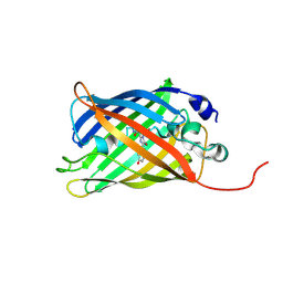
Crystal structure of rsEGFP2 in its off-state determined by SFX
Descriptor: Green fluorescent protein
Authors:Woodhouse, J, Coquelle, N, Adam, V, Barends, T.R.M, De La Mora, E, Bourgeois, D, Colletier, J.P, Schlichting, I, Weik, M.
Deposit date:2019-10-10
Release date:2020-02-19
Last modified:2024-01-24
Method:X-RAY DIFFRACTION (1.6 Å)
Cite:Photoswitching mechanism of a fluorescent protein revealed by time-resolved crystallography and transient absorption spectroscopy.
Nat Commun, 11, 2020
5AZB
DownloadVisualize
BU of 5azb by Molmil
Crystal structure of Escherichia coli Lgt in complex with phosphatidylglycerol and the inhibitor palmitic acid
Descriptor: (1S)-2-{[{[(2R)-2,3-DIHYDROXYPROPYL]OXY}(HYDROXY)PHOSPHORYL]OXY}-1-[(PALMITOYLOXY)METHYL]ETHYL STEARATE, PALMITIC ACID, Prolipoprotein diacylglyceryl transferase, ...
Authors:Zhang, X.C, Mao, G, Zhao, Y.
Deposit date:2015-09-30
Release date:2016-01-27
Last modified:2024-03-20
Method:X-RAY DIFFRACTION (1.6 Å)
Cite:Crystal structure of E. coli lipoprotein diacylglyceryl transferase
Nat Commun, 7, 2016
4DXI
DownloadVisualize
BU of 4dxi by Molmil
Crystal Structure of an Ancestor of All Faviina Proteins
Descriptor: GREEN FLUORESCENT PROTEIN, MAGNESIUM ION
Authors:Kim, H, Wachter, R.M.
Deposit date:2012-02-27
Release date:2013-02-27
Last modified:2023-11-15
Method:X-RAY DIFFRACTION (1.6 Å)
Cite:A hinge migration mechanism unlocks the evolution of green-to-red photoconversion in GFP-like proteins.
Structure, 23, 2015
7Z7Q
DownloadVisualize
BU of 7z7q by Molmil
Structure of the T207D single-point mutant of the fluorescent protein NeonCyan1 at pH 6.5
Descriptor: NeonCyan1
Authors:Duarte, K, Dupuy, J, Royant, A.
Deposit date:2022-03-16
Release date:2022-05-04
Last modified:2024-01-31
Method:X-RAY DIFFRACTION (1.6 Å)
Cite:Cyan fluorescent proteins derived from mNeonGreen.
Protein Eng.Des.Sel., 35, 2022
3WUP
DownloadVisualize
BU of 3wup by Molmil
Crystal Structure of the Ubiquitin-Binding Zinc Finger (UBZ) Domain of the Human DNA Polymerase Eta
Descriptor: CHLORIDE ION, DNA polymerase eta, GLYCEROL, ...
Authors:Suzuki, N, Wakatsuki, S, Kawasaki, S.
Deposit date:2014-05-01
Release date:2015-06-17
Last modified:2022-08-24
Method:X-RAY DIFFRACTION (1.6 Å)
Cite:A novel mode of ubiquitin recognition by the ubiquitin-binding zinc finger domain of WRNIP1.
Febs J., 283, 2016
5T3I
DownloadVisualize
BU of 5t3i by Molmil
cyan fluorescence protein soaked with selenourea for 5 min
Descriptor: Green fluorescent protein, PHOSPHATE ION, selenourea
Authors:Luo, Z, Dauter, Z.
Deposit date:2016-08-25
Release date:2016-11-30
Last modified:2019-12-04
Method:X-RAY DIFFRACTION (1.6 Å)
Cite:Selenourea: a convenient phasing vehicle for macromolecular X-ray crystal structures.
Sci Rep, 6, 2016
3AI4
DownloadVisualize
BU of 3ai4 by Molmil
Crystal structure of yeast enhanced green fluorescent protein - mouse polymerase iota ubiquitin binding motif fusion protein
Descriptor: SULFATE ION, yeast enhanced green fluorescent protein,DNA polymerase iota
Authors:Suzuki, N, Wakatsuki, S, Kawasaki, M.
Deposit date:2010-05-10
Release date:2010-09-29
Last modified:2023-11-15
Method:X-RAY DIFFRACTION (1.6 Å)
Cite:Crystallization of small proteins assisted by green fluorescent protein
Acta Crystallogr.,Sect.D, 66, 2010
8AAB
DownloadVisualize
BU of 8aab by Molmil
S148F mutant of blue-to-red fluorescent timer mRubyFT
Descriptor: mRubyFT S148F mutant of blue-to-red fluorescent timer
Authors:Boyko, K.M, Nikolaeva, A.Y, Vlaskina, A.V, Dorovatovskii, P.V, Subach, O.M, Popov, V.O, Subach, F.V.
Deposit date:2022-06-30
Release date:2022-08-17
Last modified:2024-02-07
Method:X-RAY DIFFRACTION (1.6 Å)
Cite:mRubyFT/S147I, a mutant of blue-to-red fluorescent timer
Crystallography Reports, 2022
4KEX
DownloadVisualize
BU of 4kex by Molmil
Crystal structure analysis of a single amino acid deletion mutation in EGFP
Descriptor: Green fluorescent protein
Authors:Arpino, J.A.J, Rizkallah, P.J.
Deposit date:2013-04-26
Release date:2014-08-06
Last modified:2023-12-06
Method:X-RAY DIFFRACTION (1.6 Å)
Cite:Structural and dynamic changes associated with beneficial engineered single-amino-acid deletion mutations in enhanced green fluorescent protein.
Acta Crystallogr.,Sect.D, 70, 2014
5Y00
DownloadVisualize
BU of 5y00 by Molmil
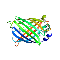
Acid-tolerant monomeric GFP, Gamillus, fluorescence (ON) state
Descriptor: CHLORIDE ION, GLYCEROL, Green fluorescent protein, ...
Authors:Nakashima, R, Sakurai, K, Shinoda, H, Matsuda, T, Nagai, T.
Deposit date:2017-07-14
Release date:2018-01-17
Last modified:2023-11-15
Method:X-RAY DIFFRACTION (1.6 Å)
Cite:Acid-Tolerant Monomeric GFP from Olindias formosa.
Cell Chem Biol, 25, 2018
3GEX
DownloadVisualize
BU of 3gex by Molmil
1.6 angstrom crystal structure of fluorescent protein Cypet
Descriptor: Green fluorescent protein
Authors:Hu, X, Liu, R.
Deposit date:2009-02-26
Release date:2010-03-09
Last modified:2023-11-22
Method:X-RAY DIFFRACTION (1.6 Å)
Cite:1.6 angstrom crystal structure of fluorescent protein Cypet
To be Published
4W6T
DownloadVisualize
BU of 4w6t by Molmil
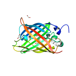
Crystal Structure of Full-Length Split GFP Mutant E115H/T118H With Copper Mediated Crystal Contacts, P 43 21 2 Space Group
Descriptor: 1,2-ETHANEDIOL, BROMIDE ION, COPPER (II) ION, ...
Authors:Leibly, D.J, Waldo, G.S, Yeates, T.O.
Deposit date:2014-08-20
Release date:2015-02-18
Last modified:2023-11-15
Method:X-RAY DIFFRACTION (1.604 Å)
Cite:A Suite of Engineered GFP Molecules for Oligomeric Scaffolding.
Structure, 23, 2015
6HOK
DownloadVisualize
BU of 6hok by Molmil
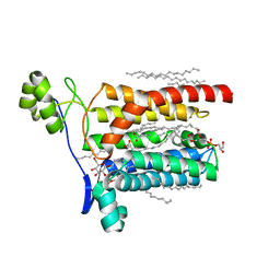
Structure of Beclin1 LIR (S96E) motif bound to GABARAP
Descriptor: 1,2-ETHANEDIOL, Beclin-1,Gamma-aminobutyric acid receptor-associated protein
Authors:Mouilleron, S, Birgisdottir, A.B, Bhujbal, Z, Wirth, M, Sjottem, E, Evjen, G, Zhang, W, Lee, R, O'Reilly, N, Tooze, S, Lamark, T, Johansen, T.
Deposit date:2018-09-17
Release date:2019-02-27
Last modified:2024-01-24
Method:X-RAY DIFFRACTION (1.61 Å)
Cite:Members of the autophagy class III phosphatidylinositol 3-kinase complex I interact with GABARAP and GABARAPL1 via LIR motifs.
Autophagy, 15, 2019
3G9A
DownloadVisualize
BU of 3g9a by Molmil
Green fluorescent protein bound to minimizer nanobody
Descriptor: Green fluorescent protein, Minimizer
Authors:Kirchhofer, A, Helma, J, Schmidthals, K, Frauer, C, Cui, S, Karcher, A, Pellis, M, Muyldermans, S, Delucci, C.C, Cardoso, M.C, Leonhardt, H, Hopfner, K.-P, Rothbauer, U.
Deposit date:2009-02-13
Release date:2009-12-08
Last modified:2023-11-15
Method:X-RAY DIFFRACTION (1.614 Å)
Cite:Modulation of protein properties in living cells using nanobodies
Nat.Struct.Mol.Biol., 17, 2010
6FPB
DownloadVisualize
BU of 6fpb by Molmil
Crystal structure of anti-mTFP1 DARPin 1238_G01 in space group I4
Descriptor: CHLORIDE ION, DARPin 1238_G01
Authors:Jakob, R.P, Vigano, M.A, Bieli, D, Matsuda, S, Schaefer, J.V, Pluckthun, A, Affolter, M, Maier, T.
Deposit date:2018-02-09
Release date:2018-10-03
Last modified:2024-01-17
Method:X-RAY DIFFRACTION (1.617 Å)
Cite:DARPins recognizing mTFP1 as novel reagents forin vitroandin vivoprotein manipulations.
Biol Open, 7, 2018
6PFU
DownloadVisualize
BU of 6pfu by Molmil
rsEGFP2 with a chlorinated chromophore in the non-fluorescent off-state in a contracted unit cell
Descriptor: Green fluorescent protein, SULFATE ION
Authors:Chang, J, Romei, M.G, Boxer, S.G.
Deposit date:2019-06-22
Release date:2019-08-07
Last modified:2023-11-15
Method:X-RAY DIFFRACTION (1.619 Å)
Cite:Structural Evidence of Photoisomerization Pathways in Fluorescent Proteins.
J.Am.Chem.Soc., 141, 2019
8I4J
DownloadVisualize
BU of 8i4j by Molmil
Structure of wild-type Azami Green from Galaxea fascicularis
Descriptor: Azami-Green
Authors:Otsubo, S, Takekawa, N, Imamura, H, Imada, K.
Deposit date:2023-01-19
Release date:2023-11-01
Last modified:2023-11-15
Method:X-RAY DIFFRACTION (1.62 Å)
Cite:Red fluorescent proteins engineered from green fluorescent proteins.
Proc.Natl.Acad.Sci.USA, 120, 2023
4EN1
DownloadVisualize
BU of 4en1 by Molmil
The 1.62A structure of a FRET-optimized Cerulean Fluorescent Protein
Descriptor: ACETATE ION, DI(HYDROXYETHYL)ETHER, GLYCEROL, ...
Authors:Watkins, J.L.
Deposit date:2012-04-12
Release date:2013-04-24
Last modified:2023-12-06
Method:X-RAY DIFFRACTION (1.62 Å)
Cite:The 1.6 A resolution structure of a FRET-optimized Cerulean fluorescent protein.
Acta Crystallogr.,Sect.D, 69, 2013
6DEJ
DownloadVisualize
BU of 6dej by Molmil
The structure of HcRed7, a brighter and red-shifted HcRed variant
Descriptor: CHLORIDE ION, DODECAETHYLENE GLYCOL, GFP-like non-fluorescent chromoprotein, ...
Authors:Wannier, T.M, Mayo, S.L.
Deposit date:2018-05-12
Release date:2018-05-23
Last modified:2023-11-15
Method:X-RAY DIFFRACTION (1.6279 Å)
Cite:Monomerization of far-red fluorescent proteins.
Proc. Natl. Acad. Sci. U.S.A., 115, 2018
3SSP
DownloadVisualize
BU of 3ssp by Molmil
Engineered low-affinity halide-binding protein derived from YFP: halide-free
Descriptor: Green fluorescent protein
Authors:Wang, W, Grimley, J.S, Beese, L.S, Hellinga, H.W.
Deposit date:2011-07-08
Release date:2012-07-11
Last modified:2023-11-15
Method:X-RAY DIFFRACTION (1.628 Å)
Cite:Determination of engineered chloride-binding site structures in fluorescent proteins reveals principles of halide recognition
To be Published
8A6O
DownloadVisualize
BU of 8a6o by Molmil
Room temperature rsEGFP2 with a chlorinated chromophore 600 fs after Photoexcitation
Descriptor: Green fluorescent protein
Authors:Fadini, A, van Thor, J.
Deposit date:2022-06-18
Release date:2023-07-19
Last modified:2024-02-07
Method:X-RAY DIFFRACTION (1.63 Å)
Cite:Serial Femtosecond Crystallography Reveals that Photoactivation in a Fluorescent Protein Proceeds via the Hula Twist Mechanism.
J.Am.Chem.Soc., 145, 2023

218500

PDB entries from 2024-04-17

PDB statisticsPDBj update infoContact PDBjnumon