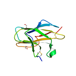
3COO
 
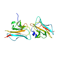 | |
2ZOU
 | | Crystal structure of human F-spondin reeler domain (fragment 2) | | 分子名称: | 1,2-ETHANEDIOL, Spondin-1 | | 著者 | Nagae, M, Nogi, T, Takagi, J. | | 登録日 | 2008-06-07 | | 公開日 | 2008-10-14 | | 最終更新日 | 2023-11-01 | | 実験手法 | X-RAY DIFFRACTION (1.45 Å) | | 主引用文献 | Structure of the F-spondin reeler domain reveals a unique beta-sandwich fold with a deformable disulfide-bonded loop
Acta Crystallogr.,Sect.D, 64, 2008
|
|
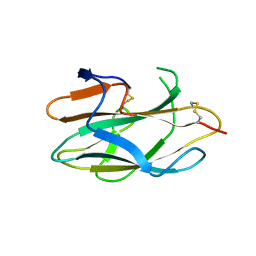
2ZOT
 | |


