 Movie
Movie Controller

[English] 日本語
 Yorodumi
- PDB-5upw: CryoEM Structure Refinement by Integrating NMR Chemical Shifts wi... -
+ Open data

- Basic information
| Entry | Database: PDB / ID: 5upw | |||||||||||||||||||||
|---|---|---|---|---|---|---|---|---|---|---|---|---|---|---|---|---|---|---|---|---|---|---|
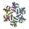
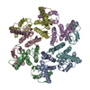
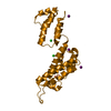
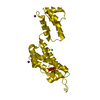
| Title | CryoEM Structure Refinement by Integrating NMR Chemical Shifts with Molecular Dynamics Simulations | |||||||||||||||||||||
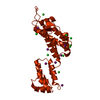
 Components Components | Gag polyprotein Group-specific antigen Group-specific antigen | |||||||||||||||||||||
 Keywords Keywords | VIRAL PROTEIN / Cryo-EM / HIV capsid / Chemical shift / Molecular Dynamics / hydrolase | |||||||||||||||||||||
| Function / homology |  Function and homology information Function and homology informationviral budding via host ESCRT complex / ISG15 antiviral mechanism / host multivesicular body / viral nucleocapsid / host cell nucleus / structural molecule activity / host cell plasma membrane / virion membrane / RNA binding / zinc ion binding / membraneSimilarity search - Function | |||||||||||||||||||||
| Biological species |   Human immunodeficiency virus type 1 Human immunodeficiency virus type 1 | |||||||||||||||||||||
| Method | ELECTRON MICROSCOPY / helical reconstruction / cryo EM / Resolution: 5 Å | |||||||||||||||||||||
 Authors Authors | Perilla, J.R. | |||||||||||||||||||||
| Funding support |  United States, 6items United States, 6items
| |||||||||||||||||||||
 Citation Citation | Journal: J Phys Chem B / Year: 2017 Title: CryoEM Structure Refinement by Integrating NMR Chemical Shifts with Molecular Dynamics Simulations. Authors: Juan R Perilla / Gongpu Zhao / Manman Lu / Jiying Ning / Guangjin Hou / In-Ja L Byeon / Angela M Gronenborn / Tatyana Polenova / Peijun Zhang /  Abstract: Single particle cryoEM has emerged as a powerful method for structure determination of proteins and complexes, complementing X-ray crystallography and NMR spectroscopy. Yet, for many systems, the ...Single particle cryoEM has emerged as a powerful method for structure determination of proteins and complexes, complementing X-ray crystallography and NMR spectroscopy. Yet, for many systems, the resolution of cryoEM density map has been limited to 4-6 Å, which only allows for resolving bulky amino acids side chains, thus hindering accurate model building from the density map. On the other hand, experimental chemical shifts (CS) from solution and solid state MAS NMR spectra provide atomic level data for each amino acid within a molecule or a complex; however, structure determination of large complexes and assemblies based on NMR data alone remains challenging. Here, we present a novel integrated strategy to combine the highly complementary experimental data from cryoEM and NMR computationally by molecular dynamics simulations to derive an atomistic model, which is not attainable by either approach alone. We use the HIV-1 capsid protein (CA) C-terminal domain as well as the large capsid assembly to demonstrate the feasibility of this approach, termed NMR CS-biased cryoEM structure refinement. | |||||||||||||||||||||
| History |
|
- Structure visualization
| Movie |
Movie viewer |
|---|---|
| Structure viewer | Molecule: MolmilJmol/JSmol |
- Downloads & links
-Download
| PDBx/mmCIF format | 5upw.cif.gz | 220.4 KB | Display | PDBx/mmCIF format |
|---|---|---|---|---|
| PDB format | pdb5upw.ent.gz | 181.4 KB | Display | PDB format |
| PDBx/mmJSON format | 5upw.json.gz | Tree view | PDBx/mmJSON format | |
| Others |  Other downloads Other downloads |
-Validation report
| Arichive directory | https://data.pdbj.org/pub/pdb/validation_reports/up/5upwftp://data.pdbj.org/pub/pdb/validation_reports/up/5upw | HTTPS FTP |
|---|
-Related structure data
| Related structure data | 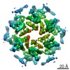 8595MC M: map data used to model this data C: citing same article ( |
|---|---|
| Similar structure data |

-Links
 PDBj
PDBj

- Assembly
| Deposited unit | 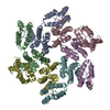
|
|---|---|
| 1 |
|
-Components
| #1: Protein | Group-specific antigen / Pr55Gag Mass: 24654.268 Da / Num. of mol.: 6 / Fragment: UNP residues 133-353 / Mutation: A92E Source method: isolated from a genetically manipulated source Source: (gene. exp.) Human immunodeficiency virus type 1 (NEW YORK-5 ISOLATE)Gene: gag / Production host:  Escherichia coli (E. coli) / References: UniProt: P12493 Escherichia coli (E. coli) / References: UniProt: P12493 |
|---|
-Experimental details
-Experiment
| Experiment | Method: ELECTRON MICROSCOPY |
|---|---|
| EM experiment | Aggregation state: HELICAL ARRAY / 3D reconstruction method: helical reconstruction |
- Sample preparation
| Component | Name: HIV-1 Capsid Protein Assembly / Type: COMPLEX / Entity ID: all / Source: RECOMBINANT | |||||||||||||||
|---|---|---|---|---|---|---|---|---|---|---|---|---|---|---|---|---|
| Molecular weight | Value: 24 kDa/nm / Experimental value: NO | |||||||||||||||
| Source (natural) | Organism: Human immunodeficiency virus type 1 (NEW YORK-5 ISOLATE) | |||||||||||||||
| Source (recombinant) | Organism: Escherichia coli (E. coli) | |||||||||||||||
| Buffer solution | pH: 8 | |||||||||||||||
| Buffer component |
| |||||||||||||||
| Specimen | Conc.: 2 mg/ml / Embedding applied: NO / Shadowing applied: NO / Staining applied: NO / Vitrification applied: YES | |||||||||||||||
| Specimen support | Grid material: COPPER / Grid mesh size: 300 divisions/in. / Grid type: Quantifoil R2/1 | |||||||||||||||
| Vitrification | Instrument: HOMEMADE PLUNGER / Cryogen name: ETHANE / Humidity: 100 % / Chamber temperature: 295 K Details: The assembled sample (1.5 microliter) was applied to the carbon side of a glow discharged perforated Quantifoil grid, followed by application of 3 microliter of low salt buffer (100 ...Details: The assembled sample (1.5 microliter) was applied to the carbon side of a glow discharged perforated Quantifoil grid, followed by application of 3 microliter of low salt buffer (100 milimolar NaCl, 50 milimolar Tris pH 8.0) on the back side of the grid, and blotting, from the back side, with a filter paper, before plunge-freezing in liquid ethane |
- Electron microscopy imaging
| Experimental equipment |  Model: Tecnai Polara / Image courtesy: FEI Company |
|---|---|
| Microscopy | Model: FEI POLARA 300 |
| Electron gun | Electron source: FIELD EMISSION GUN / Accelerating voltage: 300 kV / Illumination mode: FLOOD BEAM |
| Electron lens | Mode: BRIGHT FIELDBright-field microscopy / Nominal magnification: 31000 X / Nominal defocus max: 2200 nm / Nominal defocus min: 700 nm / Cs: 2.2 mm / C2 aperture diameter: 100 µm |
| Specimen holder | Cryogen: NITROGEN / Specimen holder model: SIDE ENTRY, EUCENTRIC |
| Image recording | Average exposure time: 6 sec. / Electron dose: 41 e/Å2 / Detector mode: SUPER-RESOLUTION / Film or detector model: GATAN K2 SUMMIT (4k x 4k) / Num. of real images: 523 |
| Image scans | Movie frames/image: 30 |
- Processing
| EM software |
| ||||||||||||||||||||||||||||||||
|---|---|---|---|---|---|---|---|---|---|---|---|---|---|---|---|---|---|---|---|---|---|---|---|---|---|---|---|---|---|---|---|---|---|
| CTF correction | Type: PHASE FLIPPING AND AMPLITUDE CORRECTION | ||||||||||||||||||||||||||||||||
| Helical symmerty | Angular rotation/subunit: -31.13 ° / Axial rise/subunit: 6.94 Å / Axial symmetry: C1 | ||||||||||||||||||||||||||||||||
| Particle selection | Num. of particles selected: 39712 | ||||||||||||||||||||||||||||||||
| 3D reconstruction | Resolution: 5 Å / Resolution method: FSC 0.143 CUT-OFF / Num. of particles: 38452 / Algorithm: FOURIER SPACE / Num. of class averages: 3 / Symmetry type: HELICAL | ||||||||||||||||||||||||||||||||
| Atomic model building | Protocol: FLEXIBLE FIT / Space: REAL | ||||||||||||||||||||||||||||||||
| Atomic model building | PDB-ID: 4XFX Pdb chain residue range: 1-220 |

