 Movie
Movie Controller


+ Open data

- Basic information
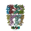
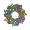
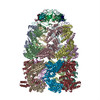
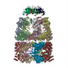
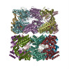
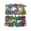
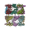
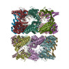
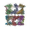
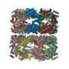
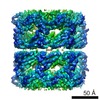
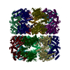
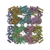
| Entry | Database: PDB / ID: 1gr5 | ||||||
|---|---|---|---|---|---|---|---|
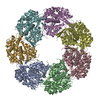
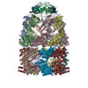
| Title | Solution Structure of apo GroEL by Cryo-Electron microscopy | ||||||
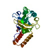
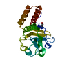
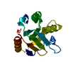
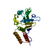
 Components Components | 60 KDA CHAPERONIN | ||||||
 Keywords Keywords |  CHAPERONE CHAPERONE | ||||||
| Function / homology |  Function and homology information Function and homology informationGroEL-GroES complex / chaperonin ATPase / virion assembly / chaperone cofactor-dependent protein refolding / isomerase activity / ATP-dependent protein folding chaperone / response to radiation / unfolded protein binding / protein folding / response to heat ...GroEL-GroES complex / chaperonin ATPase / virion assembly / chaperone cofactor-dependent protein refolding / isomerase activity / ATP-dependent protein folding chaperone / response to radiation / unfolded protein binding / protein folding / response to heat / protein refolding / cell cycle / cell division / magnesium ion binding / ATP hydrolysis activity / ATP binding / membrane / identical protein binding / cytosol / cytoplasmSimilarity search - Function | ||||||
| Biological species |  ESCHERICHIA COLI (E. coli) ESCHERICHIA COLI (E. coli) | ||||||
| Method | ELECTRON MICROSCOPY / single particle reconstruction / cryo EM / Resolution: 7.9 Å | ||||||
 Authors Authors | Ranson, N.A. / Farr, G.W. / Roseman, A.M. / Gowen, B. / Fenton, W.A. / Horwich, A.L. / Saibil, H.R. | ||||||
 Citation Citation | Journal: Cell / Year: 2001 Title: ATP-bound states of GroEL captured by cryo-electron microscopy. Authors: N A Ranson / G W Farr / A M Roseman / B Gowen / W A Fenton / A L Horwich / H R Saibil /  Abstract: The chaperonin GroEL drives its protein-folding cycle by cooperatively binding ATP to one of its two rings, priming that ring to become folding-active upon GroES binding, while simultaneously ...The chaperonin GroEL drives its protein-folding cycle by cooperatively binding ATP to one of its two rings, priming that ring to become folding-active upon GroES binding, while simultaneously discharging the previous folding chamber from the opposite ring. The GroEL-ATP structure, determined by cryo-EM and atomic structure fitting, shows that the intermediate domains rotate downward, switching their intersubunit salt bridge contacts from substrate binding to ATP binding domains. These observations, together with the effects of ATP binding to a GroEL-GroES-ADP complex, suggest structural models for the ATP-induced reduction in affinity for polypeptide and for cooperativity. The model for cooperativity, based on switching of intersubunit salt bridge interactions around the GroEL ring, may provide general insight into cooperativity in other ring complexes and molecular machines. #1: Journal: Nature / Year: 1997Title: The crystal structure of the asymmetric GroEL-GroES-(ADP)7 chaperonin complex. Authors: Z Xu / A L Horwich / P B Sigler /  Abstract: Chaperonins assist protein folding with the consumption of ATP. They exist as multi-subunit protein assemblies comprising rings of subunits stacked back to back. In Escherichia coli, asymmetric ...Chaperonins assist protein folding with the consumption of ATP. They exist as multi-subunit protein assemblies comprising rings of subunits stacked back to back. In Escherichia coli, asymmetric intermediates of GroEL are formed with the co-chaperonin GroES and nucleotides bound only to one of the seven-subunit rings (the cis ring) and not to the opposing ring (the trans ring). The structure of the GroEL-GroES-(ADP)7 complex reveals how large en bloc movements of the cis ring's intermediate and apical domains enable bound GroES to stabilize a folding chamber with ADP confined to the cis ring. Elevation and twist of the apical domains double the volume of the central cavity and bury hydrophobic peptide-binding residues in the interface with GroES, as well as between GroEL subunits, leaving a hydrophilic cavity lining that is conducive to protein folding. An inward tilt of the cis equatorial domain causes an outward tilt in the trans ring that opposes the binding of a second GroES. When combined with new functional results, this negative allosteric mechanism suggests a model for an ATP-driven folding cycle that requires a double toroid. #2: Journal: Nat Struct Biol / Year: 1996 Title: The 2.4 A crystal structure of the bacterial chaperonin GroEL complexed with ATP gamma S. Authors: D C Boisvert / J Wang / Z Otwinowski / A L Horwich / P B Sigler / Abstract: GroEL is a bacterial chaperonin of 14 identical subunits required to help fold newly synthesized proteins. The crystal structure of GroEL with ATP gamma S bound to each subunit shows that ATP binds ...GroEL is a bacterial chaperonin of 14 identical subunits required to help fold newly synthesized proteins. The crystal structure of GroEL with ATP gamma S bound to each subunit shows that ATP binds to a novel pocket, whose primary sequence is highly conserved among chaperonins. Interaction of Mg2+ and ATP involves phosphate oxygens of the alpha-, beta- and gamma-phosphates, which is unique for known structures of nucleotide-binding proteins. Although bound ATP induces modest conformational shifts in the equatorial domain, the stereochemistry that functionally coordinates GroEL's affinity for nucleotides, polypeptide, and GroES remains uncertain. #3: Journal: Nature / Year: 1994Title: The crystal structure of the bacterial chaperonin GroEL at 2.8 A. Authors: K Braig / Z Otwinowski / R Hegde / D C Boisvert / A Joachimiak / A L Horwich / P B Sigler / Abstract: The crystal structure of Escherichia coli GroEL shows a porous cylinder of 14 subunits made of two nearly 7-fold rotationally symmetrical rings stacked back-to-back with dyad symmetry. The subunits ...The crystal structure of Escherichia coli GroEL shows a porous cylinder of 14 subunits made of two nearly 7-fold rotationally symmetrical rings stacked back-to-back with dyad symmetry. The subunits consist of three domains: a large equatorial domain that forms the foundation of the assembly at its waist and holds the rings together; a large loosely structured apical domain that forms the ends of the cylinder; and a small slender intermediate domain that connects the two, creating side windows. The three-dimensional structure places most of the mutationally defined functional sites on the channel walls and its outward invaginations, and at the ends of the cylinder. | ||||||
| History |
| ||||||
| Remark 700 | SHEET THE SHEET STRUCTURE OF THIS MOLECULE IS BIFURCATED. IN ORDER TO REPRESENT THIS FEATURE IN ... SHEET THE SHEET STRUCTURE OF THIS MOLECULE IS BIFURCATED. IN ORDER TO REPRESENT THIS FEATURE IN THE SHEET RECORDS BELOW, TWO SHEETS ARE DEFINED. |
- Structure visualization
| Movie |
Movie viewer |
|---|---|
| Structure viewer | Molecule: MolmilJmol/JSmol |
- Downloads & links
-Download
| PDBx/mmCIF format | 1gr5.cif.gz | 1.2 MB | Display | PDBx/mmCIF format |
|---|---|---|---|---|
| PDB format | pdb1gr5.ent.gz | 1 MB | Display | PDB format |
| PDBx/mmJSON format | 1gr5.json.gz | Tree view | PDBx/mmJSON format | |
| Others |  Other downloads Other downloads |
-Validation report
| Arichive directory | https://data.pdbj.org/pub/pdb/validation_reports/gr/1gr5ftp://data.pdbj.org/pub/pdb/validation_reports/gr/1gr5 | HTTPS FTP |
|---|
-Related structure data
| Related structure data | 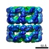 1042MC  1046C 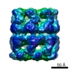 1047C 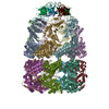 1gruC 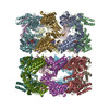 2c7eC M: map data used to model this data C: citing same article ( |
|---|---|
| Similar structure data |


-Links
 PDBj
PDBj
- Assembly
| Deposited unit | 
|
|---|---|
| 1 |
|
-Components
| #1: Protein | Mass: 57188.410 Da / Num. of mol.: 14 / Mutation: YES Source method: isolated from a genetically manipulated source Source: (gene. exp.) ESCHERICHIA COLI (E. coli) / Production host: ESCHERICHIA COLI (E. coli) / References: UniProt: P0A6F6, UniProt: P0A6F5*PLUSCompound details | GROEL IS A HOMOOLIGOMER OF FOURTEEN SUBUNITS ARRANGED IN A DOUBLE RING STRUCTURE. ENGINEERED ...GROEL IS A HOMOOLIGOM | Sequence details | MET 1 IN ALL CHAINS HAS BEEN POST-TRANSLATIO | |
|---|
-Experimental details
-Experiment
| Experiment | Method: ELECTRON MICROSCOPY |
|---|---|
| EM experiment | Aggregation state: PARTICLE / 3D reconstruction method: single particle reconstruction |
- Sample preparation
| Component | Name: MOLECULAR CHAPERONEChaperone (protein) / Type: COMPLEX |
|---|---|
| Buffer solution | Name: HEPES / pH: 7.5 / Details: HEPES |
| Specimen | Conc.: 0.7 mg/ml / Embedding applied: NO / Shadowing applied: NO / Staining applied: NO / Vitrification applied: YES |
| Specimen support | Details: HOLEY CARBON |
| Vitrification | Instrument: HOMEMADE PLUNGER / Cryogen name: ETHANE / Details: LIQUID ETHANE |
| Crystal grow | *PLUS Method: cryo-electron microscopy |
- Electron microscopy imaging
| Microscopy | Model: FEI/PHILIPS CM200T / Date: Feb 1, 1997 |
|---|---|
| Electron gun | Electron source: FIELD EMISSION GUN / Accelerating voltage: 200 kV / Illumination mode: FLOOD BEAM |
| Electron lens | Mode: BRIGHT FIELDBright-field microscopy / Nominal magnification: 38000 X / Calibrated magnification: 40200 X / Nominal defocus max: 1900 nm / Nominal defocus min: 800 nm |
| Specimen holder | Temperature: 95 K / Tilt angle max: 0 ° / Tilt angle min: 0 ° |
| Image recording | Electron dose: 20 e/Å2 / Film or detector model: KODAK SO-163 FILM |
| Image scans | Num. digital images: 50 |
| Radiation wavelength | Relative weight: 1 |
- Processing
| EM software |
| ||||||||||||
|---|---|---|---|---|---|---|---|---|---|---|---|---|---|
| CTF correction | Details: CLASS AVERAGES | ||||||||||||
| Symmetry | Point symmetry: D7 (2x7 fold dihedral) | ||||||||||||
| 3D reconstruction | Method: MODEL-BASED ANGULAR REFINEMENT / Resolution: 7.9 Å / Resolution method: FSC 0.5 CUT-OFF / Num. of particles: 8728 / Nominal pixel size: 1.82 Å / Actual pixel size: 1.74 Å Details: THE THREE DOMAINS FROM EACH SUBUNIT (PDB 1DER) WERE FITTED SEPARATELY INTO EM DENSITY OF WILD TYPE UNLIGANDED GROEL. THE MUTATIONS LISTED ARE PRESENT IN THE FITTED COORDINATES AND NOT IN THE ...Details: THE THREE DOMAINS FROM EACH SUBUNIT (PDB 1DER) WERE FITTED SEPARATELY INTO EM DENSITY OF WILD TYPE UNLIGANDED GROEL. THE MUTATIONS LISTED ARE PRESENT IN THE FITTED COORDINATES AND NOT IN THE MOLECULE WHICH GENERATED THE EM MAP. THE QUOTED RESOLUTION IS AT 3SIGMA - THE RESOLUTION AT FSC=0.5 IS 10.8A THIS ENTRY REPRESENTS THE COMPLETE BIOMOLECULE. Symmetry type: POINT | ||||||||||||
| Atomic model building | Protocol: OTHER / Space: REAL Details: METHOD--LOCAL CORRELATION REFINEMENT PROTOCOL--X-RAY | ||||||||||||
| Atomic model building | PDB-ID: 1DER 1der | ||||||||||||
| Refinement | Highest resolution: 7.9 Å | ||||||||||||
| Refinement step | Cycle: LAST / Highest resolution: 7.9 Å
| ||||||||||||
| Refinement | *PLUS Highest resolution: 7.9 Å | ||||||||||||
| Solvent computation | *PLUS | ||||||||||||
| Displacement parameters | *PLUS |
