 Movie
Movie Controller

[English] 日本語
 Yorodumi
- EMDB-8900: Negative stain reconstruction of human autophagy associated prote... -
+ Open data

- Basic information
| Entry | Database: EMDB / ID: EMD-8900 | |||||||||
|---|---|---|---|---|---|---|---|---|---|---|
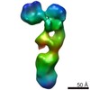
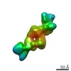
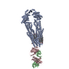
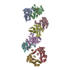
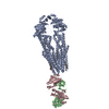
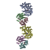
| Title | Negative stain reconstruction of human autophagy associated protein, ATG2A | |||||||||
 Map data Map data | Negative stain reconstruction of human ATG2A protein | |||||||||
 Sample Sample |
| |||||||||
| Function / homology |  Function and homology information Function and homology informationorganelle membrane contact site / lipid transfer activity / positive regulation of autophagosome assembly / nucleophagy / piecemeal microautophagy of the nucleus / phagophore assembly site membrane /  phosphatidylinositol-3-phosphate binding / phagophore assembly site / reticulophagy / autophagosome assembly ...organelle membrane contact site / lipid transfer activity / positive regulation of autophagosome assembly / nucleophagy / piecemeal microautophagy of the nucleus / phagophore assembly site membrane / phosphatidylinositol-3-phosphate binding / phagophore assembly site / reticulophagy / autophagosome assembly / lipid droplet / endoplasmic reticulum membrane phosphatidylinositol-3-phosphate binding / phagophore assembly site / reticulophagy / autophagosome assembly ...organelle membrane contact site / lipid transfer activity / positive regulation of autophagosome assembly / nucleophagy / piecemeal microautophagy of the nucleus / phagophore assembly site membrane / phosphatidylinositol-3-phosphate binding / phagophore assembly site / reticulophagy / autophagosome assembly / lipid droplet / endoplasmic reticulum membraneSimilarity search - Function | |||||||||
| Biological species |  Homo sapiens (human) Homo sapiens (human) | |||||||||
| Method | single particle reconstruction / negative staining / Resolution: 29.4 Å | |||||||||
 Authors Authors | Chowdhury S / Otomo C / Leitner A / Ohashi K / Aebersold R / Lander GC / Otomo T | |||||||||
| Funding support |  United States, European Union, 2 items United States, European Union, 2 items
| |||||||||
 Citation Citation | Journal: Proc Natl Acad Sci U S A / Year: 2018 Title: Insights into autophagosome biogenesis from structural and biochemical analyses of the ATG2A-WIPI4 complex. Authors: Saikat Chowdhury / Chinatsu Otomo / Alexander Leitner / Kazuto Ohashi / Ruedi Aebersold / Gabriel C Lander / Takanori Otomo /  Abstract: Autophagy is an enigmatic cellular process in which double-membrane compartments, called "autophagosomes, form de novo adjacent to the endoplasmic reticulum (ER) and package cytoplasmic contents for ...Autophagy is an enigmatic cellular process in which double-membrane compartments, called "autophagosomes, form de novo adjacent to the endoplasmic reticulum (ER) and package cytoplasmic contents for delivery to lysosomes. Expansion of the precursor membrane phagophore requires autophagy-related 2 (ATG2), which localizes to the PI3P-enriched ER-phagophore junction. We combined single-particle electron microscopy, chemical cross-linking coupled with mass spectrometry, and biochemical analyses to characterize human ATG2A in complex with the PI3P effector WIPI4. ATG2A is a rod-shaped protein that can bridge neighboring vesicles through interactions at each of its tips. WIPI4 binds to one of the tips, enabling the ATG2A-WIPI4 complex to tether a PI3P-containing vesicle to another PI3P-free vesicle. These data suggest that the ATG2A-WIPI4 complex mediates ER-phagophore association and/or tethers vesicles to the ER-phagophore junction, establishing the required organization for phagophore expansion via the transfer of lipid membranes from the ER and/or the vesicles to the phagophore. | |||||||||
| History |
|
- Structure visualization
| Movie |
Movie viewer |
|---|---|
| Structure viewer | EM map: SurfViewMolmilJmol/JSmol |
| Supplemental images |

- Downloads & links
-EMDB archive
| Map data | emd_8900.map.gz | 249.7 KB | EMDB map data format | |
|---|---|---|---|---|
| Header (meta data) | emd-8900-v30.xmlemd-8900.xml | 14.9 KB 14.9 KB | Display Display | EMDB header |
| Images |  emd_8900.png emd_8900.png | 26.4 KB | ||
| Others | emd_8900_additional.map.gz | 2.4 MB | ||
| Archive directory |  http://ftp.pdbj.org/pub/emdb/structures/EMD-8900ftp://ftp.pdbj.org/pub/emdb/structures/EMD-8900 http://ftp.pdbj.org/pub/emdb/structures/EMD-8900ftp://ftp.pdbj.org/pub/emdb/structures/EMD-8900 | HTTPS FTP |
-Related structure data
-Links
| EMDB pages | EMDB (EBI/PDBe) / EMDataResource |
|---|
-Map
| File | Download / File: emd_8900.map.gz / Format: CCP4 / Size: 3.4 MB / Type: IMAGE STORED AS FLOATING POINT NUMBER (4 BYTES) | ||||||||||||||||||||||||||||||||||||||||||||||||||||||||||||||||||||
|---|---|---|---|---|---|---|---|---|---|---|---|---|---|---|---|---|---|---|---|---|---|---|---|---|---|---|---|---|---|---|---|---|---|---|---|---|---|---|---|---|---|---|---|---|---|---|---|---|---|---|---|---|---|---|---|---|---|---|---|---|---|---|---|---|---|---|---|---|---|
| Annotation | Negative stain reconstruction of human ATG2A protein | ||||||||||||||||||||||||||||||||||||||||||||||||||||||||||||||||||||
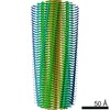
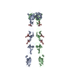
| Projections & slices | Image control
Images are generated by Spider. | ||||||||||||||||||||||||||||||||||||||||||||||||||||||||||||||||||||
| Voxel size | X=Y=Z: 4.1 Å | ||||||||||||||||||||||||||||||||||||||||||||||||||||||||||||||||||||
| Density |
| ||||||||||||||||||||||||||||||||||||||||||||||||||||||||||||||||||||
| Symmetry | Space group: 1 | ||||||||||||||||||||||||||||||||||||||||||||||||||||||||||||||||||||
| Details | EMDB XML:
CCP4 map header:
| ||||||||||||||||||||||||||||||||||||||||||||||||||||||||||||||||||||
 Z (Sec.)
 Y (Row.)
 X (Col.)


-Supplemental data
-Additional map: Unsharpened negative stain EM map of human ATG2A protein
| File | emd_8900_additional.map | ||||||||||||
|---|---|---|---|---|---|---|---|---|---|---|---|---|---|
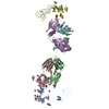
| Annotation | Unsharpened negative stain EM map of human ATG2A protein | ||||||||||||
| Projections & Slices |
| ||||||||||||
| Density Histograms |


- Sample components
-Entire : Human ATG2A protein
| Entire | Name: Human ATG2A protein |
|---|---|
| Components |
|
-Supramolecule #1: Human ATG2A protein
| Supramolecule | Name: Human ATG2A protein / type: complex / ID: 1 / Parent: 0 / Details: Full length protein |
|---|---|
| Source (natural) | Organism: Homo sapiens (human) |
| Recombinant expression | Organism:   Spodoptera frugiperda (fall armyworm) / Recombinant strain: 9 Spodoptera frugiperda (fall armyworm) / Recombinant strain: 9 |
| Molecular weight | Theoretical: 200 KDa |
-Experimental details
-Structure determination
| Method | negative staining |
|---|---|
 Processing Processing | single particle reconstruction |
| Aggregation state | particle |
-Sample preparation
| Concentration | 0.01 mg/mL |
|---|---|
| Buffer | pH: 7.5 |
| Staining | Type: NEGATIVE / Material: Uranyl Formate |
| Grid | Model: Maxtaform / Material: COPPER/RHODIUM / Mesh: 400 / Support film - Material: CARBON / Support film - topology: CONTINUOUS / Pretreatment - Type: GLOW DISCHARGE / Pretreatment - Atmosphere: AIR |
- Electron microscopy
| Microscope | FEI TECNAI SPIRIT |
|---|---|
| Electron beam | Acceleration voltage: 120 kV / Electron source: LAB6 |
| Electron optics | C2 aperture diameter: 100.0 µm / Calibrated defocus max: 2.0 µm / Calibrated defocus min: 0.62 µm / Calibrated magnification: 52000 / Illumination mode: FLOOD BEAM / Imaging mode: BRIGHT FIELDBright-field microscopy / Cs: 2.2 mm / Nominal defocus max: 1.5 µm / Nominal defocus min: 1.0 µm / Nominal magnification: 52000 |
| Sample stage | Specimen holder model: SIDE ENTRY, EUCENTRIC |
| Temperature | Min: 273.15 K / Max: 298.15 K |
| Image recording | Film or detector model: TVIPS TEMCAM-F416 (4k x 4k) / Digitization - Dimensions - Width: 4096 pixel / Digitization - Dimensions - Height: 4096 pixel / Digitization - Sampling interval: 15.6 µm / Number grids imaged: 1 / Number real images: 590 / Average exposure time: 0.42 sec. / Average electron dose: 20.0 e/Å2 |
| Experimental equipment |  Model: Tecnai Spirit / Image courtesy: FEI Company |
-Image processing
| Particle selection | Number selected: 37021 |
|---|---|
| CTF correction | Software - Name: RELION (ver. 1.4) |
| Startup model | Type of model: EMDB MAP EMDB ID: Details: A negative stain reconstruction of human ATG2-WIPI4 complex was low pass filtered to 60 Angstrom and used as initial model. |
| Initial angle assignment | Type: PROJECTION MATCHING |
| Final 3D classification | Software - Name: RELION (ver. 1.4) |
| Final angle assignment | Type: PROJECTION MATCHING / Software - Name: RELION (ver. 1.4) |
| Final reconstruction | Applied symmetry - Point group: C1 (asymmetric) / Resolution.type: BY AUTHOR / Resolution: 29.4 Å / Resolution method: FSC 0.143 CUT-OFF / Software - Name: RELION (ver. 1.4) / Number images used: 6386 |
