 Movie
Movie Controller

[English] 日本語
 Yorodumi
- EMDB-7072: RagA/RagC:Ragulator complex structure determined by single partic... -
+ Open data

- Basic information
| Entry | Database: EMDB / ID: EMD-7072 | |||||||||
|---|---|---|---|---|---|---|---|---|---|---|
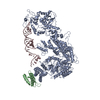
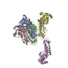
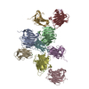
| Title | RagA/RagC:Ragulator complex structure determined by single particle negative stain electron microscopy | |||||||||
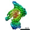
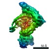
 Map data Map data | RagA/RagC Ragulator complex determined by single particle negative stain electron microscopy | |||||||||
 Sample Sample |
| |||||||||
| Biological species |   Homo sapiens (human) Homo sapiens (human) | |||||||||
| Method | single particle reconstruction / negative staining / Resolution: 16.2 Å | |||||||||
 Authors Authors | Morris KL / Su M / Kim DJ / Fu Y / Lawrence R / Stjepanovic G / Zoncu R / Hurley JH | |||||||||
| Funding support |  United States, 1 items United States, 1 items
| |||||||||
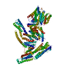
 Citation Citation | Journal: Mol Cell / Year: 2017 Title: Hybrid Structure of the RagA/C-Ragulator mTORC1 Activation Complex. Authors: Ming-Yuan Su / Kyle L Morris / Do Jin Kim / Yangxue Fu / Rosalie Lawrence / Goran Stjepanovic / Roberto Zoncu / James H Hurley / Abstract: The lysosomal membrane is the locus for sensing cellular nutrient levels, which are transduced to mTORC1 via the Rag GTPases and the Ragulator complex. The crystal structure of the five-subunit human ...The lysosomal membrane is the locus for sensing cellular nutrient levels, which are transduced to mTORC1 via the Rag GTPases and the Ragulator complex. The crystal structure of the five-subunit human Ragulator at 1.4 Å resolution was determined. Lamtor1 wraps around the other four subunits to stabilize the assembly. The Lamtor2:Lamtor3 dimer stacks upon Lamtor4:Lamtor5 to create a platform for Rag binding. Hydrogen-deuterium exchange was used to map the Rag binding site to the outer face of the Lamtor2:Lamtor3 dimer and to the N-terminal intrinsically disordered region of Lamtor1. EM was used to reconstruct the assembly of the full-length RagA:RagC dimer bound to Ragulator at 16 Å resolution, revealing that the G-domains of the Rags project away from the Ragulator core. The combined structural model shows how Ragulator functions as a platform for the presentation of active Rags for mTORC1 recruitment, and might suggest an unconventional mechanism for Rag GEF activity. | |||||||||
| History |
|
- Structure visualization
| Movie |
 Movie viewer Movie viewer |
|---|---|
| Structure viewer | EM map: SurfViewMolmilJmol/JSmol |
| Supplemental images |

- Downloads & links
-EMDB archive
| Map data | emd_7072.map.gz | 13.2 MB | EMDB map data format | |
|---|---|---|---|---|
| Header (meta data) | emd-7072-v30.xmlemd-7072.xml | 9.2 KB 9.2 KB | Display Display | EMDB header |
| FSC (resolution estimation) | emd_7072_fsc.xml | 7 KB | Display | FSC data file |
| Images | 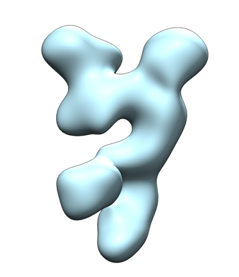 emd_7072.png emd_7072.png | 55 KB | ||
| Archive directory |  http://ftp.pdbj.org/pub/emdb/structures/EMD-7072ftp://ftp.pdbj.org/pub/emdb/structures/EMD-7072 http://ftp.pdbj.org/pub/emdb/structures/EMD-7072ftp://ftp.pdbj.org/pub/emdb/structures/EMD-7072 | HTTPS FTP |
-Related structure data


-Links
| EMDB pages | EMDB (EBI/PDBe) / EMDataResource |
|---|
-Map
| File | Download / File: emd_7072.map.gz / Format: CCP4 / Size: 27 MB / Type: IMAGE STORED AS FLOATING POINT NUMBER (4 BYTES) | ||||||||||||||||||||||||||||||||||||||||||||||||||||||||||||||||||||
|---|---|---|---|---|---|---|---|---|---|---|---|---|---|---|---|---|---|---|---|---|---|---|---|---|---|---|---|---|---|---|---|---|---|---|---|---|---|---|---|---|---|---|---|---|---|---|---|---|---|---|---|---|---|---|---|---|---|---|---|---|---|---|---|---|---|---|---|---|---|
| Annotation | RagA/RagC Ragulator complex determined by single particle negative stain electron microscopy | ||||||||||||||||||||||||||||||||||||||||||||||||||||||||||||||||||||
| Voxel size | X=Y=Z: 1.5 Å | ||||||||||||||||||||||||||||||||||||||||||||||||||||||||||||||||||||
| Density |
| ||||||||||||||||||||||||||||||||||||||||||||||||||||||||||||||||||||
| Symmetry | Space group: 1 | ||||||||||||||||||||||||||||||||||||||||||||||||||||||||||||||||||||
| Details | EMDB XML:
CCP4 map header:
| ||||||||||||||||||||||||||||||||||||||||||||||||||||||||||||||||||||
-Supplemental data
- Sample components
-Entire : RagA/RagC:Ragulator
| Entire | Name: RagA/RagC:Ragulator |
|---|---|
| Components |
|
-Supramolecule #1: RagA/RagC:Ragulator
| Supramolecule | Name: RagA/RagC:Ragulator / type: complex / ID: 1 / Parent: 0 |
|---|---|
| Source (natural) | Organism: Homo sapiens (human) |
| Recombinant expression | Organism: Insect cell expression vector pTIE1 (others) |
| Molecular weight | Theoretical: 150 KDa |
-Experimental details
-Structure determination
| Method | negative staining |
|---|---|
 Processing Processing | single particle reconstruction |
| Aggregation state | particle |
-Sample preparation
| Buffer | pH: 7.5 |
|---|---|
| Staining | Type: NEGATIVE / Material: Uranyl Formate |
- Electron microscopy
| Microscope | FEI TECNAI F20 |
|---|---|
| Electron beam | Acceleration voltage: 120 kV / Electron source: FIELD EMISSION GUN |
| Electron optics | Illumination mode: FLOOD BEAM / Imaging mode: BRIGHT FIELDBright-field microscopy |
| Image recording | Film or detector model: GATAN ULTRASCAN 4000 (4k x 4k) / Average electron dose: 30.0 e/Å2 |
| Experimental equipment |  Model: Tecnai F20 / Image courtesy: FEI Company |
-Image processing
| CTF correction | Software - Name: Gctf |
|---|---|
| Startup model | Type of model: OTHER / Details: cryosparc ab-initio |
| Initial angle assignment | Type: PROJECTION MATCHING |
| Final angle assignment | Type: PROJECTION MATCHING / Software - Name: cryoSPARC |
| Final reconstruction | Applied symmetry - Point group: C1 (asymmetric) / Resolution.type: BY AUTHOR / Resolution: 16.2 Å / Resolution method: FSC 0.143 CUT-OFF / Software - Name: cryoSPARC / Number images used: 24061 |
| FSC plot (resolution estimation) | 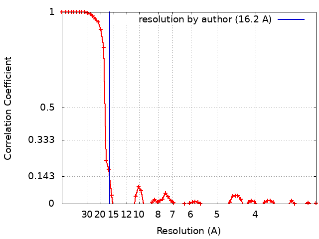 |
