 Movie
Movie Controller


+ Open data

- Basic information
| Entry | Database: PDB / ID: 5xsy | |||||||||
|---|---|---|---|---|---|---|---|---|---|---|
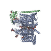
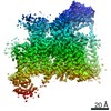
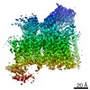
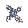
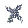
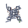
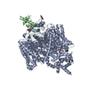
| Title | Structure of the Nav1.4-beta1 complex from electric eel | |||||||||
 Components Components |
| |||||||||
 Keywords Keywords |  MEMBRANE PROTEIN / voltage gated sodium channel MEMBRANE PROTEIN / voltage gated sodium channel | |||||||||
| Function / homology |  Function and homology information Function and homology informationvoltage-gated monoatomic ion channel activity / voltage-gated sodium channel complex / sodium channel activity / voltage-gated sodium channel activity / regulation of monoatomic ion transmembrane transport / sodium channel regulator activitySimilarity search - Function | |||||||||
| Biological species |  Electrophorus electricus (electric eel) Electrophorus electricus (electric eel) | |||||||||
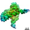
| Method | ELECTRON MICROSCOPY / single particle reconstruction / cryo EM / Resolution: 4 Å | |||||||||
 Authors Authors | Yan, Z. / Zhou, Q. / Wu, J.P. / Yan, N. | |||||||||
| Funding support |  China, 2items China, 2items
| |||||||||
 Citation Citation | Journal: Cell / Year: 2017 Title: Structure of the Na1.4-β1 Complex from Electric Eel. Authors: Zhen Yan / Qiang Zhou / Lin Wang / Jianping Wu / Yanyu Zhao / Gaoxingyu Huang / Wei Peng / Huaizong Shen / Jianlin Lei / Nieng Yan / Abstract: Voltage-gated sodium (Na) channels initiate and propagate action potentials. Here, we present the cryo-EM structure of EeNa1.4, the Na channel from electric eel, in complex with the β1 subunit at 4. ...Voltage-gated sodium (Na) channels initiate and propagate action potentials. Here, we present the cryo-EM structure of EeNa1.4, the Na channel from electric eel, in complex with the β1 subunit at 4.0 Å resolution. The immunoglobulin domain of β1 docks onto the extracellular L5 and L6 loops of EeNa1.4 via extensive polar interactions, and the single transmembrane helix interacts with the third voltage-sensing domain (VSD). The VSDs exhibit "up" conformations, while the intracellular gate of the pore domain is kept open by a digitonin-like molecule. Structural comparison with closed NaPaS shows that the outward transfer of gating charges is coupled to the iris-like pore domain dilation through intricate force transmissions involving multiple channel segments. The IFM fast inactivation motif on the III-IV linker is plugged into the corner enclosed by the outer S4-S5 and inner S6 segments in repeats III and IV, suggesting a potential allosteric blocking mechanism for fast inactivation. | |||||||||
| History |
|
- Structure visualization
| Movie |
Movie viewer |
|---|---|
| Structure viewer | Molecule: MolmilJmol/JSmol |
- Downloads & links
-Download
| PDBx/mmCIF format | 5xsy.cif.gz | 285.1 KB | Display | PDBx/mmCIF format |
|---|---|---|---|---|
| PDB format | pdb5xsy.ent.gz | 223.5 KB | Display | PDB format |
| PDBx/mmJSON format | 5xsy.json.gz | Tree view | PDBx/mmJSON format | |
| Others |  Other downloads Other downloads |
-Validation report
| Arichive directory | https://data.pdbj.org/pub/pdb/validation_reports/xs/5xsyftp://data.pdbj.org/pub/pdb/validation_reports/xs/5xsy | HTTPS FTP |
|---|
-Related structure data
| Related structure data |  6770MC M: map data used to model this data C: citing same article ( |
|---|---|
| Similar structure data |
-Links
 PDBj
PDBj
- Assembly
| Deposited unit | 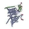
|
|---|---|
| 1 |
|
-Components
| #1: Protein | / Na(+) channel Mass: 208519.797 Da / Num. of mol.: 1 / Source method: isolated from a natural source / Source: (natural) Electrophorus electricus (electric eel) / References: UniProt: P02719 | ||
|---|---|---|---|
| #2: Protein | Mass: 23657.322 Da / Num. of mol.: 1 / Source method: isolated from a natural source / Source: (natural) Electrophorus electricus (electric eel) / References: UniProt: A0A1L3MZ94 | ||
| #3: Polysaccharide | beta-D-mannopyranose-(1-3)-[beta-D-mannopyranose-(1-6)]beta-D-mannopyranose-(1-4)-2-acetamido-2- ...beta-D-mannopyranose-(1-3)-[beta-D-mannopyranose-(1-6)]beta-D-mannopyranose-(1-4)-2-acetamido-2-deoxy-beta-D-glucopyranose-(1-4)-2-acetamido-2-deoxy-beta-D-glucopyranose / Mass: 910.823 Da / Num. of mol.: 1 Source method: isolated from a genetically manipulated source | ||
| #4: Polysaccharide | / Mass: 586.542 Da / Num. of mol.: 3 Source method: isolated from a genetically manipulated source #5: Polysaccharide | 2-acetamido-2-deoxy-beta-D-glucopyranose-(1-4)-2-acetamido-2-deoxy-beta-D-glucopyranose | / Mass: 424.401 Da / Num. of mol.: 1Source method: isolated from a genetically manipulated source |
-Experimental details
-Experiment
| Experiment | Method: ELECTRON MICROSCOPY |
|---|---|
| EM experiment | Aggregation state: PARTICLE / 3D reconstruction method: single particle reconstruction |
- Sample preparation
| Component | Name: voltage gated sodium channel EeNav1.4 / Type: ORGANELLE OR CELLULAR COMPONENT / Entity ID: #1-#2 / Source: NATURAL |
|---|---|
| Source (natural) | Organism: Electrophorus electricus (electric eel) |
| Buffer solution | pH: 7.4 |
| Specimen | Conc.: 1 mg/ml / Embedding applied: NO / Shadowing applied: NO / Staining applied: NO / Vitrification applied: YES |
| Vitrification | Cryogen name: ETHANE |
- Electron microscopy imaging
| Experimental equipment |  Model: Titan Krios / Image courtesy: FEI Company |
|---|---|
| Microscopy | Model: FEI TITAN KRIOS |
| Electron gun | Electron source: FIELD EMISSION GUN / Accelerating voltage: 300 kV / Illumination mode: FLOOD BEAM |
| Electron lens | Mode: BRIGHT FIELDBright-field microscopy / Calibrated defocus min: 1200 nm / Calibrated defocus max: 2200 nm |
| Image recording | Electron dose: 1.5 e/Å2 / Film or detector model: GATAN K2 SUMMIT (4k x 4k) |
| Image scans | Movie frames/image: 32 |
- Processing
| Software | Name: PHENIX / Version: dev_2405: / Classification: refinement | ||||||||||||||||||||||||
|---|---|---|---|---|---|---|---|---|---|---|---|---|---|---|---|---|---|---|---|---|---|---|---|---|---|
| CTF correction | Type: PHASE FLIPPING AND AMPLITUDE CORRECTION | ||||||||||||||||||||||||
| 3D reconstruction | Resolution: 4 Å / Resolution method: FSC 0.143 CUT-OFF / Num. of particles: 123431 / Symmetry type: POINT | ||||||||||||||||||||||||
| Refinement | Highest resolution: 4 Å | ||||||||||||||||||||||||
| Refine LS restraints |
|
