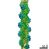
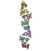
PROTEIN TRANSPORT / helical polymer / bacterial secretion / cryo-EM
Function / homology
Function and homology information
protein secretion by the type II secretion system / type II protein secretion system complex / membrane => GO:0016020 / plasma membrane Similarity search - Function
Type II secretion system protein GspG / Type II secretion system protein GspG, C-terminal / Type II secretion system (T2SS), protein G / Bacterial general secretion pathway protein G-type pilin / Prokaryotic N-terminal methylation site. / Prokaryotic N-terminal methylation motif / Prokaryotic N-terminal methylation site / Pilin-like Similarity search - Domain/homology
National Institutes of Health/National Institute of General Medical Sciences (NIH/NIGMS)
GM122510
United States
Agence Nationale de la Recherche
ANR-14-CE09-0004
France
European Union (EU)
FP7-IDEAS-ERC 294809
European Union
Citation
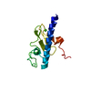
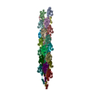
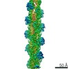
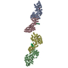
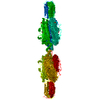
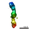
Journal: Nat Microbiol / Year: 2017 Title: Structure of the calcium-dependent type 2 secretion pseudopilus. Authors: Aracelys López-Castilla / Jenny-Lee Thomassin / Benjamin Bardiaux / Weili Zheng / Mangayarkarasi Nivaskumar / Xiong Yu / Michael Nilges / Edward H Egelman / Nadia Izadi-Pruneyre / Olivera Francetic / Abstract: Many Gram-negative bacteria use type 2 secretion systems (T2SSs) to secrete proteins involved in virulence and adaptation. Transport of folded proteins via T2SS nanomachines requires the assembly of ...Many Gram-negative bacteria use type 2 secretion systems (T2SSs) to secrete proteins involved in virulence and adaptation. Transport of folded proteins via T2SS nanomachines requires the assembly of inner membrane-anchored fibres called pseudopili. Although efficient pseudopilus assembly is essential for protein secretion, structure-based functional analyses are required to unravel the mechanistic link between these processes. Here, we report an atomic model for a T2SS pseudopilus from Klebsiella oxytoca, obtained by fitting the NMR structure of its calcium-bound subunit PulG into the ~5-Å-resolution cryo-electron microscopy reconstruction of assembled fibres. This structure reveals the comprehensive network of inter-subunit contacts and unexpected features, including a disordered central region of the PulG helical stem, and highly flexible C-terminal residues on the fibre surface. NMR, mutagenesis and functional analyses highlight the key role of calcium in PulG folding and stability. Fibre disassembly in the absence of calcium provides a basis for pseudopilus length control, essential for protein secretion, and supports the Archimedes screw model for the type 2 secretion mechanism.
A: General secretion pathway protein G B: General secretion pathway protein G C: General secretion pathway protein G D: General secretion pathway protein G E: General secretion pathway protein G F: General secretion pathway protein G G: General secretion pathway protein G H: General secretion pathway protein G I: General secretion pathway protein G J: General secretion pathway protein G K: General secretion pathway protein G L: General secretion pathway protein G M: General secretion pathway protein G N: General secretion pathway protein G O: General secretion pathway protein G P: General secretion pathway protein G Q: General secretion pathway protein G R: General secretion pathway protein G S: General secretion pathway protein G T: General secretion pathway protein G U: General secretion pathway protein G V: General secretion pathway protein G W: General secretion pathway protein G X: General secretion pathway protein G Y: General secretion pathway protein G hetero molecules
Mass: 40.078 Da / Num. of mol.: 25 / Source method: obtained synthetically / Formula: Ca
-
Experimental details
-
Experiment
Experiment
Method: ELECTRON MICROSCOPY
EM experiment
Aggregation state: FILAMENT / 3D reconstruction method: helical reconstruction
-
Sample preparation
Component
Name: PulG pseudopilus / Type: ORGANELLE OR CELLULAR COMPONENT / Entity ID: all / Source: RECOMBINANT
Molecular weight
Experimental value: NO
Source (natural)
Organism: Klebsiella oxytoca (bacteria)
Source (recombinant)
Organism: Escherichia coli (E. coli)
Buffer solution
pH: 7.5
Specimen
Embedding applied: NO / Shadowing applied: NO / Staining applied: NO / Vitrification applied: YES
Vitrification
Instrument: FEI VITROBOT MARK IV / Cryogen name: ETHANE
-
Electron microscopy imaging
Experimental equipment
Model: Titan Krios / Image courtesy: FEI Company
Microscopy
Model: FEI TITAN KRIOS
Electron gun
Electron source: FIELD EMISSION GUN / Accelerating voltage: 300 kV / Illumination mode: FLOOD BEAM
Electron lens
Mode: BRIGHT FIELDBright-field microscopy
Image recording
Electron dose: 20 e/Å2 / Detector mode: INTEGRATING / Film or detector model: FEI FALCON II (4k x 4k) / Num. of grids imaged: 1 / Num. of real images: 1819
In the structure databanks used in Yorodumi, some data are registered as the other names, "COVID-19 virus" and "2019-nCoV". Here are the details of the virus and the list of structure data.
Jan 31, 2019. EMDB accession codes are about to change! (news from PDBe EMDB page)
EMDB accession codes are about to change! (news from PDBe EMDB page)
The allocation of 4 digits for EMDB accession codes will soon come to an end. Whilst these codes will remain in use, new EMDB accession codes will include an additional digit and will expand incrementally as the available range of codes is exhausted. The current 4-digit format prefixed with “EMD-” (i.e. EMD-XXXX) will advance to a 5-digit format (i.e. EMD-XXXXX), and so on. It is currently estimated that the 4-digit codes will be depleted around Spring 2019, at which point the 5-digit format will come into force.
The EM Navigator/Yorodumi systems omit the EMD- prefix.
Related info.:Q: What is EMD? / ID/Accession-code notation in Yorodumi/EM Navigator
Yorodumi is a browser for structure data from EMDB, PDB, SASBDB, etc.
This page is also the successor to EM Navigator detail page, and also detail information page/front-end page for Omokage search.
The word "yorodu" (or yorozu) is an old Japanese word meaning "ten thousand". "mi" (miru) is to see.
Related info.:EMDB / PDB / SASBDB / Comparison of 3 databanks / Yorodumi Search / Aug 31, 2016. New EM Navigator & Yorodumi / Yorodumi Papers / Jmol/JSmol / Function and homology information / Changes in new EM Navigator and Yorodumi
 Movie
Movie Controller


 Open data
 Basic information
 Components
 Keywords
 PROTEIN TRANSPORT / helical polymer / bacterial secretion /
  Function and homology information
 Authors
 United States,
  France, European Union, 3items
  Citation
 Structure visualization
 Downloads & links
 Other downloads

 PDBj
PDBj
 Assembly

 Mass: 40.078 Da / Num. of mol.: 25 / Source method: obtained synthetically / Formula: Ca
 Sample preparation
 Electron microscopy imaging
 Processing