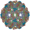
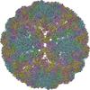
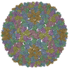
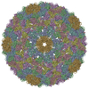
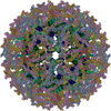
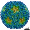
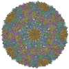
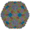
Journal: Proc Natl Acad Sci U S A / Year: 2017 Title: Virus found in a boreal lake links ssDNA and dsDNA viruses. Authors: Elina Laanto / Sari Mäntynen / Luigi De Colibus / Jenni Marjakangas / Ashley Gillum / David I Stuart / Janne J Ravantti / Juha T Huiskonen / Lotta-Riina Sundberg / Abstract: Viruses have impacted the biosphere in numerous ways since the dawn of life. However, the evolution, genetic, structural, and taxonomic diversity of viruses remain poorly understood, in part because ...Viruses have impacted the biosphere in numerous ways since the dawn of life. However, the evolution, genetic, structural, and taxonomic diversity of viruses remain poorly understood, in part because sparse sampling of the virosphere has concentrated mostly on exploring the abundance and diversity of dsDNA viruses. Furthermore, viral genomes are highly diverse, and using only the current sequence-based methods for classifying viruses and studying their phylogeny is complicated. Here we describe a virus, FLiP (-infecting, lipid-containing phage), with a circular ssDNA genome and an internal lipid membrane enclosed in the icosahedral capsid. The 9,174-nt-long genome showed limited sequence similarity to other known viruses. The genetic data imply that this virus might use replication mechanisms similar to those found in other ssDNA replicons. However, the structure of the viral major capsid protein, elucidated at near-atomic resolution using cryo-electron microscopy, is strikingly similar to that observed in dsDNA viruses of the PRD1-adenovirus lineage, characterized by a major capsid protein bearing two β-barrels. The strong similarity between FLiP and another member of the structural lineage, bacteriophage PM2, extends to the capsid organization (pseudo = 21 ) despite the difference in the genetic material packaged and the lack of significant sequence similarity.
A: Major capsid protein B: Major capsid protein C: Major capsid protein D: Major capsid protein E: Major capsid protein F: Major capsid protein G: Major capsid protein H: Major capsid protein I: Major capsid protein J: Major capsid protein
A: Major capsid protein B: Major capsid protein C: Major capsid protein D: Major capsid protein E: Major capsid protein F: Major capsid protein G: Major capsid protein H: Major capsid protein I: Major capsid protein J: Major capsid protein
Idetical with deposited unit in distinct coordinate
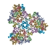
icosahedral asymmetric unit
Type
Name
Symmetry operation
Number
point symmetry operation
1
3
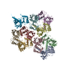
A: Major capsid protein B: Major capsid protein C: Major capsid protein D: Major capsid protein E: Major capsid protein F: Major capsid protein G: Major capsid protein H: Major capsid protein I: Major capsid protein J: Major capsid protein
x 5
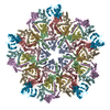
icosahedral pentamer
1.73 MDa, 50 polymers
Theoretical mass
Number of molelcules
Total (without water)
1,726,697
50
Polymers
1,726,697
50
Non-polymers
0
0
Water
0
Type
Name
Symmetry operation
Number
point symmetry operation
5
4
A: Major capsid protein B: Major capsid protein C: Major capsid protein D: Major capsid protein E: Major capsid protein F: Major capsid protein G: Major capsid protein H: Major capsid protein I: Major capsid protein J: Major capsid protein
x 6
icosahedral 23 hexamer
2.07 MDa, 60 polymers
Theoretical mass
Number of molelcules
Total (without water)
2,072,036
60
Polymers
2,072,036
60
Non-polymers
0
0
Water
0
Type
Name
Symmetry operation
Number
point symmetry operation
6
5
Idetical with deposited unit in distinct coordinate
icosahedral asymmetric unit, std point frame
Type
Name
Symmetry operation
Number
transform to point frame
1
Symmetry
Point symmetry: (Schoenflies symbol: I (icosahedral))
-
Components
#1: Protein
Majorcapsidprotein
Mass: 34533.934 Da / Num. of mol.: 10 / Source method: isolated from a natural source / Source: (natural) unidentified phage (virus) / References: UniProt: A0A2D0TC94*PLUS
-
Experimental details
-
Experiment
Experiment
Method: ELECTRON MICROSCOPY
EM experiment
Aggregation state: PARTICLE / 3D reconstruction method: single particle reconstruction
In the structure databanks used in Yorodumi, some data are registered as the other names, "COVID-19 virus" and "2019-nCoV". Here are the details of the virus and the list of structure data.
Jan 31, 2019. EMDB accession codes are about to change! (news from PDBe EMDB page)
EMDB accession codes are about to change! (news from PDBe EMDB page)
The allocation of 4 digits for EMDB accession codes will soon come to an end. Whilst these codes will remain in use, new EMDB accession codes will include an additional digit and will expand incrementally as the available range of codes is exhausted. The current 4-digit format prefixed with “EMD-” (i.e. EMD-XXXX) will advance to a 5-digit format (i.e. EMD-XXXXX), and so on. It is currently estimated that the 4-digit codes will be depleted around Spring 2019, at which point the 5-digit format will come into force.
The EM Navigator/Yorodumi systems omit the EMD- prefix.
Related info.:Q: What is EMD? / ID/Accession-code notation in Yorodumi/EM Navigator
Yorodumi is a browser for structure data from EMDB, PDB, SASBDB, etc.
This page is also the successor to EM Navigator detail page, and also detail information page/front-end page for Omokage search.
The word "yorodu" (or yorozu) is an old Japanese word meaning "ten thousand". "mi" (miru) is to see.
Related info.:EMDB / PDB / SASBDB / Comparison of 3 databanks / Yorodumi Search / Aug 31, 2016. New EM Navigator & Yorodumi / Yorodumi Papers / Jmol/JSmol / Function and homology information / Changes in new EM Navigator and Yorodumi
 Movie
Movie Controller


 Open data
 Basic information
 Components
 Keywords
 VIRUS /
  Function and homology information
 Authors
 Citation

 Structure visualization
 Downloads & links
 Other downloads
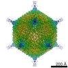
 PDBj
PDBj Assembly
 Sample preparation
 Electron microscopy imaging
 Processing