 Movie
Movie Controller


+ Open data

- Basic information
| Entry | Database: PDB / ID: 5oa9 | ||||||
|---|---|---|---|---|---|---|---|
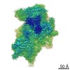
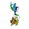
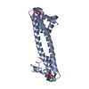
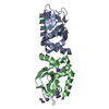
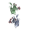
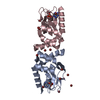
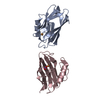
| Title | Human translation re-initiation complex containing eIF2D | ||||||
 Components Components | Eukaryotic translation initiation factor 2D | ||||||
 Keywords Keywords |  TRANSLATION / translation re-initiation complex / translation initiation factor / RNA binding protein / small ribosomal subunit TRANSLATION / translation re-initiation complex / translation initiation factor / RNA binding protein / small ribosomal subunit | ||||||
| Function / homology |  Function and homology information Function and homology informationIRES-dependent viral translational initiation / ribosome disassembly / formation of translation preinitiation complex / translation initiation factor activity / intracellular protein transport / signaling receptor activity / nuclear body / RNA binding / cytosol / cytoplasmSimilarity search - Function | ||||||
| Biological species |  Homo sapiens (human) Homo sapiens (human) | ||||||
| Method | X-RAY DIFFRACTION / SYNCHROTRON / SAD / Resolution: 1.8 Å | ||||||
 Authors Authors | Weisser, M. / Schaefer, T. / Leibundgut, M. / Boehringer, D. / Aylett, C.H.S. / Ban, N. | ||||||
 Citation Citation | Journal: Mol Cell / Year: 2017 Title: Structural and Functional Insights into Human Re-initiation Complexes. Authors: Melanie Weisser / Tanja Schäfer / Marc Leibundgut / Daniel Böhringer / Christopher Herbert Stanley Aylett / Nenad Ban /  Abstract: After having translated short upstream open reading frames, ribosomes can re-initiate translation on the same mRNA. This process, referred to as re-initiation, controls the translation of a large ...After having translated short upstream open reading frames, ribosomes can re-initiate translation on the same mRNA. This process, referred to as re-initiation, controls the translation of a large fraction of mammalian cellular mRNAs, many of which are important in cancer. Key ribosomal binding proteins involved in re-initiation are the eukaryotic translation initiation factor 2D (eIF2D) or the homologous complex of MCT-1/DENR. We determined the structures of these factors bound to the human 40S ribosomal subunit in complex with initiator tRNA positioned on an mRNA start codon in the P-site using a combination of cryoelectron microscopy and X-ray crystallography. The structures, supported by biochemical experiments, reveal how eIF2D emulates the function of several canonical translation initiation factors by using three independent, flexibly connected RNA binding domains to simultaneously monitor codon-anticodon interactions in the ribosomal P-site and position the initiator tRNA. | ||||||
| History |
|
- Structure visualization
| Structure viewer | Molecule: MolmilJmol/JSmol |
|---|
- Downloads & links
-Download
| PDBx/mmCIF format | 5oa9.cif.gz | 97.7 KB | Display | PDBx/mmCIF format |
|---|---|---|---|---|
| PDB format | pdb5oa9.ent.gz | 79.7 KB | Display | PDB format |
| PDBx/mmJSON format | 5oa9.json.gz | Tree view | PDBx/mmJSON format | |
| Others |  Other downloads Other downloads |
-Validation report
| Arichive directory | https://data.pdbj.org/pub/pdb/validation_reports/oa/5oa9ftp://data.pdbj.org/pub/pdb/validation_reports/oa/5oa9 | HTTPS FTP |
|---|
-Related structure data


-Links
 PDBj
PDBj


- Assembly
| Deposited unit | 
| ||||||||
|---|---|---|---|---|---|---|---|---|---|
| 1 |
| ||||||||
| Unit cell |
|
-Components
| #1: Protein | Mass: 23277.051 Da / Num. of mol.: 1 Source method: isolated from a genetically manipulated source Source: (gene. exp.) Homo sapiens (human) / Gene: EIF2D, HCA56, LGTN / Production host:  Escherichia coli BL21(DE3) (bacteria) / Variant (production host): Codon Plus pRIL / References: UniProt: P41214 Escherichia coli BL21(DE3) (bacteria) / Variant (production host): Codon Plus pRIL / References: UniProt: P41214 |
|---|---|
| #2: Water | ChemComp-HOH / Water Mass: 18.015 Da / Num. of mol.: 134 / Source method: isolated from a natural source / Formula: H2O Mass: 18.015 Da / Num. of mol.: 134 / Source method: isolated from a natural source / Formula: H2O |
-Experimental details
-Experiment
| Experiment | Method: X-RAY DIFFRACTION / Number of used crystals: 1 |
|---|
- Sample preparation
| Crystal | Density Matthews: 2.73 Å3/Da / Density % sol: 54.89 % |
|---|---|
| Crystal grow | Temperature: 292 K / Method: vapor diffusion, sitting drop / pH: 7.6 Details: 1:1 mix protein (50 mM HEPES-KOH pH 7.6, 150 mM KCl, 1 mM TCEP) with reservoir solution (100 mM HEPES-KOH pH 7.6, 25 mM sodium formate, 25 mM ammonium acetate, 25 mM sodium citrate tribasic, ...Details: 1:1 mix protein (50 mM HEPES-KOH pH 7.6, 150 mM KCl, 1 mM TCEP) with reservoir solution (100 mM HEPES-KOH pH 7.6, 25 mM sodium formate, 25 mM ammonium acetate, 25 mM sodium citrate tribasic, 25 mM sodium potassium tartrate, 10.7% PEG3350, 10.7% MPD, 10.7% PEG1000) |
-Data collection
| Diffraction | Mean temperature: 273 K |
|---|---|
| Diffraction source | Source: SYNCHROTRON / Site: SLS / Beamline: X06SA / Wavelength: 1 Å |
| Detector | Type: DECTRIS EIGER X 16M / Detector: PIXEL / Date: Sep 3, 2016 |
| Radiation | Protocol: SINGLE WAVELENGTH / Monochromatic (M) / Laue (L): M / Scattering type: x-ray |
| Radiation wavelength | Wavelength: 1 Å / Relative weight: 1 |
| Reflection | Resolution: 1.8→49.35 Å / Num. obs: 24936 / % possible obs: 99.85 % / Redundancy: 6.9 % / CC1/2: 0.999 / Rmerge(I) obs: 0.057 / Net I/σ(I): 14.9 |
| Reflection shell | Resolution: 1.8→1.91 Å / Rmerge(I) obs: 1.026 / CC1/2: 0.71 / % possible all: 99.9 |
- Processing
| Software |
| ||||||||||||||||||||||||||||||||||||||||||||||||||||||||||||||||||||||||||||||||||||||||||||||||||||||||||||||||||||||||||||||||||||||||||||||||||||||||||||||||||||||||||||||||||||||||||||||||||||||||
|---|---|---|---|---|---|---|---|---|---|---|---|---|---|---|---|---|---|---|---|---|---|---|---|---|---|---|---|---|---|---|---|---|---|---|---|---|---|---|---|---|---|---|---|---|---|---|---|---|---|---|---|---|---|---|---|---|---|---|---|---|---|---|---|---|---|---|---|---|---|---|---|---|---|---|---|---|---|---|---|---|---|---|---|---|---|---|---|---|---|---|---|---|---|---|---|---|---|---|---|---|---|---|---|---|---|---|---|---|---|---|---|---|---|---|---|---|---|---|---|---|---|---|---|---|---|---|---|---|---|---|---|---|---|---|---|---|---|---|---|---|---|---|---|---|---|---|---|---|---|---|---|---|---|---|---|---|---|---|---|---|---|---|---|---|---|---|---|---|---|---|---|---|---|---|---|---|---|---|---|---|---|---|---|---|---|---|---|---|---|---|---|---|---|---|---|---|---|---|---|---|---|
| Refinement | Method to determine structure: SAD / Resolution: 1.8→24.67 Å / SU ML: 0.19 / Cross valid method: FREE R-VALUE / σ(F): 0 / Phase error: 25.04 / Stereochemistry target values: ML
| ||||||||||||||||||||||||||||||||||||||||||||||||||||||||||||||||||||||||||||||||||||||||||||||||||||||||||||||||||||||||||||||||||||||||||||||||||||||||||||||||||||||||||||||||||||||||||||||||||||||||
| Solvent computation | Shrinkage radii: 0.9 Å / VDW probe radii: 1.11 Å / Solvent model: FLAT BULK SOLVENT MODEL | ||||||||||||||||||||||||||||||||||||||||||||||||||||||||||||||||||||||||||||||||||||||||||||||||||||||||||||||||||||||||||||||||||||||||||||||||||||||||||||||||||||||||||||||||||||||||||||||||||||||||
| Refinement step | Cycle: LAST / Resolution: 1.8→24.67 Å
| ||||||||||||||||||||||||||||||||||||||||||||||||||||||||||||||||||||||||||||||||||||||||||||||||||||||||||||||||||||||||||||||||||||||||||||||||||||||||||||||||||||||||||||||||||||||||||||||||||||||||
| Refine LS restraints |
| ||||||||||||||||||||||||||||||||||||||||||||||||||||||||||||||||||||||||||||||||||||||||||||||||||||||||||||||||||||||||||||||||||||||||||||||||||||||||||||||||||||||||||||||||||||||||||||||||||||||||
| LS refinement shell |
| ||||||||||||||||||||||||||||||||||||||||||||||||||||||||||||||||||||||||||||||||||||||||||||||||||||||||||||||||||||||||||||||||||||||||||||||||||||||||||||||||||||||||||||||||||||||||||||||||||||||||
| Refinement TLS params. | Method: refined / Refine-ID: X-RAY DIFFRACTION
| ||||||||||||||||||||||||||||||||||||||||||||||||||||||||||||||||||||||||||||||||||||||||||||||||||||||||||||||||||||||||||||||||||||||||||||||||||||||||||||||||||||||||||||||||||||||||||||||||||||||||
| Refinement TLS group |
|
