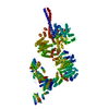
 Movie
Movie Controller


+ Open data

- Basic information
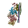
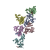
| Entry | Database: PDB / ID: 5np1 | |||||||||
|---|---|---|---|---|---|---|---|---|---|---|
| Title | Open protomer of human ATM (Ataxia telangiectasia mutated) | |||||||||
 Components Components | Serine-protein kinase ATM | |||||||||
 Keywords Keywords |  TRANSFERASE / FAT / MRN / DNA-repair / HEAT-repeats TRANSFERASE / FAT / MRN / DNA-repair / HEAT-repeats | |||||||||
| Function / homology |  Function and homology information Function and homology informationpositive regulation of DNA catabolic process / establishment of RNA localization to telomere / positive regulation of telomerase catalytic core complex assembly / cellular response to nitrosative stress / negative regulation of telomere capping / positive regulation of DNA damage response, signal transduction by p53 class mediator / establishment of protein-containing complex localization to telomere / regulation of microglial cell activation / Sensing of DNA Double Strand Breaks / positive regulation of telomere maintenance via telomere lengthening ...positive regulation of DNA catabolic process / establishment of RNA localization to telomere / positive regulation of telomerase catalytic core complex assembly / cellular response to nitrosative stress / negative regulation of telomere capping / positive regulation of DNA damage response, signal transduction by p53 class mediator / establishment of protein-containing complex localization to telomere / regulation of microglial cell activation / Sensing of DNA Double Strand Breaks / positive regulation of telomere maintenance via telomere lengthening / meiotic telomere clustering / lipoprotein catabolic process / pre-B cell allelic exclusion / DNA-dependent protein kinase activity / histone H2AXS139 kinase activity / male meiotic nuclear division / histone mRNA catabolic process / female meiotic nuclear division / regulation of telomere maintenance via telomerase / pexophagy / cellular response to X-ray / peptidyl-serine autophosphorylation / V(D)J recombination / oocyte development / Impaired BRCA2 binding to PALB2 / reciprocal meiotic recombination / DNA repair complex / Defective homologous recombination repair (HRR) due to BRCA1 loss of function / Defective HDR through Homologous Recombination Repair (HRR) due to PALB2 loss of BRCA1 binding function / Defective HDR through Homologous Recombination Repair (HRR) due to PALB2 loss of BRCA2/RAD51/RAD51C binding function / Homologous DNA Pairing and Strand Exchange / Resolution of D-loop Structures through Synthesis-Dependent Strand Annealing (SDSA) / Resolution of D-loop Structures through Holliday Junction Intermediates / HDR through Single Strand Annealing (SSA) / Impaired BRCA2 binding to RAD51 / 1-phosphatidylinositol-3-kinase activity / response to ionizing radiation / mitotic spindle assembly checkpoint signaling / TP53 Regulates Transcription of Caspase Activators and Caspases / negative regulation of B cell proliferation / mitotic G2 DNA damage checkpoint signaling / Presynaptic phase of homologous DNA pairing and strand exchange / peroxisomal matrix / TP53 Regulates Transcription of Genes Involved in Cytochrome C Release / replicative senescence / positive regulation of cell adhesion / Regulation of HSF1-mediated heat shock response / somitogenesis / regulation of cellular response to heat / DNA damage response, signal transduction by p53 class mediator resulting in cell cycle arrest / cellular response to retinoic acid / signal transduction in response to DNA damage / ovarian follicle development / negative regulation of TORC1 signaling / positive regulation of telomere maintenance via telomerase / Pexophagy / telomere maintenance / post-embryonic development / thymus development / regulation of signal transduction by p53 class mediator / DNA damage checkpoint signaling / regulation of autophagy / determination of adult lifespan / TP53 Regulates Transcription of DNA Repair Genes / Nonhomologous End-Joining (NHEJ) / Stabilization of p53 / double-strand break repair via homologous recombination / Autodegradation of the E3 ubiquitin ligase COP1 / brain development / multicellular organism growth / cellular response to gamma radiation / HDR through Homologous Recombination (HRR) / G2/M DNA damage checkpoint / Regulation of TP53 Activity through Methylation / DNA Damage/Telomere Stress Induced Senescence / spindle / Meiotic recombination / cellular response to reactive oxygen species / double-strand break repair via nonhomologous end joining / positive regulation of neuron apoptotic process / double-strand break repair / intrinsic apoptotic signaling pathway in response to DNA damage / cellular senescence / Regulation of TP53 Degradation / Recruitment and ATM-mediated phosphorylation of repair and signaling proteins at DNA double strand breaks / heart development / Processing of DNA double-strand break ends / cytoplasmic vesicle / peptidyl-serine phosphorylation / neuron apoptotic process / regulation of apoptotic process / Regulation of TP53 Activity through Phosphorylation / protein autophosphorylation / response to hypoxia / regulation of cell cycle / non-specific serine/threonine protein kinase / positive regulation of cell migration / positive regulation of apoptotic process / protein phosphorylation / protein serine kinase activitySimilarity search - Function | |||||||||
| Biological species |  Homo sapiens (human) Homo sapiens (human) | |||||||||
| Method | ELECTRON MICROSCOPY / single particle reconstruction / cryo EM / Resolution: 5.7 Å | |||||||||
 Authors Authors | Baretic, D. / Pollard, H.K. / Fisher, D.I. / Johnson, C.M. / Santhanam, B. / Truman, C.M. / Kouba, T. / Fersht, A.R. / Phillips, C. / Williams, R.L. | |||||||||
| Funding support |  United Kingdom, 2items United Kingdom, 2items
| |||||||||
 Citation Citation | Journal: Sci Adv / Year: 2017 Title: Structures of closed and open conformations of dimeric human ATM. Authors: Domagoj Baretić / Hannah K Pollard / David I Fisher / Christopher M Johnson / Balaji Santhanam / Caroline M Truman / Tomas Kouba / Alan R Fersht / Christopher Phillips / Roger L Williams / Abstract: ATM (ataxia-telangiectasia mutated) is a phosphatidylinositol 3-kinase-related protein kinase (PIKK) best known for its role in DNA damage response. ATM also functions in oxidative stress response, ...ATM (ataxia-telangiectasia mutated) is a phosphatidylinositol 3-kinase-related protein kinase (PIKK) best known for its role in DNA damage response. ATM also functions in oxidative stress response, insulin signaling, and neurogenesis. Our electron cryomicroscopy (cryo-EM) suggests that human ATM is in a dynamic equilibrium between closed and open dimers. In the closed state, the PIKK regulatory domain blocks the peptide substrate-binding site, suggesting that this conformation may represent an inactive or basally active enzyme. The active site is held in this closed conformation by interaction with a long helical hairpin in the TRD3 (tetratricopeptide repeats domain 3) domain of the symmetry-related molecule. The open dimer has two protomers with only a limited contact interface, and it lacks the intermolecular interactions that block the peptide-binding site in the closed dimer. This suggests that the open conformation may be more active. The ATM structure shows the detailed topology of the regulator-interacting N-terminal helical solenoid. The ATM conformational dynamics shown by the structures represent an important step in understanding the enzyme regulation. | |||||||||
| History |
|
- Structure visualization
| Movie |
Movie viewer |
|---|---|
| Structure viewer | Molecule: MolmilJmol/JSmol |
- Downloads & links
-Download
| PDBx/mmCIF format | 5np1.cif.gz | 374.4 KB | Display | PDBx/mmCIF format |
|---|---|---|---|---|
| PDB format | pdb5np1.ent.gz | 250.9 KB | Display | PDB format |
| PDBx/mmJSON format | 5np1.json.gz | Tree view | PDBx/mmJSON format | |
| Others |  Other downloads Other downloads |
-Validation report
| Arichive directory | https://data.pdbj.org/pub/pdb/validation_reports/np/5np1ftp://data.pdbj.org/pub/pdb/validation_reports/np/5np1 | HTTPS FTP |
|---|
-Related structure data
| Related structure data | 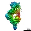 3672MC 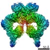 3668C  3669C 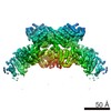 3670C 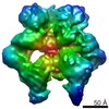 3671C 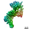 3673C 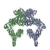 5np0C M: map data used to model this data C: citing same article ( |
|---|---|
| Similar structure data |


-Links
 PDBj
PDBj


- Assembly
| Deposited unit | 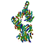
|
|---|---|
| 1 |
|
-Components
| #1: Protein | Mass: 352393.969 Da / Num. of mol.: 1 Source method: isolated from a genetically manipulated source Source: (gene. exp.) Homo sapiens (human) / Cell line: HEK293 / Gene: ATM / Plasmid: pDEST12.2-OriP / Cell line (production host): HEK293 / Production host: Homo sapiens (human)References: UniProt: Q13315, non-specific serine/threonine protein kinase |
|---|
-Experimental details
-Experiment
| Experiment | Method: ELECTRON MICROSCOPY |
|---|---|
| EM experiment | Aggregation state: PARTICLE / 3D reconstruction method: single particle reconstruction |
- Sample preparation
| Component | Name: Dimeric human ATM (Ataxia telangiectasia mutated) kinase Type: ORGANELLE OR CELLULAR COMPONENT / Entity ID: all / Source: RECOMBINANT | ||||||||||||||||||||||||||||||
|---|---|---|---|---|---|---|---|---|---|---|---|---|---|---|---|---|---|---|---|---|---|---|---|---|---|---|---|---|---|---|---|
| Molecular weight | Value: 0.705 MDa / Experimental value: YES | ||||||||||||||||||||||||||||||
| Source (natural) | Organism: Homo sapiens (human) | ||||||||||||||||||||||||||||||
| Source (recombinant) | Organism: Homo sapiens (human) / Cell: HEK293 / Plasmid: pDEST12.2-OriP | ||||||||||||||||||||||||||||||
| Buffer solution | pH: 8 | ||||||||||||||||||||||||||||||
| Buffer component |
| ||||||||||||||||||||||||||||||
| Specimen | Conc.: 0.6 mg/ml / Embedding applied: NO / Shadowing applied: NO / Staining applied: NO / Vitrification applied: YES Details: The sample was purified by anti-FLAG affinity chromatography followed by overnight dialysis and a final gel-filtration. | ||||||||||||||||||||||||||||||
| Specimen support |
| ||||||||||||||||||||||||||||||
| Vitrification | Instrument: HOMEMADE PLUNGER / Cryogen name: ETHANE / Humidity: 100 % / Chamber temperature: 277.15 K Details: 3 uL sample/grid blotted for 12 s before plunge-freezing |
- Electron microscopy imaging
| Experimental equipment |  Model: Titan Krios / Image courtesy: FEI Company |
|---|---|
| Microscopy | Model: FEI TITAN KRIOS |
| Electron gun | Electron source: FIELD EMISSION GUN / Accelerating voltage: 300 kV / Illumination mode: FLOOD BEAM |
| Electron lens | Mode: BRIGHT FIELDBright-field microscopy / Nominal magnification: 97902 X / Calibrated magnification: 35714 X / Nominal defocus max: 4000 nm / Nominal defocus min: 2500 nm / Cs: 2.7 mm / C2 aperture diameter: 70 µm / Alignment procedure: COMA FREE |
| Specimen holder | Cryogen: NITROGEN / Specimen holder model: FEI TITAN KRIOS AUTOGRID HOLDER / Temperature (min): 80.15 K |
| Image recording | Average exposure time: 0.8 sec. / Electron dose: 2.1 e/Å2 / Detector mode: SUPER-RESOLUTION / Film or detector model: GATAN K2 SUMMIT (4k x 4k) / Num. of grids imaged: 4 / Num. of real images: 2720 |
| EM imaging optics | Energyfilter name: GIF / Energyfilter upper: 20 eV / Energyfilter lower: 0 eV |
| Image scans | Movie frames/image: 20 / Used frames/image: 1-20 |
- Processing
| Software | Name: PHENIX / Version: 1.10.1_2155: / Classification: refinement | ||||||||||||||||||||||||||||||||||||||||||||||||||
|---|---|---|---|---|---|---|---|---|---|---|---|---|---|---|---|---|---|---|---|---|---|---|---|---|---|---|---|---|---|---|---|---|---|---|---|---|---|---|---|---|---|---|---|---|---|---|---|---|---|---|---|
| EM software |
| ||||||||||||||||||||||||||||||||||||||||||||||||||
| CTF correction | Type: PHASE FLIPPING ONLY | ||||||||||||||||||||||||||||||||||||||||||||||||||
| Particle selection | Num. of particles selected: 371671 | ||||||||||||||||||||||||||||||||||||||||||||||||||
| Symmetry | Point symmetry: C1 (asymmetric) | ||||||||||||||||||||||||||||||||||||||||||||||||||
| 3D reconstruction | Resolution: 5.7 Å / Resolution method: FSC 0.143 CUT-OFF / Num. of particles: 60556 / Num. of class averages: 2 / Symmetry type: POINT | ||||||||||||||||||||||||||||||||||||||||||||||||||
| Atomic model building | Space: REAL | ||||||||||||||||||||||||||||||||||||||||||||||||||
| Refine LS restraints |
|
