- PDB-5n8o: Cryo EM structure of the conjugative relaxase TraI of the F/R1 pl... -
+
Open data
ID or keywords:
Loading...
-
Basic information
Entry
Database: PDB / ID: 5n8o
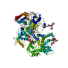
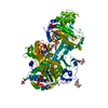
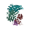
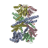
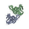
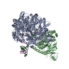
Title
Cryo EM structure of the conjugative relaxase TraI of the F/R1 plasmid system
Components
DNA (5'-D(P*TP*TP*TP*TP*TP*TP*TP*TP*TP*TP*TP*TP*TP*TP*TP*TP*TP*T)-3')
DNA helicase IHelicase
Keywords
TRANSFERASE / Relaxase / Cryo EM / Helicase / translocase
Function / homology
Function and homology information
DNA topoisomerase / DNA topoisomerase type I (single strand cut, ATP-independent) activity / hydrolase activity, acting on acid anhydrides, in phosphorus-containing anhydrides / metabolic process / DNA helicase activity / DNA helicase / ATP hydrolysis activity / DNA binding / ATP binding / metal ion binding / cytoplasm Similarity search - Function
DNA helicase, TraI type / Conjugative transfer relaxase protein TraI / TraI, 2B/2B-like domain / TraI, N-terminal subdomain / DNA helicase TraI, C-terminal / single-stranded DNA binding TraI N-terminal subdomain / DNA relaxase TraI 2B/2B-like domain / Conjugative relaxase, N-terminal / TrwC relaxase / TrwC relaxase ...DNA helicase, TraI type / Conjugative transfer relaxase protein TraI / TraI, 2B/2B-like domain / TraI, N-terminal subdomain / DNA helicase TraI, C-terminal / single-stranded DNA binding TraI N-terminal subdomain / DNA relaxase TraI 2B/2B-like domain / Conjugative relaxase, N-terminal / TrwC relaxase / TrwC relaxase / AAA domain / P-loop containing nucleoside triphosphate hydrolase Similarity search - Domain/homology
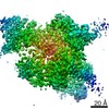
Journal: Cell / Year: 2017 Title: Cryo-EM Structure of a Relaxase Reveals the Molecular Basis of DNA Unwinding during Bacterial Conjugation. Authors: Aravindan Ilangovan / Christopher W M Kay / Sandro Roier / Hassane El Mkami / Enrico Salvadori / Ellen L Zechner / Giulia Zanetti / Gabriel Waksman / Abstract: Relaxases play essential roles in conjugation, the main process by which bacteria exchange genetic material, notably antibiotic resistance genes. They are bifunctional enzymes containing a trans- ...Relaxases play essential roles in conjugation, the main process by which bacteria exchange genetic material, notably antibiotic resistance genes. They are bifunctional enzymes containing a trans-esterase activity, which is responsible for nicking the DNA strand to be transferred and for covalent attachment to the resulting 5'-phosphate end, and a helicase activity, which is responsible for unwinding the DNA while it is being transported to a recipient cell. Here we show that these two activities are carried out by two conformers that can both load simultaneously on the origin of transfer DNA. We solve the structure of one of these conformers by cryo electron microscopy to near-atomic resolution, elucidating the molecular basis of helicase function by relaxases and revealing insights into the mechanistic events taking place in the cell prior to substrate transport during conjugation.
Average exposure time: 0.4 sec. / Electron dose: 2.5 e/Å2 / Detector mode: COUNTING / Film or detector model: GATAN K2 SUMMIT (4k x 4k) / Num. of grids imaged: 1 / Num. of real images: 2900 / Details: Total exposure 8 sec for a total dose of 50 e-
EM imaging optics
Energyfilter name: GIF / Energyfilter upper: 20 eV / Energyfilter lower: 0 eV
Image scans
Sampling size: 5 µm / Width: 3838 / Height: 3710 / Movie frames/image: 20 / Used frames/image: 1-20
In the structure databanks used in Yorodumi, some data are registered as the other names, "COVID-19 virus" and "2019-nCoV". Here are the details of the virus and the list of structure data.
Jan 31, 2019. EMDB accession codes are about to change! (news from PDBe EMDB page)
EMDB accession codes are about to change! (news from PDBe EMDB page)
The allocation of 4 digits for EMDB accession codes will soon come to an end. Whilst these codes will remain in use, new EMDB accession codes will include an additional digit and will expand incrementally as the available range of codes is exhausted. The current 4-digit format prefixed with “EMD-” (i.e. EMD-XXXX) will advance to a 5-digit format (i.e. EMD-XXXXX), and so on. It is currently estimated that the 4-digit codes will be depleted around Spring 2019, at which point the 5-digit format will come into force.
The EM Navigator/Yorodumi systems omit the EMD- prefix.
Related info.:Q: What is EMD? / ID/Accession-code notation in Yorodumi/EM Navigator
Yorodumi is a browser for structure data from EMDB, PDB, SASBDB, etc.
This page is also the successor to EM Navigator detail page, and also detail information page/front-end page for Omokage search.
The word "yorodu" (or yorozu) is an old Japanese word meaning "ten thousand". "mi" (miru) is to see.
Related info.:EMDB / PDB / SASBDB / Comparison of 3 databanks / Yorodumi Search / Aug 31, 2016. New EM Navigator & Yorodumi / Yorodumi Papers / Jmol/JSmol / Function and homology information / Changes in new EM Navigator and Yorodumi
 Movie
Movie Controller

 Yorodumi
 Open data
 Basic information
 Components
 Keywords
 TRANSFERASE /
  Function and homology information
 Authors
 United Kingdom, 1items
  Citation
 Structure visualization
 Downloads & links
 Other downloads


 PDBj
PDBj


 Assembly
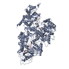
 Sample preparation
 Electron microscopy imaging
 Processing