 Movie
Movie Controller

[English] 日本語
 Yorodumi
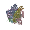
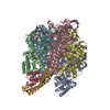
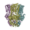
- PDB-5m3l: Single-particle cryo-EM using alignment by classification (ABC): ... -
+ Open data

- Basic information
| Entry | Database: PDB / ID: 5m3l | |||||||||||||||||||||
|---|---|---|---|---|---|---|---|---|---|---|---|---|---|---|---|---|---|---|---|---|---|---|
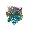
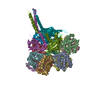
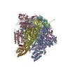
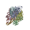
| Title | Single-particle cryo-EM using alignment by classification (ABC): the structure of Lumbricus terrestris hemoglobin | |||||||||||||||||||||
 Components Components |
| |||||||||||||||||||||
 Keywords Keywords |  OXYGEN TRANSPORT / Lumbricus terrestris / hemoglobin / oxygen carrier / erythrocruorin OXYGEN TRANSPORT / Lumbricus terrestris / hemoglobin / oxygen carrier / erythrocruorin | |||||||||||||||||||||
| Function / homology |  Function and homology informationhemoglobin complex / oxygen carrier activity / oxygen binding / iron ion binding / heme binding / extracellular region / metal ion binding Function and homology informationhemoglobin complex / oxygen carrier activity / oxygen binding / iron ion binding / heme binding / extracellular region / metal ion bindingSimilarity search - Function | |||||||||||||||||||||
| Biological species |  Lumbricus terrestris (common earthworm) Lumbricus terrestris (common earthworm) | |||||||||||||||||||||
| Method | ELECTRON MICROSCOPY / single particle reconstruction / cryo EM / Resolution: 3.8 Å | |||||||||||||||||||||
 Authors Authors | Afanasyev, P. / Linnemayr-Seer, C. / Ravelli, R.B.G. / Matadeen, R. / De Carlo, S. / Alewijnse, B. / Portugal, R.V. / Pannu, N.S. / Schatz, M. / van Heel, M. | |||||||||||||||||||||
| Funding support |  Netherlands, Netherlands,  United Kingdom, United Kingdom,  Brazil, 6items Brazil, 6items
| |||||||||||||||||||||
 Citation Citation | Journal: IUCrJ / Year: 2017 Title: Single-particle cryo-EM using alignment by classification (ABC): the structure of haemoglobin. Authors: Pavel Afanasyev / Charlotte Seer-Linnemayr / Raimond B G Ravelli / Rishi Matadeen / Sacha De Carlo / Bart Alewijnse / Rodrigo V Portugal / Navraj S Pannu / Michael Schatz / Marin van Heel /   Abstract: Single-particle cryogenic electron microscopy (cryo-EM) can now yield near-atomic resolution structures of biological complexes. However, the reference-based alignment algorithms commonly used in ...Single-particle cryogenic electron microscopy (cryo-EM) can now yield near-atomic resolution structures of biological complexes. However, the reference-based alignment algorithms commonly used in cryo-EM suffer from reference bias, limiting their applicability (also known as the 'Einstein from random noise' problem). Low-dose cryo-EM therefore requires robust and objective approaches to reveal the structural information contained in the extremely noisy data, especially when dealing with small structures. A reference-free pipeline is presented for obtaining near-atomic resolution three-dimensional reconstructions from heterogeneous ('four-dimensional') cryo-EM data sets. The methodologies integrated in this pipeline include camera correction, movie-based full-data-set contrast transfer function determination, movie-alignment algorithms, (Fourier-space) multivariate statistical data compression and unsupervised classification, 'random-startup' three-dimensional reconstructions, four-dimensional structural refinements and Fourier shell correlation criteria for evaluating anisotropic resolution. The procedures exclusively use information emerging from the data set itself, without external 'starting models'. Euler-angle assignments are performed by angular reconstitution rather than by the inherently slower projection-matching approaches. The comprehensive 'ABC-4D' pipeline is based on the two-dimensional reference-free 'alignment by classification' (ABC) approach, where similar images in similar orientations are grouped by unsupervised classification. Some fundamental differences between X-ray crystallography single-particle cryo-EM data collection and data processing are discussed. The structure of the giant haemoglobin from at a global resolution of ∼3.8 Å is presented as an example of the use of the ABC-4D procedure. | |||||||||||||||||||||
| History |
|
- Structure visualization
| Movie |
Movie viewer |
|---|---|
| Structure viewer | Molecule: MolmilJmol/JSmol |
- Downloads & links
-Download
| PDBx/mmCIF format | 5m3l.cif.gz | 484.9 KB | Display | PDBx/mmCIF format |
|---|---|---|---|---|
| PDB format | pdb5m3l.ent.gz | 402.1 KB | Display | PDB format |
| PDBx/mmJSON format | 5m3l.json.gz | Tree view | PDBx/mmJSON format | |
| Others |  Other downloads Other downloads |
-Validation report
| Arichive directory | https://data.pdbj.org/pub/pdb/validation_reports/m3/5m3lftp://data.pdbj.org/pub/pdb/validation_reports/m3/5m3l | HTTPS FTP |
|---|
-Related structure data
| Related structure data |  3434MC M: map data used to model this data C: citing same article ( |
|---|---|
| Similar structure data |

-Links
 PDBj
PDBj


- Assembly
| Deposited unit | 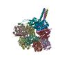
| ||||||||||||||||||||||||||||||||||||||||||||||||||||||||||||||||||||||||||||||||||||||||||||||||||||||||||||||||||||||||||||||||||||||||||||||||||||||||||||||||||||||||||||||||||||||||||||||||||||||||||||||||||||||||||||||||||||||||||||||
|---|---|---|---|---|---|---|---|---|---|---|---|---|---|---|---|---|---|---|---|---|---|---|---|---|---|---|---|---|---|---|---|---|---|---|---|---|---|---|---|---|---|---|---|---|---|---|---|---|---|---|---|---|---|---|---|---|---|---|---|---|---|---|---|---|---|---|---|---|---|---|---|---|---|---|---|---|---|---|---|---|---|---|---|---|---|---|---|---|---|---|---|---|---|---|---|---|---|---|---|---|---|---|---|---|---|---|---|---|---|---|---|---|---|---|---|---|---|---|---|---|---|---|---|---|---|---|---|---|---|---|---|---|---|---|---|---|---|---|---|---|---|---|---|---|---|---|---|---|---|---|---|---|---|---|---|---|---|---|---|---|---|---|---|---|---|---|---|---|---|---|---|---|---|---|---|---|---|---|---|---|---|---|---|---|---|---|---|---|---|---|---|---|---|---|---|---|---|---|---|---|---|---|---|---|---|---|---|---|---|---|---|---|---|---|---|---|---|---|---|---|---|---|---|---|---|---|---|---|---|---|---|---|---|---|---|---|---|---|---|
| 1 |
| ||||||||||||||||||||||||||||||||||||||||||||||||||||||||||||||||||||||||||||||||||||||||||||||||||||||||||||||||||||||||||||||||||||||||||||||||||||||||||||||||||||||||||||||||||||||||||||||||||||||||||||||||||||||||||||||||||||||||||||||
| Symmetry | Point symmetry: (Schoenflies symbol: D6 (2x6 fold dihedral)) | ||||||||||||||||||||||||||||||||||||||||||||||||||||||||||||||||||||||||||||||||||||||||||||||||||||||||||||||||||||||||||||||||||||||||||||||||||||||||||||||||||||||||||||||||||||||||||||||||||||||||||||||||||||||||||||||||||||||||||||||
| Noncrystallographic symmetry (NCS) | NCS domain:
NCS domain segments:
NCS ensembles :
|
-Components
-Extracellular globin- ... , 3 types, 9 molecules AEIBFJCGK
| #1: Protein | Mass: 17566.990 Da / Num. of mol.: 3 / Source method: isolated from a natural source / Source: (natural) Lumbricus terrestris (common earthworm) / References: UniProt: P13579#2: Protein | Mass: 16268.229 Da / Num. of mol.: 3 / Source method: isolated from a natural source / Source: (natural) Lumbricus terrestris (common earthworm) / References: UniProt: P02218#3: Protein | Mass: 17331.793 Da / Num. of mol.: 3 / Source method: isolated from a natural source / Source: (natural) Lumbricus terrestris (common earthworm) / References: UniProt: P11069 |
|---|
-Protein , 2 types, 4 molecules DHLM
| #4: Protein | Mass: 15988.263 Da / Num. of mol.: 3 / Source method: isolated from a natural source / Source: (natural) Lumbricus terrestris (common earthworm) / References: UniProt: O61233#5: Protein | | Mass: 24936.734 Da / Num. of mol.: 1 / Source method: isolated from a natural source / Source: (natural) Lumbricus terrestris (common earthworm) / References: UniProt: Q9GV76 |
|---|
-Extracellular hemoglobin linker ... , 2 types, 2 molecules NO
| #6: Protein | Mass: 25085.037 Da / Num. of mol.: 1 / Source method: isolated from a natural source / Source: (natural) Lumbricus terrestris (common earthworm) / References: UniProt: Q2I743 |
|---|---|
| #7: Protein | Mass: 24486.023 Da / Num. of mol.: 1 / Source method: isolated from a natural source / Source: (natural) Lumbricus terrestris (common earthworm) / References: UniProt: Q2I742 |
-Non-polymers , 1 types, 12 molecules 
| #8: Chemical | ChemComp-HEM / Heme B Mass: 616.487 Da / Num. of mol.: 12 / Source method: obtained synthetically / Formula: C34H32FeN4O4 Mass: 616.487 Da / Num. of mol.: 12 / Source method: obtained synthetically / Formula: C34H32FeN4O4 |
|---|
-Experimental details
-Experiment
| Experiment | Method: ELECTRON MICROSCOPY |
|---|---|
| EM experiment | Aggregation state: PARTICLE / 3D reconstruction method: single particle reconstruction |
- Sample preparation
| Component | Name: Giant worm hemoglobin / Type: COMPLEX / Entity ID: all / Source: NATURAL | ||||||||||||
|---|---|---|---|---|---|---|---|---|---|---|---|---|---|
| Molecular weight | Value: 3.6 MDa / Experimental value: NO | ||||||||||||
| Source (natural) | Organism: Lumbricus terrestris (common earthworm) / Organ: Hemolymph | ||||||||||||
| Buffer solution | pH: 7 | ||||||||||||
| Buffer component |
| ||||||||||||
| Specimen | Embedding applied: NO / Shadowing applied: NO / Staining applied: NO / Vitrification applied: YES | ||||||||||||
| Specimen support | Grid material: COPPER / Grid type: Quantifoil R2/2 | ||||||||||||
| Vitrification | Instrument: FEI VITROBOT MARK IV / Cryogen name: ETHANE / Humidity: 100 % / Chamber temperature: 298 K |
- Electron microscopy imaging
| Experimental equipment |  Model: Titan Krios / Image courtesy: FEI Company |
|---|---|
| Microscopy | Model: FEI TITAN KRIOS |
| Electron gun | Electron source: FIELD EMISSION GUN / Accelerating voltage: 300 kV / Illumination mode: FLOOD BEAM |
| Electron lens | Mode: BRIGHT FIELDBright-field microscopy / Calibrated magnification: 59000 X / Nominal defocus max: 1200 nm / Nominal defocus min: 1000 nm / Cs: 0.02 mm / Alignment procedure: ZEMLIN TABLEAU |
| Specimen holder | Cryogen: NITROGEN / Specimen holder model: FEI TITAN KRIOS AUTOGRID HOLDER |
| Image recording | Electron dose: 6 e/Å2 / Detector mode: INTEGRATING / Film or detector model: FEI FALCON II (4k x 4k) / Num. of real images: 5235 |
| Image scans | Movie frames/image: 7 / Used frames/image: 2-5 |
- Processing
| Software | Name: REFMAC / Version: 5.8.0155 / Classification: refinement | ||||||||||||||||||||||||||||||||||||||||||||||||||||||||||||||||||||||||||||||||||||||||||||||||||||||||||
|---|---|---|---|---|---|---|---|---|---|---|---|---|---|---|---|---|---|---|---|---|---|---|---|---|---|---|---|---|---|---|---|---|---|---|---|---|---|---|---|---|---|---|---|---|---|---|---|---|---|---|---|---|---|---|---|---|---|---|---|---|---|---|---|---|---|---|---|---|---|---|---|---|---|---|---|---|---|---|---|---|---|---|---|---|---|---|---|---|---|---|---|---|---|---|---|---|---|---|---|---|---|---|---|---|---|---|---|
| EM software |
| ||||||||||||||||||||||||||||||||||||||||||||||||||||||||||||||||||||||||||||||||||||||||||||||||||||||||||
| CTF correction | Details: Unsupervised MSA classification of amplitude spectra throughout the full data set ("Full data set CTF correction") Type: PHASE FLIPPING ONLY | ||||||||||||||||||||||||||||||||||||||||||||||||||||||||||||||||||||||||||||||||||||||||||||||||||||||||||
| Particle selection | Num. of particles selected: 319746 | ||||||||||||||||||||||||||||||||||||||||||||||||||||||||||||||||||||||||||||||||||||||||||||||||||||||||||
| Symmetry | Point symmetry: D6 (2x6 fold dihedral) | ||||||||||||||||||||||||||||||||||||||||||||||||||||||||||||||||||||||||||||||||||||||||||||||||||||||||||
| 3D reconstruction | Resolution: 3.8 Å / Resolution method: FSC 1/2 BIT CUT-OFF / Num. of particles: 75000 / Algorithm: EXACT BACK PROJECTION Details: FSC as per the original definition (Harauz & van Heel 1986). Num. of class averages: 75000 / Symmetry type: POINT | ||||||||||||||||||||||||||||||||||||||||||||||||||||||||||||||||||||||||||||||||||||||||||||||||||||||||||
| Atomic model building | Protocol: RIGID BODY FIT / Space: REAL / Target criteria: Cross-correlation coefficient | ||||||||||||||||||||||||||||||||||||||||||||||||||||||||||||||||||||||||||||||||||||||||||||||||||||||||||
| Refinement | Resolution: 3.8→146.67 Å / Cor.coef. Fo:Fc: 0.833 / Cor.coef. Fo:Fc free: 0.841 / SU B: 36.387 / SU ML: 0.487 / Cross valid method: THROUGHOUT / ESU R: 1.633 / ESU R Free: 0.598 Stereochemistry target values: MAXIMUM LIKELIHOOD WITH PHASES Details: HYDROGENS HAVE BEEN ADDED IN THE RIDING POSITIONS
| ||||||||||||||||||||||||||||||||||||||||||||||||||||||||||||||||||||||||||||||||||||||||||||||||||||||||||
| Solvent computation | Ion probe radii: 0.8 Å / Shrinkage radii: 0.8 Å / VDW probe radii: 1.2 Å / Solvent model: MASK | ||||||||||||||||||||||||||||||||||||||||||||||||||||||||||||||||||||||||||||||||||||||||||||||||||||||||||
| Displacement parameters | Biso mean: 129.873 Å2
| ||||||||||||||||||||||||||||||||||||||||||||||||||||||||||||||||||||||||||||||||||||||||||||||||||||||||||
| Refinement step | Cycle: 1 / Total: 19015 | ||||||||||||||||||||||||||||||||||||||||||||||||||||||||||||||||||||||||||||||||||||||||||||||||||||||||||
| Refine LS restraints |
|