stringent response / transcriptional attenuation / endoribonuclease inhibitor activity / RNA-binding transcription regulator activity / positive regulation of ribosome biogenesis / negative regulation of cytoplasmic translation / translational termination / DnaA-L2 complex / translation repressor activity / negative regulation of DNA-templated DNA replication initiation ...stringent response / transcriptional attenuation / endoribonuclease inhibitor activity / RNA-binding transcription regulator activity / positive regulation of ribosome biogenesis / negative regulation of cytoplasmic translation / translational termination / DnaA-L2 complex / translation repressor activity / negative regulation of DNA-templated DNA replication initiation / ribosome assembly / mRNA regulatory element binding translation repressor activity / response to reactive oxygen species / assembly of large subunit precursor of preribosome / cytosolic ribosome assembly / : / regulation of cell growth / DNA-templated transcription termination / response to radiation / mRNA 5'-UTR binding / ribosomal large subunit assembly / large ribosomal subunit rRNA binding / ribosome binding / large ribosomal subunit / 5S rRNA binding / cytoplasmic translation / cytosolic large ribosomal subunit / transferase activity / tRNA binding / negative regulation of translation / rRNA binding / ribosome / structural constituent of ribosome / translation / response to antibiotic / mRNA binding / negative regulation of DNA-templated transcription / DNA binding / RNA binding / zinc ion binding / cytosol / cytoplasm Similarity search - Function
Ribosomal protein L25, short-form / Ribosomal protein L11, bacterial-type / Ribosomal protein L11, conserved site / Ribosomal protein L21, conserved site / Ribosomal protein L21 signature. / Ribosomal protein L11 signature. / Ribosomal protein L16 signature 1. / : / Ribosomal protein L6, conserved site / Ribosomal protein L6 signature 1. ...Ribosomal protein L25, short-form / Ribosomal protein L11, bacterial-type / Ribosomal protein L11, conserved site / Ribosomal protein L21, conserved site / Ribosomal protein L21 signature. / Ribosomal protein L11 signature. / Ribosomal protein L16 signature 1. / : / Ribosomal protein L6, conserved site / Ribosomal protein L6 signature 1. / Ribosomal protein L16, conserved site / Ribosomal protein L16 signature 2. / Ribosomal protein L11, N-terminal / Ribosomal protein L17 signature. / Ribosomal protein L9 signature. / Ribosomal protein L9, bacteria/chloroplast / Ribosomal protein L9, C-terminal / Ribosomal protein L9, C-terminal domain / Ribosomal protein L9, C-terminal domain superfamily / Ribosomal protein L11/L12 / Ribosomal protein L11, C-terminal / Ribosomal protein L11, C-terminal domain superfamily / Ribosomal protein L11/L12, N-terminal domain superfamily / Ribosomal protein L11/L12 / Ribosomal protein L11, N-terminal domain / Ribosomal protein L11, RNA binding domain / Ribosomal L25p family / Ribosomal protein L25 / Ribosomal protein L28/L24 superfamily / Ribosomal protein L36 signature. / Ribosomal protein L25/Gln-tRNA synthetase, N-terminal / Ribosomal protein L32p, bacterial type / Ribosomal protein L25/Gln-tRNA synthetase, anti-codon-binding domain superfamily / Ribosomal protein L9, N-terminal domain superfamily / Ribosomal protein L9 / Ribosomal protein L9, N-terminal / Ribosomal protein L9, N-terminal domain / Ribosomal protein L28 / Ribosomal protein L35, conserved site / Ribosomal protein L35 signature. / Ribosomal protein L33, conserved site / Ribosomal protein L33 signature. / Ribosomal protein L35, non-mitochondrial / Ribosomal protein L5, bacterial-type / Ribosomal protein L6, bacterial-type / Ribosomal protein L18, bacterial-type / Ribosomal protein L19, conserved site / Ribosomal protein L19 signature. / Ribosomal protein L36 / Ribosomal protein L36 superfamily / Ribosomal protein L36 / Ribosomal protein L9/RNase H1, N-terminal / Ribosomal protein L20 signature. / Ribosomal protein L27, conserved site / Ribosomal protein L27 signature. / Ribosomal protein L14P, bacterial-type / Ribosomal protein L34, conserved site / Ribosomal protein L34 signature. / Ribosomal protein L22, bacterial/chloroplast-type / Ribosomal protein L35 / Ribosomal protein L35 superfamily / Ribosomal protein L2, bacterial/organellar-type / Ribosomal protein L35 / Ribosomal L28 family / Ribosomal protein L33 / Ribosomal protein L33 / Ribosomal protein L28/L24 / Ribosomal protein L33 superfamily / : / Ribosomal protein L30, bacterial-type / Ribosomal protein L16 / Ribosomal protein L18 / Ribosomal L18 of archaea, bacteria, mitoch. and chloroplast / L28p-like / Ribosomal protein L20 / Ribosomal protein L20 / Ribosomal protein L20, C-terminal / Ribosomal protein L21 / Ribosomal protein L27 / Ribosomal L27 protein / Ribosomal protein L19 / Ribosomal protein L19 superfamily / Ribosomal protein L19 / Ribosomal proteins 50S L24/mitochondrial 39S L24 / Ribosomal protein L17 / Ribosomal protein L17 superfamily / Ribosomal protein L17 / Ribosomal protein L21-like / L21-like superfamily / Ribosomal prokaryotic L21 protein / Ribosomal L32p protein family / Ribosomal protein L24 / Ribosomal protein L32p / Ribosomal protein L34 / Ribosomal protein L34 / Ribosomal protein L13, bacterial-type / Ribosomal protein L3, bacterial/organelle-type / Ribosomal protein L15, bacterial-type / 50S ribosomal protein uL4 / Ribosomal protein L23/L25, conserved site Similarity search - Domain/homology
ERYTHROMYCIN A / Unknown ligand / : / : / RNA / RNA (> 10) / RNA (> 100) / RNA (> 1000) / Large ribosomal subunit protein uL15 / Large ribosomal subunit protein uL11 ...ERYTHROMYCIN A / Unknown ligand / : / : / RNA / RNA (> 10) / RNA (> 100) / RNA (> 1000) / Large ribosomal subunit protein uL15 / Large ribosomal subunit protein uL11 / Large ribosomal subunit protein bL19 / Large ribosomal subunit protein bL20 / Large ribosomal subunit protein bL27 / Large ribosomal subunit protein bL28 / Large ribosomal subunit protein uL29 / Large ribosomal subunit protein bL32 / Large ribosomal subunit protein bL33 / Large ribosomal subunit protein bL34 / Large ribosomal subunit protein bL35 / Large ribosomal subunit protein bL36A / Large ribosomal subunit protein bL9 / Large ribosomal subunit protein uL13 / Large ribosomal subunit protein uL14 / Large ribosomal subunit protein uL16 / Large ribosomal subunit protein uL23 / Large ribosomal subunit protein bL17 / Large ribosomal subunit protein bL21 / Large ribosomal subunit protein uL30 / Large ribosomal subunit protein uL6 / Large ribosomal subunit protein uL18 / Large ribosomal subunit protein uL2 / Large ribosomal subunit protein uL3 / Large ribosomal subunit protein uL24 / Large ribosomal subunit protein uL4 / Large ribosomal subunit protein uL22 / Large ribosomal subunit protein uL5 / Large ribosomal subunit protein bL25 Similarity search - Component
Biological species
Escherichia coli (E. coli) Streptococcus sanguinis (bacteria)
Method
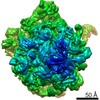
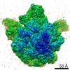
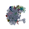
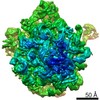
ELECTRON MICROSCOPY / single particle reconstruction / cryo EM / Resolution: 6.6 Å
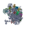
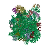
Journal: Nat Commun / Year: 2014 Title: Molecular basis for erythromycin-dependent ribosome stalling during translation of the ErmBL leader peptide. Authors: Stefan Arenz / Haripriya Ramu / Pulkit Gupta / Otto Berninghausen / Roland Beckmann / Nora Vázquez-Laslop / Alexander S Mankin / Daniel N Wilson / Abstract: In bacteria, ribosome stalling during translation of ErmBL leader peptide occurs in the presence of the antibiotic erythromycin and leads to induction of expression of the downstream macrolide ...In bacteria, ribosome stalling during translation of ErmBL leader peptide occurs in the presence of the antibiotic erythromycin and leads to induction of expression of the downstream macrolide resistance methyltransferase ErmB. The lack of structures of drug-dependent stalled ribosome complexes (SRCs) has limited our mechanistic understanding of this regulatory process. Here we present a cryo-electron microscopy structure of the erythromycin-dependent ErmBL-SRC. The structure reveals that the antibiotic does not interact directly with ErmBL, but rather redirects the path of the peptide within the tunnel. Furthermore, we identify a key peptide-ribosome interaction that defines an important relay pathway from the ribosomal tunnel to the peptidyltransferase centre (PTC). The PTC of the ErmBL-SRC appears to adopt an uninduced state that prevents accommodation of Lys-tRNA at the A-site, thus providing structural basis for understanding how the drug and the nascent peptide cooperate to inhibit peptide bond formation and induce translation arrest.
History
Deposition
Oct 23, 2013
Deposition site: RCSB / Processing site: RCSB
Revision 1.0
Mar 26, 2014
Provider: repository / Type: Initial release
Revision 1.1
Apr 2, 2014
Group: Database references
Revision 1.2
Jul 18, 2018
Group: Data collection / Category: em_software / Item: _em_software.image_processing_id
In the structure databanks used in Yorodumi, some data are registered as the other names, "COVID-19 virus" and "2019-nCoV". Here are the details of the virus and the list of structure data.
Jan 31, 2019. EMDB accession codes are about to change! (news from PDBe EMDB page)
EMDB accession codes are about to change! (news from PDBe EMDB page)
The allocation of 4 digits for EMDB accession codes will soon come to an end. Whilst these codes will remain in use, new EMDB accession codes will include an additional digit and will expand incrementally as the available range of codes is exhausted. The current 4-digit format prefixed with “EMD-” (i.e. EMD-XXXX) will advance to a 5-digit format (i.e. EMD-XXXXX), and so on. It is currently estimated that the 4-digit codes will be depleted around Spring 2019, at which point the 5-digit format will come into force.
The EM Navigator/Yorodumi systems omit the EMD- prefix.
Related info.:Q: What is EMD? / ID/Accession-code notation in Yorodumi/EM Navigator
Yorodumi is a browser for structure data from EMDB, PDB, SASBDB, etc.
This page is also the successor to EM Navigator detail page, and also detail information page/front-end page for Omokage search.
The word "yorodu" (or yorozu) is an old Japanese word meaning "ten thousand". "mi" (miru) is to see.
Related info.:EMDB / PDB / SASBDB / Comparison of 3 databanks / Yorodumi Search / Aug 31, 2016. New EM Navigator & Yorodumi / Yorodumi Papers / Jmol/JSmol / Function and homology information / Changes in new EM Navigator and Yorodumi
 Movie
Movie Controller


 Open data
 Basic information
 Components
 Keywords
 erythromycin /
  Function and homology information
 Authors
 Citation

 Structure visualization
 Downloads & links
 Other downloads
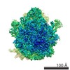
 PDBj
PDBj


 Assembly
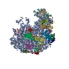
 Mass: 733.927 Da / Num. of mol.: 1 / Source method: obtained synthetically / Formula: C37H67NO13 / Comment: antibiotic*YM
 Sample preparation
 Electron microscopy imaging
 Processing
