 Movie
Movie Controller


+ Open data

- Basic information
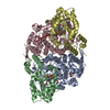
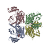
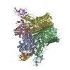
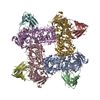
| Entry | Database: PDB / ID: 2xky | ||||||
|---|---|---|---|---|---|---|---|
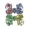
| Title | Single particle analysis of Kir2.1NC_4 in negative stain | ||||||
 Components Components | INWARD RECTIFIER POTASSIUM CHANNEL 2 Inward-rectifier potassium channel Inward-rectifier potassium channel | ||||||
 Keywords Keywords | METAL TRANSPORT / ION CHANNEL / MEMBRANE PROTEIN | ||||||
| Function / homology |  Function and homology information Function and homology informationClassical Kir channels / regulation of skeletal muscle contraction via regulation of action potential / cardiac muscle cell action potential / relaxation of skeletal muscle / Phase 4 - resting membrane potential / magnesium ion transport / voltage-gated potassium channel activity involved in cardiac muscle cell action potential repolarization / membrane repolarization during action potential / Activation of G protein gated Potassium channels / Inhibition of voltage gated Ca2+ channels via Gbeta/gamma subunits ...Classical Kir channels / regulation of skeletal muscle contraction via regulation of action potential / cardiac muscle cell action potential / relaxation of skeletal muscle / Phase 4 - resting membrane potential / magnesium ion transport / voltage-gated potassium channel activity involved in cardiac muscle cell action potential repolarization / membrane repolarization during action potential / Activation of G protein gated Potassium channels / Inhibition of voltage gated Ca2+ channels via Gbeta/gamma subunits / membrane repolarization during cardiac muscle cell action potential / regulation of membrane repolarization / positive regulation of potassium ion transmembrane transport / inward rectifier potassium channel activity / cardiac muscle cell action potential involved in contraction / regulation of cardiac muscle cell contraction / regulation of monoatomic ion transmembrane transport / relaxation of cardiac muscle / potassium ion import across plasma membrane / regulation of heart rate by cardiac conduction / intercalated disc / voltage-gated potassium channel complex / potassium ion transmembrane transport / phosphatidylinositol-4,5-bisphosphate binding / T-tubule / potassium ion transport / cellular response to mechanical stimulus / postsynaptic membrane / protein homotetramerization / dendritic spine / dendrite / neuronal cell body / glutamatergic synapse / membrane / identical protein binding / plasma membraneSimilarity search - Function | ||||||
| Biological species |  MUS MUSCULUS (house mouse) MUS MUSCULUS (house mouse) | ||||||
| Method | ELECTRON MICROSCOPY / SOLUTION SCATTERING / single particle reconstruction / negative staining / Resolution: 17.2 Å | ||||||
| Model type details | CA ATOMS ONLY, CHAIN I, J, K, L | ||||||
 Authors Authors | Fomina, S. / Howard, T.D. / Sleator, O.K. / Golovanova, M. / O'Ryan, L. / Leyland, M.L. / Grossmann, J.G. / Collins, R.F. / Prince, S.M. | ||||||
 Citation Citation | Journal: Biochim Biophys Acta / Year: 2011 Title: Self-directed assembly and clustering of the cytoplasmic domains of inwardly rectifying Kir2.1 potassium channels on association with PSD-95. Authors: Svetlana Fomina / Tina D Howard / Olivia K Sleator / Marina Golovanova / Liam O'Ryan / Mark L Leyland / J Günter Grossmann / Richard F Collins / Stephen M Prince /  Abstract: The interaction of the extra-membranous domain of tetrameric inwardly rectifying Kir2.1 ion channels (Kir2.1NC(4)) with the membrane associated guanylate kinase protein PSD-95 has been studied using ...The interaction of the extra-membranous domain of tetrameric inwardly rectifying Kir2.1 ion channels (Kir2.1NC(4)) with the membrane associated guanylate kinase protein PSD-95 has been studied using Transmission Electron Microscopy in negative stain. Three types of complexes were observed in electron micrographs corresponding to a 1:1 complex, a large self-enclosed tetrad complex and extended chains of linked channel domains. Using models derived from small angle X-ray scattering experiments in which high resolution structures from X-ray crystallographic and Nuclear Magnetic Resonance studies are positioned, the envelopes from single particle analysis can be resolved as a Kir2.1NC(4):PSD-95 complex and a tetrad of this unit (Kir2.1NC(4):PSD-95)(4). The tetrad complex shows the close association of the Kir2.1 cytoplasmic domains and the influence of PSD-95 mediated self-assembly on the clustering of these channels. #1: Journal: Nat Neurosci / Year: 2005Title: Cytoplasmic domain structures of Kir2.1 and Kir3.1 show sites for modulating gating and rectification. Authors: Scott Pegan / Christine Arrabit / Wei Zhou / Witek Kwiatkowski / Anthony Collins / Paul A Slesinger / Senyon Choe /  Abstract: N- and C-terminal cytoplasmic domains of inwardly rectifying K (Kir) channels control the ion-permeation pathway through diverse interactions with small molecules and protein ligands in the cytoplasm. ...N- and C-terminal cytoplasmic domains of inwardly rectifying K (Kir) channels control the ion-permeation pathway through diverse interactions with small molecules and protein ligands in the cytoplasm. Two new crystal structures of the cytoplasmic domains of Kir2.1 (Kir2.1(L)) and the G protein-sensitive Kir3.1 (Kir3.1(S)) channels in the absence of PIP(2) show the cytoplasmic ion-permeation pathways occluded by four cytoplasmic loops that form a girdle around the central pore (G-loop). Significant flexibility of the pore-facing G-loop of Kir2.1(L) and Kir3.1(S) suggests a possible role as a diffusion barrier between cytoplasmic and transmembrane pores. Consistent with this, mutations of the G-loop disrupted gating or inward rectification. Structural comparison shows a di-aspartate cluster on the distal end of the cytoplasmic pore of Kir2.1(L) that is important for modulating inward rectification. Taken together, these results suggest the cytoplasmic domains of Kir channels undergo structural changes to modulate gating and inward rectification. | ||||||
| History |
|
- Structure visualization
| Movie |
Movie viewer |
|---|---|
| Structure viewer | Molecule: MolmilJmol/JSmol |
- Downloads & links
-Download
| PDBx/mmCIF format | 2xky.cif.gz | 42.2 KB | Display | PDBx/mmCIF format |
|---|---|---|---|---|
| PDB format | pdb2xky.ent.gz | 27.3 KB | Display | PDB format |
| PDBx/mmJSON format | 2xky.json.gz | Tree view | PDBx/mmJSON format | |
| Others |  Other downloads Other downloads |
-Validation report
| Arichive directory | https://data.pdbj.org/pub/pdb/validation_reports/xk/2xkyftp://data.pdbj.org/pub/pdb/validation_reports/xk/2xky | HTTPS FTP |
|---|
-Related structure data
| Related structure data | 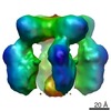 1764MC 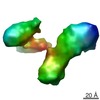 1761C 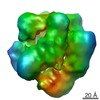 1765C 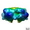 1766C  2xkxC M: map data used to model this data C: citing same article ( |
|---|---|
| Similar structure data |
-Links
 PDBj
PDBj
- Assembly
| Deposited unit | 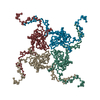
| ||||||||||||
|---|---|---|---|---|---|---|---|---|---|---|---|---|---|
| 1 |
| ||||||||||||
| Noncrystallographic symmetry (NCS) | NCS oper:
|
-Components
| #1: Protein | Inward-rectifier potassium channel / POTASSIUM CHANNEL / INWARDLY RECTIFYING SUBFAMILY J MEMBER 2 / INWARD RECTIFIER K(+) CHANNEL KIR2.1 / IRK-1 / Coordinate model: Cα atoms only Mass: 34980.273 Da / Num. of mol.: 4 / Fragment: KIR2.1 CYTOPLASMIC DOMAIN, RESIDUES 1-67,189-428 Source method: isolated from a genetically manipulated source Details: HOMOTETRAMER OF FUSED N, C TERMINI / Source: (gene. exp.) MUS MUSCULUS (house mouse) / Plasmid: PET15B / Production host:  ESCHERICHIA COLI (E. coli) / Strain (production host): BL21 / References: UniProt: P35561 ESCHERICHIA COLI (E. coli) / Strain (production host): BL21 / References: UniProt: P35561 |
|---|
-Experimental details
-Experiment
| Experiment |
| |||
|---|---|---|---|---|
| EM experiment | Aggregation state: PARTICLE / 3D reconstruction method: single particle reconstruction |
- Sample preparation
| Component | Name: MOUSE KIR2.1, CYTOPLASMIC DOMAIN / Type: COMPLEX |
|---|---|
| Buffer solution | Name: 20MM TRIS/HCL, 150MM NACL, 1MM REDUCED GSH, 1MM EDTA, 50MM L-GLUTAMIC ACID, 50MM L-ARGININE pH: 7.5 Details: 20MM TRIS/HCL, 150MM NACL, 1MM REDUCED GSH, 1MM EDTA, 50MM L-GLUTAMIC ACID, 50MM L-ARGININE |
| Specimen | Conc.: 1 mg/ml / Embedding applied: NO / Shadowing applied: NO / Staining applied: YES / Vitrification applied: NO |
| EM staining | Type: NEGATIVE / Material: Uranyl Acetate |
| Specimen support | Details: CARBON |
-Data collection
| Microscopy | Model: FEI TECNAI 10 / Details: LOW DOSE | |||||||||||||||||||||||||||||||||||||||
|---|---|---|---|---|---|---|---|---|---|---|---|---|---|---|---|---|---|---|---|---|---|---|---|---|---|---|---|---|---|---|---|---|---|---|---|---|---|---|---|---|
| Electron gun | Electron source: TUNGSTEN HAIRPIN / Accelerating voltage: 100 kV / Illumination mode: FLOOD BEAM | |||||||||||||||||||||||||||||||||||||||
| Electron lens | Mode: BRIGHT FIELDBright-field microscopy / Nominal defocus max: 1250 nm / Nominal defocus min: 600 nm / Cs: 2 mm | |||||||||||||||||||||||||||||||||||||||
| Specimen holder | Tilt angle max: 0.1 ° / Tilt angle min: 0 ° | |||||||||||||||||||||||||||||||||||||||
| Image recording | Film or detector model: GENERIC GATAN | |||||||||||||||||||||||||||||||||||||||
| Image scans | Num. digital images: 22 | |||||||||||||||||||||||||||||||||||||||
| Radiation wavelength | Relative weight: 1 | |||||||||||||||||||||||||||||||||||||||
| Soln scatter |
|
- Processing
| EM software |
| ||||||||||||
|---|---|---|---|---|---|---|---|---|---|---|---|---|---|
| CTF correction | Details: PARAMETERS DETERMINED USING SCATTERING CURVE | ||||||||||||
| Symmetry | Point symmetry: C4 (4 fold cyclic) | ||||||||||||
| 3D reconstruction | Resolution: 17.2 Å / Num. of particles: 49012 / Nominal pixel size: 2.93 Å / Actual pixel size: 2.93 Å Details: THE COORDINATES DEPOSITED ARE FROM A COMBINED SAXS/EM STUDY. THE DOMAINS IN THE PROTEINS (HIGH RESOLUTION STRUCTURES FROM THE PDB) ARE POSITIONED RELATIVE TO ONE ANOTHER USING A SAXS CURVE, ...Details: THE COORDINATES DEPOSITED ARE FROM A COMBINED SAXS/EM STUDY. THE DOMAINS IN THE PROTEINS (HIGH RESOLUTION STRUCTURES FROM THE PDB) ARE POSITIONED RELATIVE TO ONE ANOTHER USING A SAXS CURVE, THIS COMPOSITE STRUCTURE IS THEN FITTED INTO AN EM MAP. MODEL GENERATED FROM SAXS REFINEMENT USING BUNCH. PETOUKHOV, M. V. AND SVERGUN, D. I. (2005). BIOPHYS J 89, 1237-50. SUBMISSION BASED ON EXPERIMENTAL DATA FROM EMDB EMD-1764. (DEPOSITION ID: 7401). Symmetry type: POINT | ||||||||||||
| Atomic model building | Protocol: RIGID BODY FIT / Space: REAL Details: METHOD--A MAP WAS GENERATED FROM THE SAXS MODEL COORDINATES AT A RESOLUTION MATCHING THE EXPERIMENTAL MAP. T HIS CALCULATED MAP WAS FITTED INTO THE EXPERIMENTAL MAP BY MAXIMIZING THE CROSS- ...Details: METHOD--A MAP WAS GENERATED FROM THE SAXS MODEL COORDINATES AT A RESOLUTION MATCHING THE EXPERIMENTAL MAP. T HIS CALCULATED MAP WAS FITTED INTO THE EXPERIMENTAL MAP BY MAXIMIZING THE CROSS-CORRELATION WITH THE EXPERIMENTAL MAP. THE COORDINATES WERE THEN REPLACED IN THE CALCULATED MAP TO GENERATE THE FINAL ENTRY. REFINEMENT PROTOCOL--DOCKED USING CHIMERA | ||||||||||||
| Atomic model building | PDB-ID: 1U4F | ||||||||||||
| Refinement | Highest resolution: 17.2 Å | ||||||||||||
| Refinement step | Cycle: LAST / Highest resolution: 17.2 Å
| ||||||||||||
| Soln scatter model | Method: RIGID BODY MODELLING Conformer selection criteria: CONFORMERS WERE CONSISTENT, BEST AGREEMENT WITH EXPERIMENTAL DATA DEPOSITED (CHI=2.6). Details: NUMBER OF TIME FRAMES USED 25(60S, 4.25M CAMERA), 35(60S, 1M CAMERA). PROTEIN CONCENTRATION 0.5 MG/ML (4.25M CAMERA), 4.8 MG/ML (1M CAMERA) Entry fitting list: PROGRAM PRE-BUNCH, 1U4F, C_4 SYMMETRY + SEQUENCE DATA Num. of conformers calculated: 20 / Num. of conformers submitted: 1 / Software author list: PETOUKHOV, M. V. & SVERGUN, D. I. / Software list: SASREF7/BUNCH8 |
